
 Laboratorial diagnosis of biliary peritonitis secondary to cystic duct rupture in a dog - case report
ABSTRACT
Biliary peritonitis results from damage to the biliary tract, causing exposure of the peritoneal surface to bile with consequent tissue inflammation. Effusion analysis is essential, as it can provide definitive findings for diagnosis. An adult female, mixed-breed dog was attended presenting jaundice, ascites, emesis, dyspnea, hypothermia, and hypotension. The most significant laboratory findings were leukocytosis due to neutrophilia with a left shift, and serum activities of alanine aminotransferase (ALT) and alkaline phosphatase (ALP) above the reference values. Abdominal effusion analysis showed a predominance of intact neutrophils, macrophages containing bluish intracytoplasmic material, golden-yellow pigments on the background of the slide and free and intracellular golden crystals, both suggestive of biliary origin. Total bilirubin concentration in abdominal effusion showed value more than five times higher than serum concentration, indicating, in association with the other findings described above, biliary peritonitis. Abdominal ultrasound revealed a slightly distended gallbladder, with no other significant changes. The dog was euthanized, and necropsy revealed an irregularity in the cystic duct consistent with rupture. The present case highlights the relevance of analyzing abdominal effusion for the diagnosis of biliary peritonitis, especially when ultrasonographic findings are not conclusive.
Keywords:
jaundice; biliary pigments; abdominal effusion; hyperbilirubinemia; gallbladder
RESUMO
A peritonite biliar é resultante de dano no trato biliar, ocasionando a exposição da superfície peritoneal à bile com consequente inflamação tecidual. A análise da efusão é essencial, podendo apresentar achados definitivos para o diagnóstico. Uma cadela adulta, sem raça definida (SRD), foi atendida apresentando mucosas ictéricas, ascite, êmese, dispneia, hipotermia e hipotensão. Nos exames laboratoriais, as alterações de maior relevância encontradas foram leucocitose por neutrofilia com desvio à esquerda e atividade sérica de alanina aminotransferase (ALT) e fosfatase alcalina (ALP) acima dos valores de referência. Na análise da efusão abdominal, observou-se predomínio de neutrófilos íntegros, macrófagos contendo material azulado intracitoplasmático, pigmentos amarelo-ouro ao fundo da lâmina e cristais dourados livres e intracelulares, ambos sugestivos de origem biliar. A concentração de bilirrubina total da efusão abdominal apresentou valor mais de cinco vezes superior à concentração sérica, indicando, em associação aos demais achados citados anteriormente, peritonite biliar. Na ultrassonografia abdominal, observou-se a vesícula biliar minimamente repleta, sem demais alterações. Foi realizada a eutanásia do animal e, na necrópsia, foi observada alteração em ducto cístico compatível com ruptura. O presente caso destaca a relevância da análise da efusão abdominal no diagnóstico de peritonite biliar, principalmente na ausência de alterações específicas no exame ultrassonográfico.
Palavras-chave:
icterícia; pigmentos biliares; efusão abdominal; hiperbilirrubinemia; vesícula biliar
INTRODUCTION
Cases of biliary peritonitis have been reported since 1949 (Thompson and Sherman, 2021), generally caused by gallbladder rupture due to mucocele or trauma. However, causes such as obstruction of the common bile duct due to cholelithiasis, neoplasia, necrosis and cholecystitis are also reported (Watson, 2020; Pavone et al., 2023). Fever, pale stools, abdominal distension and jaundice are possible clinical presentations, which may progress to signs of hypovolemia, systemic inflammatory response syndrome and sepsis (Watson, 2020; Thompson and Sherman, 2021).
Serum alkaline phosphatase (ALP) and alanine aminotransferase (ALT) activities above reference values, leukocytosis due to neutrophilia and hyperbilirubinemia are expected laboratory findings during biliary peritonitis (Wilson et al., 2021; Thompson and Sherman, 2021; Linhares et al., 2022). However, Guess et al. (2015) and Wilson et al. (2021) observed, in their studies, the occurrence of biliary peritonitis associated with normobilirubinemia.
Physical-chemical and cytological analysis of the effusion is essential to assist in the diagnosis if the patient presents abdominal effusion. The color of the liquid varies between dark orange, yellow, or green (Watson, 2020; Wilson et al., 2021). However, Francisco et al. (2024) reported that effusions due to biliary peritonitis are dark brown, while Pavone et al. (2023) consider all the mentioned colors to be possible and highlight that the effusion may assume a reddish tone due to the presence of erythrocytes.
Total nucleated cell count (TNCC) tends to be high in the effusion, with a predominance of intact neutrophils, which may be degenerated if there is septic biliary peritonitis. In these cases, intracytoplasmic bacteria can be visualized, and serum glucose level tends to be considerably higher than effusion (Watson, 2020; Francisco et al., 2024). Bilirubin pigments can be visualized in the cytological evaluation, being characterized by greenish, golden yellow, or brown amorphous material present at the bottom of the slide and in the cytoplasm of macrophages (Thompson and Sherman, 2021; Wilson et al., 2021; Pavone et al., 2023).
The measurement of bilirubin in the effusion is the most useful tool in the diagnosis of biliary peritonitis (Linhares et al., 2022), and the bilirubin concentration in the effusion must be higher than in the blood (Rebar and Thompson, 2011; Watson, 2020). Thompson and Sherman (2021) and Pavone et al. (2023) consider that ratios greater than 1:1 and 1:2, respectively, are suggestive of biliary peritonitis.
Abdominal ultrasound is the most used diagnostic tool when there is not enough abdominal fluid for analysis, however, it presents low sensitivity. Observed signs compatible with biliary peritonitis include loss of continuity of the gallbladder wall, with increased adjacent echogenicity and the presence of fluid and free striated material in the peritoneal cavity (Thompson and Sherman, 2021).
Thus, the present study seeks to report a clinical case from a dog attended at the Veterinary Hospital of State University of Maringá, Brazil, presenting intense jaundice and abdominal effusion which, when subjected to analysis, allowed the suggestive diagnosis of biliary peritonitis.
CASUISTRY
An adult, female, mixed-breed dog, weighing 18kg, presenting weakness, dyspnea, emesis, and jaundice, was admitted on August 7, 2024, at Veterinary Hospital of State University of Maringá for clinical emergency care. The animal had been recently adopted; thus its history was unknown. Physical examination showed intense jaundice with yellowish discoloration of mucous membranes (Fig. 1), capillary refill time (CRT) greater than two seconds, hypothermia (35.9ºC), hypotension (80 mmHg systolic blood pressure measured on Delta Life Veterinary Vascular Doppler DL330), wheezing, bulging abdomen, dehydration, sialorrhea and low responsiveness to external stimuli. Based on this information, the differential diagnoses of leptospirosis, hepatitis, babesiosis, and immune-mediated hemolytic anemia were established.
Blood sample was collected to perform a hemogram (hematocrit determined by microcentrifugation, total WBC count performed using neubauer counting chamber, and differential WBC count and platelet count performed in the blood smear) and serum biochemistry analysis (Bioplus Semiautomatic Biochemical Analyzer BIO-2000), and an abdominocentesis was executed, draining approximately 3 liters of abdominal fluid, which was also analyzed. The patient was admitted to the infectious diseases sector for treatment, considering that leptospirosis was suspected. Initially, antibiotic therapy was prescribed with ceftriaxone (50.0mg/kg/TID), dexamethasone at an immunosuppressive dose (6.3mg/kg/BID), ondansetron (0.5mg/kg/BID), maropitant hydrochloride (0.1 mg/kg/SID) and dipyrone (25.0mg/kg/BID). Urethral catheterization was performed, aiming to reduce contact with the patient's urine. In addition, fluid therapy (Lactated Ringer's) was performed to replace dehydration and losses.

Oral cavity icteric mucosa (A) and icteric ocular conjunctiva (B) of a dog, SRD, diagnosed with biliary peritonitis due to rupture of the cystic duct. Source: Personal Archive (Veterinary Hospital of State University of Maringá), 2024.
Anemia (Packed cell volume - PCV: 35%), leukocytosis (52,000/μL) due to neutrophilia (48,880/μL) with left shift (2,080/μL), lymphopenia (520/μL) and thrombocytopenia (105,000/μL) were observed in the hemogram. High serum activities of ALT, and high concentrations of urea and glucose (267.1U/L, 103mg/dL and 307mg/dL, respectively) were observed. In addition, the biochemical profile also evidenced high serum activities of ALP, approximately ten times above the reference value (1,360U/L).
The abdominal fluid was also analyzed and presented a yellowish-brown color, high turbidity, density of 1.026, and TNCC of 24,100/μL, which added to the protein concentration of 4.6g/dL (measured in a semi-automatic biochemical device using the biuret method), allowed us to conclude that it was an exudate.
Cytological evaluation (Fig. 2) pointed out a predominance of intact neutrophils, with a discrete presence of lymphocytes. Also, a frequent presence of macrophages with intense cytoplasmic vacuolization and amorphous intracellular material of blue-gray coloration being phagocytosed, compatible with bile pigments, were observed. The background of the slide was composed by erythrocytes and mostly with an amorphous golden-yellow material, suggesting the presence of bile, in addition to the presence of free golden crystals or golden crystals within the cytoplasm of macrophages. No bacteria were visualized. Considering these findings, total bilirubin was measured in serum and effusion, with results of 15.8mg/dL and 80.0mg/dL, respectively, therefore concluding that the effusion was an exudate due to biliary peritonitis.
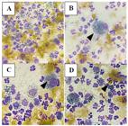
Cytological analysis of abdominal effusion from a female dog, mixed breed, diagnosed with biliary peritonitis due to cystic duct rupture, highlighting the presence of yellow amorphous material at the bottom of the slide compatible with bile (A), presence of macrophages with intensely vacuolated cytoplasm containing intracytoplasmic gray-blue material (arrowhead) (B), presence of intracytoplasmic golden crystals in macrophage (arrowhead) (C) and presence of free golden crystal on the slide (arrowhead) (D). Optical microscopy photo at 100x magnification. Source: Personal Archive (Laboratory of Veterinary Clinical Pathology, Veterinary Hospital of State University of Maringá), 2024.
These results were consistent with the ultrasound evaluation, which showed increased mesenteric echogenicity, suggesting peritonitis, and a hyperechogenic liver with slightly coarse echotexture, compatible with liver disease. The gallbladder had a thin and regular wall, filled with homogeneous and anechoic content, without morphological alterations (Fig. 3). However, despite the anorexia presented by the patient, the gallbladder was minimally filled. It was not possible to visualize the cystic duct.
During hospitalization, the patient presented unstable parameters, with dehydration refractory to fluid therapy, hypothermia and low responsiveness to external stimuli. Two days after the treatment, the patient's hemogram was repeated, which showed mild leukocytosis due to neutrophilia and lymphopenia, in addition to worsening of the anemia (PCV: 19%). Since the main suspicion was a rupture of the biliary tract, the owners were advised to perform an exploratory laparotomy to precisely locate the rupture point and repair it. However, considering the patient's unstable clinical condition and the costs of the procedure, the owners opted to euthanize the animal, which was performed on the same day.
Necropsy was performed on August 13, 2024. 250mL of orange, cloudy fluid was drained from the abdominal cavity. The liver had an evident and friable architecture, the gallbladder was slightly full and with an alteration compatible with rupture of the cystic duct. Furthermore, the mucous membranes and serous membranes were intensely icteric, and alterations compatible with renal infarction were observed (Fig. 4).

Ultrasound image of a female dog, mixed breed, diagnosed with biliary peritonitis due to cystic duct rupture, showing hepatic lobe and gallbladder (arrowhead) minimally filled with anechoic content and with a regular wall (A) and hepatic lobe surrounded by an anechoic area consistent with free fluid in the abdominal cavity (arrowheads) (B). Source: Sector of Imaging Diagnostics of the Veterinary Hospital of State University of Maringá, 2024.

Necropsy of a female dog, mixed breed, diagnosed with biliary peritonitis due to cystic duct rupture, showing the liver with icteric serosa, evident architecture and presence of irregularity in the cystic duct compatible with rupture (arrowhead) in A, while in B the left (E) and right (D) kidneys are shown, with darkened cortex and areas of pallor compatible with renal infarction. Source: Sector of Animal Pathology of the Veterinary Hospital of State University of Maringá, 2024.
DISCUSSION
The abdominal effusion analyzed presented a TNCC of 24,100/μL, protein concentration of 4.6g/dL, and density of 1.026, being classified as an exudate (Watson, 2020). The fluid presented total bilirubin concentrations 5.06 times higher than serum bilirubin, allowing the diagnosis of biliary effusion, in which the bilirubin concentration in effusion must be higher than the concentration in serum (Rebar and Thompson, 2011), also agreeing with Thompson and Sherman (2021) and Pavone et al. (2023), that consider the positive diagnosis if the ratio is greater than 1:1 and 1:2, respectively.
The yellowish-brown coloration of the abdominal fluid allowed us to consider the differential diagnosis of biliary peritonitis (Rebar and Thompson, 2011; Pavone et al., 2023; Francisco et al., 2024). Also, Watson (2020) and Francisco et al. (2024) described that biliary effusions have high cellularity, with predominance of intact neutrophils and with intense presence of macrophages, a characteristic present in the reported case.
Depending on the cause, biliary peritonitis is initially aseptic but may present bacterial development as the condition progresses. In the present case, no intracytoplasmic bacteria were found, determining non-septic peritonitis. However, the serum and effusion glucose concentrations were 307.0mg/dL and 165.0mg/dL, respectively, a characteristic that could be compatible with septic peritonitis (Francisco et al., 2024). The finding, however, corroborates the case reported by Pavone et al. (2023), in which the serum glucose concentration was higher than the concentration of the effusion, but without visualization of bacteria in the cytological analysis. Although septic cases of peritonitis are the minority, when finding bacteria in the biliary effusion, Escherichia coli is the most frequently isolated (Wilson et al., 2021; Kradangnga et al., 2023).
In addition to cellularity, an intense presence of golden-yellow pigments was also observed at the bottom of the slide, suggestive of bile pigments. Although this finding is characteristic of biliary effusion (Pavone et al., 2023), in the study by Wilson et al. (2021), only 33.3% of animals with biliary tract rupture presented bile pigments in the abdominal fluid. Furthermore, cytological analysis of abdominal fluid presented bluish-colored material in the cytoplasm of macrophages, also suggestive of bile pigments undergoing phagocytosis, corroborating the findings of Francisco et al. (2024).
According to Rebar and Thompson (2011), when the biliary effusion is persistent, bile granules may take the form of golden crystals of bile pigment, with rhomboid to amorphous morphology, and are considered an important diagnostic feature in the absence of previous hemorrhage. In the present case, crystals with the same characteristics were visualized in the cytoplasm of macrophages and free on the slide. However, this is a finding that was not reported in the cases of Wilson et al. (2021), Pavone et al. (2023) and Francisco et al. (2024).
Intense jaundice was a clinical sign that helped in the diagnosis of biliary tract rupture, in which the flow of bile from the gallbladder to the cystic duct, common bile duct, and duodenum is impaired. Thompson and Sherman (2021) stated that when serum bilirubin concentrations are higher than 4 mg/dL, there is jaundice of the sclera, skin, and mucous membranes. Wilson et al. (2021) and Linhares et al. (2022) reported the occurrence of hyperbilirubinemia in cases of biliary tract rupture. However, in the cases addressed by Pascual et al. (2022) and Pavone et al. (2023), the animals presented normobilirubinemia.
The time of progression of the disease and the cause of the rupture of the biliary tract are factors that can interfere with the serum bilirubin concentrations, presence of jaundice and presence of crystals in the cytological analysis of the effusion. In the cases reported by Guess et al. (2015), patients presented clinical signs 12 to 72 hours before veterinary care, with all cases with no jaundice or hyperbilirubinemia. Furthermore, in the report by Pavone et al. (2023), the animal presented biliary effusion on the third day of clinical signs, despite normobilirubinemia. Therefore, the clinical case we reported may have had clinical progression longer than 72 hours, considering the hyperbilirubinemia with consequent jaundice presented by the patient.
Other laboratory alterations besides hyperbilirubinemia were also observed, such as increased serum activities of ALT and ALP, hyperglycemia and leukocytosis with left shift, mostly corroborating other reported cases (Guess et al., 2015; Wilson et al., 2021; Cordella et al., 2022). However, it is worth mentioning that Guess et al. (2015) reported only non-icteric cases, while Wilson et al. (2021) observed normobilirubinemia in 40% of the reported cases.
Cholelithiasis, biliary mucocele, and cholecystitis, which are common causes of gallbladder or bile duct rupture, were dismissed as differential diagnoses after performing abdominal ultrasound. Since the history of the animal was unknown, the possibility of trauma could not be ruled out, although there were no other signs that could suggest this cause. Thompson and Sherman (2021) stated that when there is a history of trauma, ruptures of the common bile duct is more usual than of the gallbladder. However, in the reported case, necropsy showed that the point of extravasation was observed in the cystic duct. In humans, the occurrence of idiopathic gallbladder rupture has already been reported by Khanna et al. (2010), and this is also a possibility considered in the case discussed. In addition to the irregularity in the cystic duct compatible with rupture, changes suggestive of renal infarction were also observed during necropsy. In 2020, a study was performed which concluded that obstructive jaundice and hyperbilirubinemia are factors that contribute to the development of hepatorenal syndrome (Bayoumi et al., 2020) and may be a possibility considered for the observed changes.
CONCLUSIONS
This report indicates that gallbladder rupture should be considered as a differential diagnosis in cases of jaundice. Abdominal ultrasound is an important tool to help in the diagnosis, but it may present low sensitivity. In effusive cases, analysis of the cavity fluid is essential for rapid diagnosis, allowing early therapeutic measures to be adopted.
REFERENCES
- BAYOUMI, Y.H.; METWALLY, E.; EL NAGAR, E.A. et al. Hepatorenal syndrome in dogs with experimental extrahepatic cholestasis. Zagazig Vet. J., v.48, p.12-22, 2020.
- CORDELLA, A.; GIANESINI, G.; ZOIA, A. et al. Multi-phase multidetector-row computed tomographic features and laboratory findings in dogs with gallbladder rupture. Res. Vet. Sci., v.153, p.137-143, 2022.
- FRANCISCO, A.C.O.; DUTRA, G.A.; RIBEIRO, I.R.N.; CANÇADO, V.F. Análise efusões cavitárias em pequenos animais: investigação de 48 casos. Pubvet, v.18, p.e1641, 2024.
- GUESS, S.C.; HARKIN, K.R.; BILLER, D.S. Anicteric gallbladder rupture in dogs: 5 cases (2007-2013). J. Am. Vet. Med. Assoc., v.247, p.1412-1414, 2015.
- KHANNA, R.; AGARWAL, N.; SINGH, A.K. et al. Spontaneous common bile duct perforation presenting as acute abdomen. India J. Surg., v.72, p.407-408, 2010.
- KRADANGNGA, K.; KONGRATTANACHAT, N.; SUWANSUKROJ, W. et al. Outcomes in dogs undergoing surgical correction for septic peritonitis: A retrospective study in 61 cases (2019-2022). Thai J. Vet. Med., v.53, p.345-353, 2023.
- LINHARES, M.T.; MILECH, V.; MARTINS, L.R. et al. Biliary peritonitis in a dog after perforation of the gallbladder during laparoscopic cholecystectomy. Acta Sci. Vet., v.50, p.786, 2022.
- PASCUAL, M.; FAUCHON, E.; MONTI, P.; VALLS, F. Anicteric gallbladder rupture with elevated bile acids in abdominal effusion in a dog with cholecystitis. J. Am. Anim. Hosp. Assoc., v.58, n.3, p.146-151, 2022.
- PAVONE, G.; CASTELLUCCI, B.; PAVONE, S. et al. Unusual case of biliary peritonitis in a dog secondary to a gastric perforation. Vet. Sci., v.10, p.384, 2023.
- REBAR, A.H.; THOMPSON, C.A. Fluidos de cavidade corporal. In: RASKIN, R.E.; MEYER, D.J. Citologia clínica de cães e gatos: atlas colorido e guia de interpretação. Rio de Janeiro: Elsevier, 2011. p.171-191.
- THOMPSON, B.J.; SHERMAN, R.A. Comprehensive review of biliary peritonitis. Top. Companion Anim. Med., v.44, p.100532, 2021.
- WATSON, P.J. Hepatobiliary and exocrine pancreatic disorders. In: NELSON, R.W.; COUTO, C.G. Small animal internal medicine. St. Louis, Missouri: Elsevier, 2020. p.518-648.
- WILSON, K.; POWERS, D.; GRASPERGE, B. et al. Dogs with biliary rupture based on ultrasound findings may have normal total serum bilirubin values. Vet. Radiol. Ultrasound, v.62, p.236-245, 2021.


