Complications in reconstructive surgeries after resection of neoplasms in dogs and cats
ABSTRACT
Surgery is the most used modality in the control of neoplasia. In many cases, due to the size and location of the neoplasia, the use of reconstructive techniques is necessary. Based on this, the objective of this study was to establish the prevalence of the main complications of reconstructive techniques after nodulectomy in dogs and cats. This study was a retrospective non-randomized study of three institutions. A total of 153 reconstructive procedures with postoperative complications were included in the analysis. Data was obtained from medical records. The most reported postoperative complications were ischemic necrosis and suture dehiscence. The observation period for postoperative complications corresponded to a mean time of five days (range 15 to 70 days) for ischemic necrosis, seven days for suture dehiscence, three days for seroma formation, two days for hematoma and four days for the occurrence of infection. Regarding surgical technique, there was no significant difference in the occurrence of complications in axial pattern flaps and subdermal pattern flaps. Despite surgical complications, postoperative management with careful wound care leads to healing in the vast majority of patients. These findings may help in prognosis and decision-making regarding the main reconstructive complications.
Keywords:
oncology; plastic surgery; surgical technique
RESUMO
A cirurgia é a modalidade mais comumente utilizada no controle de neoplasias. Em muitos casos, devido ao tamanho e à localização da neoplasia, o uso de técnicas reconstrutivas é necessário. Com base nisso, o objetivo deste estudo foi estabelecer a prevalência das principais complicações das técnicas reconstrutivas após a nodulectomias em cães e gatos. Trata-se de um estudo retrospectivo não randomizado de três instituições. Um total de 153 procedimentos reconstrutivos com complicações pós-operatórias foi incluído na análise. Os dados foram obtidos de prontuários médicos. As complicações pós-operatórias mais relatadas foram necrose isquêmica e deiscência de sutura. O período de observação das complicações pós-operatórias correspondeu a um tempo médio de cinco dias (de 15 a 70 dias) para necrose isquêmica, sete dias para deiscência de sutura, três dias para formação de seroma, dois dias para hematoma e quatro dias para ocorrência de infecção. Em relação à técnica cirúrgica, não houve diferença significativa na ocorrência de complicações nos retalhos de padrão axial e retalhos de padrão subdérmico. Apesar das complicações cirúrgicas, o manejo pós-operatório com feridas de cuidado leva à cicatrização na grande maioria dos pacientes. Esses achados podem auxiliar no prognóstico e na tomada de decisão quanto às principais complicações reconstrutivas.
Palavras-chave:
oncologia; cirurgia plástica; técnica cirúrgica
INTRODUCTION
Once the diagnosis of a neoplasm has been established, treatment is mainly performed according to the patient's clinical staging. Among the treatment options, surgery is the most used modality in the control of neoplasms. In many cases, due to the size and location of the neoplasm and the need for a wide surgical approach (including considerations of margins), a large wound is created, which then requires the use of reconstructive techniques (Castro et al., 2015; Martins et al., 2015). Although reconstructive surgery has many benefits, it is important to note that there may be complications (Adams and Ramsey, 2005).
The causes of these complications are variable and depend on the origin and type of flap, the etiology of the wound, intrinsic factors of the patient, perioperative and postoperative management, the patient’s general condition and the recipient area. Among the types of complications, the most common include the presence of seroma, hematoma, ischemia, necrosis, edema, infection and suture dehiscence (Kirpensteijn and Haar, 2013; Wardlaw and Lanz, 2013).
Due to the related complications that are associated with the performance of reconstructive surgery, the aim of this retrospective study was to analyze the main complications of reconstructive procedures from 153 cases (131 dogs, and 22 cats) after the removal of neoplasms at three educational institutions.
MATERIALS AND METHODS
We included 153 patients that were diagnosed with neoplasms; patients included dogs and cats from three institutions: Pontifical Catholic University of Paraná (PUC), Center for Higher Education of Maringá (Unicesumar) and the São Paulo State University "Júlio de Mesquita Filho" (Unesp).).
The tissue samples were collected between January 2013 and January 2016. Only well-informed and oriented owners were able to correctly follow the care that was prescribed by the veterinarians who participated in this study.
The following inclusion criteria were used in this study: patients without distant metastasis (regional lymph node metastasis was included), primary lesions and use of reconstructive techniques performed for skin closure. All of the collected data from the dogs and cats, such as age, anatomic region, histopathologic exam, pattern flaps that were performed, post surgery complications and wound management, were extracted from the medical records.
All the patients were operated by surgeons from the oncology service, and all the procedures met the criteria necessary for margin procedures. The procedures followed the principles of reconstructive surgery, in addition to the removal of the regional lymph node based on the lymphatic drainage system.
Subdermal pattern flaps were used, including uniped and bipedicle advancement flaps, rotation flaps, transposition flaps, tubular flaps, axillary folds and inguinal folds. In addition, axillary pattern flaps were used, such as that of the caudal, temporal, superficial cervical, thoracodorsal, lateral thoracic, superficial brachial, superficial cranial epigastric, superficial caudal epigastric, deep circumflex iliac, medial genicular and superficial lateral caudal arteries.
Appropriate care of surgical wounds in the postoperative period was performed with bandages and changes every three days, with the removal of the stitches being performed on Day 15 after the procedure for dogs and on Day 21 after the procedure for cats.
Day 0 was considered the day of the surgical procedure, and regions were classified as head and neck, thoracic and pelvic members, chest and abdomen and perineum. Complications included ischemic necrosis, suture dehiscence, edema, infection, hematoma and seroma.
Histopathological reports included soft tissue sarcomas, mast cell tumors and squamous cell carcinomas. Some of the patients were receiving corticosteroids or had undergone chemotherapy procedures prior to the surgical procedure to cytoreduce the neoplasm. After the complication was identified, the possible treatments were evaluated, including a second intention wound with the use of ointments and dressings or a new surgical procedure.
The data results were analyzed by using a descriptive statistical analysis and are presented as the total number, percentage, average and standard deviation (depending on the case), and comparisons between the different parameters were performed by using the chi-square test (p < 0.05). Statistical analyses were performed by using the GraphPad Prism® program (version 6.0-GraphPad Software, Inc., San Diego, CA, USA) with a significance level of 0.05.
ETHICAL ASPECTS
All the procedures were performed in accordance with the national and international guidelines for use of animals in research (protocol number: CEUA 3882/21). This study was approved by the Ethics Committee for the Use of Animals of each institution.
RESULTS
From the 153 clinical records that were evaluated, 131 cases corresponded to dogs, and 22 cases corresponded to cats. The mean age of the patients was 2.1 ± 8.7 years. Postoperative complications were recorded in 35 cases (27%), with 30 dogs and 5 cats exhibiting complications. The most observed complications included ischemic necrosis (42.85%) (Fig. 1A and 1B), suture dehiscence (22.85%) (Fig. 1C), hematoma (14.28%) (Fig. 1D), seroma (8.57%) (Fig. 1E), infection (8.57%) and edema (2.85%). Suture dehiscence was not observed in cats.

Photographic images of complications in reconstructive surgeries performed in dogs. A) Note the presence of ischemic necrosis at the end of the axial pattern flap of the superficial brachial artery in the right forelimb (arrow). B) Note the presence of ischemic necrosis of the skin in the distal portion of the axial pattern flap of the thoracodorsal artery in the left forelimb (arrow). C) Observation of the occurrence of suture dehiscence after the performance of an axial pattern flap of the lateral thoracic artery in the left thoracic limb (arrow). D) Note the presence of hematoma in the axial pattern flap (arrow) of the superficial brachial artery in the left forelimb. E) Note the formation of seroma after the performance of the medial genicular artery skin flap in the right pelvic limb.
The observation period of complications in the postoperative period corresponded to an average time of five days (minimum of 15-21 days and maximum of 70 days) for ischemic necrosis, seven days for suture dehiscence, three days for seroma formation, two days for the occurrence of hematoma and four days for the occurrence of infection.
Regarding the anatomical region, 28.57% of the complications occurred in the head and neck, 22.85% occurred in the thoracic limbs, 20% occurred in the pelvic limbs, 17.15% occurred in the chest and abdomen and 11.43% occurred in the perineum. Regarding the histopathological examination of the excised neoplasms, we observed that the three most frequent histological types were soft tissue sarcomas (38.56%), mast cell tumors (22.87%) and squamous cell carcinomas (16.99%). Regarding the surgical technique, there were no significant differences in the occurrence of complications in axial pattern flaps (51.43%) and subdermal pattern flaps (48.57%).
It was observed that a higher proportion of subdermal flaps corresponded to higher rates of complications in the postoperative period. During follow-up of patients with axial pattern flaps, a higher rate of necrosis was observed for those flaps of the caudal auricular arteries (60%) (Fig. 2) and superficial brachial arteries (50%) (Figure 1A and 1D). Conversely, a low rate of necrosis (5% and 6.6%) was observed in flaps of the thoracodorsal and caudal superficial epigastric arteries (p <0.05), respectively.

Photographic images of complications in reconstructive surgery in a cat. A) Immediate postoperative period of reconstructive surgery using the caudal auricular artery axial pattern flap technique. B) Note the presence of necrosis at the edge of the flap (arrow), which is the main postoperative complication.
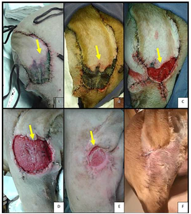
In patients who presented with postoperative complications, quantitative and qualitative differences were observed between the healing processes of dogs and cats. In the inflammatory phase, the wounds in dogs were more exudative, edematous and erythematous compared to wounds with the same dimensions in cats. In this study, the approaches varied for wound treatment according to second intention, because it was conducted at three different universities. However, the same goals were shared by the institutions; specifically, they strived to remove necrotic tissue when it existed, to prevent or control bacterial infections and to stimulate the formation of granulation tissue as early as possible, thus contributing to the process of re-epithelialization (Fig.3).

Photographic images of complications in reconstructive surgery in a dog after resection of a mast cell tumor on the lateral aspect of the proximal portion of the right femur. A) Blackened appearance at the end of the axial pattern flap of the lateral caudal arteries at three days after the surgical procedure (arrow). B) Presence of necrosis at the end of the axial pattern flap of the lateral caudal arteries at six days after the surgical procedure (arrow). C) Wound (10 days) after the surgical procedure (arrow). D) Appearance of the surgical wound 20 days after the surgical procedure (arrow). E) Aspect of the surgical wound 30 days after the surgical procedure (arrow). F) Complete synthesis of the surgical wound 60 days after the surgical procedure.
The bruises that formed in subdermal and axial pattern flaps were mainly treated with the use of topical escin (Reparil® Gel) and/or mucopolysaccharide polysulfate (Hirudoid® Gel), and an improvement in the absorption of the hematomas was observed, thus avoiding major postoperative complications.
In the three patients, the wound resulting from the surgical complication was initially treated until granulation tissue appeared, thus allowing for the surgical approach of the lesion edges to subsequently occur. In two cases, the complication was corrected by using a reconstruction procedure. This type of approach can be exemplified in the sequence images in Figure 4 and 5, in which the patient had a neoplasm close to the right radio-carpal joint; however, before the removal of this tumor, a tubular flap of subdermal pattern was created with the purpose of correcting the surgical wound resulting from the removal of the injury. Afterwards, the tube could not be used due to the occurrence of necrosis, thus requiring a change in technique; specifically, a graft was chosen to cover the wound resulting from the removal of the neoplasm.

Photographic images of the complication that occurred after the performance of a tubular flap with a subdermal pattern in a dog. A) Low-grade neurofibrosarcoma near the right radiocarpal joint (arrow). B) Confection of a tubular flap with a subdermal pattern, which was used to close the surgical defect after the removal of the neurofibrosarcoma (arrow). C) Final appearance of the tube (arrow). D) Five days after reconstructive surgery with the use of the subdermal pattern flap technique. Note the presence of necrosis in the middle third of the tube (arrow). E) Eight postoperative days. Note the occurrence of hematoma and necrosis in the tubular flap with a subdermal pattern (arrow). F) Rupture of the tube due to the occurrence of necrosis (arrow) at twelve days after the operation.

Photographic images of neoplasm excision and surgical wound closure with grafts in dogs. G) Excision of the low-grade neurofibrosarcoma near the right radiocarpal joint (arrow). H) Surgical wound after resection of the neoplasm (arrow). I) Donor bed for obtaining the skin graft (arrow). J) Skin graft ready to be implanted in the recipient bed. K) Final aspect of skin graft synthesis (arrow). L) Final aspect of the skin graft synthesis in the recipient bed (black arrow) and of the site where the first surgical intervention that resulted in the occurrence of a complication (yellow arrow) was performed.
DISCUSSION
Based on a literature review, some authors have observed that cats are more subject to complications than dogs after the performance of reconstruction techniques (Castro et al., 2015; Bohling and Handerson, 2006). However, in this study, there was no significant difference in the occurrence of postsurgical complications between the evaluated species.
Complications were more frequently observed in middle-aged and elderly dogs and cats, as these two groups are more predisposed to the occurrence of neoplasms (Daleck and Nardi, 2016) consequently, they were inclined to receive oncological and reconstructive surgeries. In addition, older animals have organic dysfunctions, which delay the recovery from surgical trauma and contribute to the occurrence of complications (Clayes, 2015; Darouiche et al., 2010).
According to the literature (Bohling and Handerson, 2006), there are microscopic differences in the cutaneous anatomy of cats and dogs, thus resulting in differences in cutaneous repair between these species. One of the factors described is that intact feline skin exhibits less perfusion than canine skin. For this reason, many of the complications result from vascular impairment, thus causing hypoxia secondary to hypoperfusion, which more frequently predisposes cats to necrosis at the end of the flaps when compared to skin flaps that are used in canines. Although we did not observe major complications in the feline species in this study, it was observed that one of the observed complications was necrosis of the flap edge.
Suture dehiscence is another possible complication that occurs in feline species, which is especially due to the shorter and longer deposition time of collagen in the suture line when compared to dogs, which favors the occurrence of less scar resistance in cats (Field et al., 2015; Riggs et al., 2015). In the present study, this complication was not observed because the removal of the skin sutures occurred after three weeks in all of the cats, whereas for dogs, the suture was removed after two weeks.
In this retrospective study, the most common postoperative complications were ischemic necrosis (42.85%) and suture dehiscence (22.85%), followed by hematoma (14.28%), seroma (8.57%), infection (8.57%) and edema (2.85%). According to Hedlund (2007), the use of reconstructive surgery techniques is an optimal alternative in the correction of extensive defects resulting from the excision of neoplasms; however, complications in the postoperative period can occur, such as ischemic necrosis in the extremities of the wound, suture dehiscence, seroma, hematoma, edema and infection (Cook and Thomson, 2014).
The occurrence of postsurgical complications was higher in the head, followed by the neck, thoracic and pelvic limbs. One of the explanations for the higher incidence of complications in these areas is related to a larger number of neoplasms that were observed in these anatomical areas in the present study. Greater movement and less blood perfusion were observed in the limb evaluation when compared to the chest, abdomen and perineum, which has been observed in other studies, in wherein greater complications were also observed in the limb region (Cook and Thomson, 2014).
Knowledge of the basic principles of plastic surgery as a nontraumatic surgical technique, as well as the correct positioning of the incisions and preservation of the regional blood supply, are important factors that can minimize postoperative complications (Chehelcheraghi et al., 2016). Based on this concept, during the initial care of patients in this study, a previously planned management for each case was used to reduce the percentage of complications.
The axial pattern flap is better irrigated than the subdermal pattern (Mota et al., 2012); however, no significant difference was observed between subdermal pattern flaps (51.43%) and axial pattern flaps (48.57%). In subdermal pattern flaps, ischemic necrosis was the most observed complication (p <0.05), whereas suture dehiscence was the main complication of axial pattern flaps (p <0.05). Subdermal flaps are more prone to the occurrence of complications (especially the presence of necrosis) because they do not have large-caliber vessels in their structures; however, they have smaller blood vessels, which make up the subdermal plexus. In addition to this issue, a ratio of 1:1 or 1:2 should always be respected between the width and the length, respectively, to minimize the occurrence of necrosis (Bohling and Handerson, 2006). In the present study, a greater proportion of subdermal flaps had higher rates of complications in the postoperative period (Montinavaro et al., 2015).
In contrast, axial pattern flaps have a direct cutaneous artery that irrigates the skin segment, thus reducing the chances of complications when they are used. However, excessive manipulation resulting in impaired vascularity, excessively long flaps and tight closure are factors that contribute to the occurrence of complications, such as suture dehiscence (Montinavaro et al., 2015).
In the current study, after analyzing the axial pattern flaps that were used, we observed that flaps of the caudal and superficial brachial arteries presented with a higher rate of necrosis when compared to the other skin flaps. This fact likely occurred because these two arteries are fragile (with reduced lengths and diameters), thus making the blood supply of these flaps insufficient (Riggs et al., 2015).
In practice, the safety of these flaps allows them to be used within a 360° field, in consideration of the respective bases. In the present study, the skin of the tail was used to close the surgical wounds; however, the length of skin that was used never exceeded two-thirds of the original size of the tail, thus contributing to a lower rate of complications (13.33%). In 7.40% of the patients in the current study, antineoplastic chemotherapy was instituted as neoadjuvant therapy with the aim of promoting tumor cytoreduction, thus facilitating tumor resection (especially in round cell neoplasms). However, there was no statistically significant difference compared to patients who did not receive antineoplastic chemotherapy (data not shown).
When there was a need for the administration of adjuvant antineoplastic chemotherapy to minimize the possibility of recurrence or progression of metastatic lesions, this treatment modality was instituted at least three weeks after the surgery to avoid delays in tissue repair due to these drugs. At the time of the first appointment, it was observed that some patients (13.07%) had been using corticosteroids for several weeks, especially as a treatment modality for mast cell tumors. This fact may have contributed to the observation that a greater number of patients (45%) with this neoplasm had suture dehiscence due to the delay in healing. Another fact that drew our attention during this study was the high incidence (45%) of necrosis in flaps that were used in patients with mast cell tumors (Figure 4). Treatment complications occurred in 88.57% of the patients. Due to the organic impairment of some of these cases or the severity of the complication, the treatments mainly consisted of the management of wounds and conduction of healing by second intention in 61.29% (n= 19) of the patients or by performing a new surgical intervention in 16.13% (n= 5) of the cases.
The use of the superficial brachial axial pattern flap was associated with a high rate of complications (as was observed in this study), and even in flaps with an axial pattern, complications related to flap extension can be observed (Montinavaro et al., 2015; Villedieu et al., 2022).
However, in the axial pattern flaps of the dorsal thoracic and superficial epigastric artery, a low rate of necrosis was observed (5% and 6.6%, respectively). The success of these flaps is undoubtedly related to the caliber and efficient blood flow of the arteries that irrigate these skin segments (Amsellem, 2011). This allows for 360° rotation with respect to the base of the flap, as well as allowing for tissue reconstruction and minimal postoperative complications. In a study with 13 evaluated cases in which the axial flap of the lateral caudal arteries was used, complications were observed in four procedures (30.76%). Among these, two patients presented with complications associated with flap extension, because the length was equivalent to 80% of the tail length (Montinavaro et al., 2015).
In the present study, the axial pattern flap of the lateral caudal arteries was implemented in the closure of surgical wounds, with a complication rate of 13.33%. In addition to the issue related to the environment in which the flap is used, there are complications that are directly linked to the patient. According to the literature (Amsellem, 2011), factors such as the use of corticosteroids, the use of antineoplastic chemotherapy or the use of radiotherapy prior to surgery contribute to delayed repair and the presence of complications in the postoperative period. However, no significant difference regarding the use of neoadjuvant chemotherapy and the occurrence of postoperative complications was observed in this study. Furthermore, in the evaluation of patients with mast cell tumors who received corticosteroids (13.07%) at the time of the first consultation, these patients had higher rates (45%) of suture dehiscence.
Although complications are associated with resection and reconstruction of defects due to mast cell tumors (Serafim et al., 2014), these complications can be caused by the release of proteolytic enzymes, vasoactive amines and stimulation of macrophages, thus reducing fibroplasia and collagen synthesis. This scenario explains the higher rate of complications that were observed in mast cell tumor patients. Furthermore, the literature does not indicate the actual reason for the occurrence of this tumor. It is speculated that mast cell tumors locally favor the occurrence of thrombi, which contribute to the development of thrombosis and, consequently, ischemic necrosis (Dernell, 2006).
In this study, 88.57% of the patients who presented with postoperative complications received two therapies: 1) healing by second intention, which was implemented in 61.29% of the cases; and 2) in 16.13% of the patients, a new surgical intervention was performed, as observed by Amsellen (2011).
It is important to note that the formation of granulation tissue in cats is different from that in dogs (Bohling and Handerson, 2006; Field et al., 2015; Riggs et al., 2015). This formation is delayed and visually differs, presenting with a paler color when compared to the granulation tissue of dogs. Another relevant factor is the origin of the formation of granulation tissue in both species; in dogs, the origin is subcutaneous, whereas in cats, the proliferation begins at the cutaneous edge of the wound (Pavletic, 2010). The fact that the granulation tissue emerges from the cutaneous border may explain the longer time for cutaneous epithelialization in this species (Pavletic, 2010).
The bruises that formed in subdermal and axial flaps were primarily treated with the use of topical escin (Reparil® Gel). When there is an increase in permeability resulting from the inflammatory process, escin inhibits exudation, thus reducing the leakage of liquid into the tissue spaces and accelerating the resorption of both edema and existing blood. Another active ingredient that is used in the treatment of hematomas is mucopolysaccharide polysulfate (Hirudoid® Gel), which intervenes in the process of tissue degradation and improves the absorption of bruises and edema (Martins et al., 2015).
Edema control was performed with the oral administration of rutoside (Venoruton® at a dosage of 50mg/kg for 3-5 days). This active ingredient can stabilize the vessel wall, which leads to a reduction in permeability and, consequently, a decrease in edema. In the case of more aggressive surgeries with the need for extensive lymphadenectomy, the administration of rutoside was initiated two or three days before the procedure to minimize the occurrence of this complication in the postoperative period.
The management of surgical complications is a major technical challenge; in addition, surgical reinterventions, which may be necessary, result in a less favorable prognosis than initial procedures (Clayes, 2015). The option for a new surgical intervention, which occurred in 16.13% of the cases, was mainly performed to reduce the healing time by second intention (Pavletic, 2010). In the three patients, the wound resulting from the surgical complication was initially treated until the formation of granulation tissue, thus allowing for the surgical approach of the edges of the lesion to occur (Slatter, 2007).
CONCLUSIONS
In conclusion, complications in reconstructive surgical techniques occurred in approximately 23% of patients. Surgical complications are minimized with proper postoperative management, and special attention should be focused on making and changing bandages. In this scenario, with the knowledge of the complication rates resulting from the performance of reconstructive surgery, further research would be essential to improve the applicability of substances that stimulate angiogenesis and contribute to healing, thus minimizing the occurrence of complications.
REFERENCES
- ADAMS, C.D.; RAMSEY, L.M. Grafts in dermatologic surgery: review and update on full- and split thickness skin grafts, free cartilage grafts, and composite grafts. Dermatol. Surg., v.31, p.1055-1067, 2005.
- AMSELLEM, P. Complications of reconstructive surgery in companion animals. Vet. Clin. North Am. Small Anim. Pract., v.41, p.995-1006, 2011.
- BOHLING, M.W.; HANDERSON, R.A. Differences in cutaneous wound healing between dogs and cats. Vet. Clin. North Am. Small Anim. Pract., v.36, p.687-692, 2006.
- CASTRO, J.L.C.; HUPPES, R.R.; NARDI, A.B. et al. Introdução à anatomia. In: _____. Princípios e técnicas de cirurgias reconstrutivas da pele de cães e gatos. Curitiba: Medvep, 2015. cap.1, p.10-16.
- CHEHELCHERAGHI, F.; EIMANI, H.; HOMAYOONSADRAEI, S. et al. Effects of acellular amniotic membrane matrix and bone marrow-derived mesenchymal stem cells in improving random skin flap survival in rats. Iran Red. Crescent. Med. J., v.16, p.1-16, 2016.
- CLAYES, S. Dehiscence. In: GRIFFON, D.J.; HAMAIDE, A. (Eds.). Complications in small animal surgery. [New York]: John Wiley & Sons, 2015. p.968.
- COOK, D.A.; THOMSON, D.J. Complications of the angularis oris axial pattern buccal flap for reconstruction of palatine defects in two dogs. Aust. Vet. J., v.92, p.156-160, 2014.
- DALECK, C.R.; NARDI, A.B. Oncologia de cães e gatos. 2.ed. Rio de Janeiro: Roca, 2016. 766p.
- DAROUICHE, R.O.; WALL, J.R.; ITANI, K.M. et al. Chlorhexidine-alcohol versus povidone-iodine for surgical-site antisepsis. New Engl. J. Med., v.362, p.18-26, 2010.
- DERNELL, W.S. Initial wound management. Vet. Clin. North Am. Small Anim. Pract., v.36, p.713-738, 2006.
- FIELD, E.J.; KELLY, G.; PLEUVRY, G.; Demetriou, J. Indications, outcome and complications with axial pattern skin flaps in dogs and cats: 73 cases. J. Small Anim. Pract., v.56, p.698-706, 2015.
- HEDLUND, C.S. Surgery of the integumentary system. In: FOSSUM, T.W. Small animal Surgery. 3.ed. St. Louis, MO: Mosby Elsevier, 2007. p.161-259.
- KIRPENSTEIJN, J.; HAAR, G.T. Reconstructive surgery and wound management of the dog and cat. [Boca Raton]: CRC Press, 2013.
- MARTINS, M.I.M.; ELIAS, B.C.; JUSTINO, R.C. Cirurgia reconstrutiva com retalho cutâneo de avanço como técnica alternativa para tratamento de carcinoma de células escamosas em cães: relato de caso. Rev. Bras. Cienc. Vet., v.22, p.131-136, 2015.
- MONTINAVARO, V.; MASSARI, F.; VEZZONI, L. et al. Lateral caudal axial pattern flap in 13 dogs. Vet. Surg., v.44, p.642-647, 2015.
- MOTA, A.K.R.; LACERDA, M.A.; ALEIXO, G.A. et al. Integração de retalhos de plexo subdérmico para o recobrimento de feridas traumáticas em membros de cães - Avaliação clínica. Med. Vet., v.6, p.1-9, 2012.
- PAVLETIC, M.M. Atlas of small animal wound management and re-constructive surgery. 3.ed. Cambridge: Wiley-Blackwell, 2010. p.127-158.
- RIGGS, J.L.; FRAZER, J.; FRIEND, B. et al. Outcome of full-thickness skin grafts used to close skin defects involving the distal aspects of the limbs in cats and dogs: 52 cases (2005-2012). J. Am. Vet. Med. Assoc., v.247, p.1042-1047, 2015.
- SERAFIM, E.L.; HUPPES, R.R.; PAZZINI, J.M. et al. Mastocitoma: síndromes paraneoplasicas. J. Bras. Cir. Vet., v.7, p.93-106, 2014.
- SLATTER, D. Manual de cirurgia de pequenos animais: pele e órgãos anexos. 3.ed. São Paulo: Manole. 2007. p.304-309.
- VILLEDIEU, E.; NOLFF, M.C.; DEL MAGNO, S.; EMMERSON, T. et al. Outcome of superficial brachial axial pattern flaps used to close skin defects in dogs: 16 cases (1996-2019). J. Small Anim. Pract., v.63, p.136-141, 2022.
- WARDLAW, J.L.; LANZ, O.I. Axial pattern and myocutaneous flap In. TOBIAS, K.M.; JOHNSTON, S.A. Veterinary surgery: small animal. [Oxford]: Elsevier Health Sciences, 2013. v.2, cap.79.
-
DATA AVAILABILITY STATEMENT
Data-available-upon-request - The research data is only available upon request of medical records from the Universities described in the methodology (Pontifical Catholic University of Paraná (PUC), Center for Higher Education of Maringá (Unicesumar) and the São Paulo State University "Júlio de Mesquita Filho" (Unesp).
Data-available-upon-request - The research data is only available upon request of medical records from the Universities described in the methodology (Pontifical Catholic University of Paraná (PUC), Center for Higher Education of Maringá (Unicesumar) and the São Paulo State University "Júlio de Mesquita Filho" (Unesp).