Characterization and Ki67 cut-off evaluation in canine indolent T-zone lymphoma: epidemiological and immunohistochemical aspects
ABSTRACT
This study aimed to characterize the epidemiological and immunohistochemical aspects of canine indolent T-zone lymphoma and to evaluate the Ki67 cut-off point for differentiating T-zone lymphomas from high-grade lymphomas using paraffin-embedded samples. A total of 39 indolent T-zone lymphoma samples were analyzed. Epidemiological data were gathered from the medical records of affected dogs, and the corresponding tissue samples underwent immunohistochemical analysis using anti-CD3, CD45, CD79a, and Ki67 antibodies. The epidemiological assessment revealed that the most affected breed was the Golden Retriever (44%), followed by the Shih Tzu (13%). The most frequently noted clinical alteration was generalized lymphadenomegaly (52%). Seven animals (30%) exhibited lymphocytosis at the time of diagnosis. Immunohistochemical staining for CD45 was negative in all samples, consistent with flow cytometry analyses. The Ki67 cell proliferation index in T-zone lymphomas ranged from 5% to 40%, with a mean of 13.32% (±7.49). A Ki67 cut-off value of 42.5% demonstrated 100% sensitivity and specificity for differentiating T-zone lymphomas from high-grade lymphomas, including high-grade T-cell lymphomas. The overall survival was 256 days (±179), with specific survival at 334 days (±226). However, 14 out of 23 animals remained alive at the conclusion of the study, which had a follow-up period extending to 565 days. The epidemiological profile of T-zone lymphoma in this study resembled that found in American research, with a high prevalence among Golden Retrievers. The absence of CD45 staining and the low Ki67 value can be utilized in routine practice to differentiate T-zone lymphomas from high-grade T immunophenotype lymphomas, as these conditions have distinct prognoses and treatment protocols.
Keywords:
dog; T-zone lymphoma; cell proliferation; immunohistochemistry
RESUMO
Este estudo teve como objetivo a caracterização epidemiológica e imuno-histoquímica, bem como a avaliação do ponto de corte de Ki67 para diferenciar linfomas de zona T de linfomas de alto grau, utilizando amostras incluídas em parafina. Para isso, foram analisadas 39 amostras de linfoma indolente de zona T. Os dados epidemiológicos foram coletados dos prontuários dos cães acometidos, e as respectivas amostras de tecido foram submetidas à imuno-histoquímica, empregando-se os anticorpos anti-CD3, CD45, CD79a e Ki67. Nos resultados da avaliação epidemiológica, a raça mais acometida foi o Golden Retriever (44%), seguido pelo Shih Tzu (13%). A alteração clínica mais relatada foi a linfadenomegalia generalizada (52%). Sete (30%) animais apresentavam linfocitose no momento do diagnóstico. A marcação imuno-histoquímica para CD45 foi negativa em todas as amostras, assim como nos exames de citometria de fluxo. O índice de proliferação celular Ki67 nos linfomas de zona T variou de 5% a 40%, com média de 13,32% (±7,49). O ponto de corte do Ki67 de 42,5% foi 100% sensível e específico para diferenciar linfomas de zona T de linfomas de alto grau e linfomas de alto grau T. A sobrevida global foi de 256 dias (±179), e a sobrevida específica de 334 dias (±226), embora 14/23 animais estivessem vivos até o término do estudo, com acompanhamento de até 565 dias. O perfil epidemiológico do linfoma de zona T neste estudo foi semelhante ao encontrado em trabalhos norte-americanos, com alta prevalência em cães da raça Golden Retriever. A perda de marcação para CD45 e o baixo valor de Ki67 podem ser utilizados na rotina para a diferenciação entre linfomas de zona T e linfomas de alto grau de imunofenótipo T, uma vez que são doenças com prognósticos e tratamentos distintos.
Palavras-chave:
cão; linfoma de zona T; proliferação celular; imuno-histoquímica
INTRODUCTION
T-zone lymphoma is a nodal T-cell lymphoma characterized by the expansion of neoplastic cells into the paracortical and medullary regions without altering the lymph node architecture in the early stages. In its nodal/multicentric form, T-zone lymphoma is considered the most common indolent lymphoma in dogs (Valli et al., 2011; Aresu et al., 2015).
T-zone lymphomas in dogs display distinct epidemiological patterns, with a particularly high prevalence among Golden Retrievers. This indolent form of lymphoma is frequently linked to prolonged survival (Seelig et al., 2014; Martini et al., 2015). However, this trend has not been consistently observed globally (Comazzi et al., 2018).
From an immunophenotypic perspective, T-zone lymphoma is characterized by the absence of the pan-leukocyte marker CD45, a feature that sets it apart from other T-cell lymphomas. CD45 plays a crucial role in lymphocyte development and immune signaling, and its loss in neoplastic cells indicates an altered regulatory mechanism. (Dahlke et al., 2004; Seelig et al., 2014; Martini et al., 2015)
Although the immunophenotypic profile of T-zone lymphomas is well established in flow cytometry, limited information exists regarding these markers in immunohistochemistry (Stein et al., 2021). The advantage of this technique lies in its ability to visualize the antigen-antibody reaction and its localization within the tissue, aiding in distinguishing hyperplastic reactions (Kojima et al., 2022). Unlike immunohistochemistry, techniques such as flow cytometry and Western blot detect and quantify antigens without identifying the specific cell type that expresses them (Amorim et al., 2016).
Immunohistochemistry enables the determination of the cell proliferation rate using monoclonal antibodies that recognize epitopes of antigens expressed by proliferating cells. Among these antibodies, Ki67 is a non-histone nuclear protein associated with replication, expressed in all phases of the cell cycle except G0 (the resting phase), and has been reported as an important prognostic marker for human non-Hodgkin lymphoma (Bacchi and Gown, 1993; Kiupel et al., 1999). Evaluating the Ki67 index in canine non-Hodgkin lymphomas can be relevant for determining individual variability of the growth fraction among subtypes, as well as assisting in the classification of high- and low-grade lymphomas in dogs (Fournel-Fleury et al., 1997).
Therefore, this study aims to characterize the epidemiological profile, establish the immunohistochemical characterization, and determine the Ki67 cut-off value in a group of dogs with multicentric T-zone lymphoma in Brazil, correlating these findings with prognosis, overall and specific survival, disease-free interval, and clinicopathological characteristics.
MATERIALS AND METHODS
The study was conducted in collaboration with the Veterinary Pathology Laboratory VETPAT, located in Campinas, São Paulo, Brazil.
TZL cases were selected retrospectively from those submitted to the Veterinary Pathology Laboratory VETPAT in Campinas, Brazil, between January 2017 and December 2019. The diagnoses were based on morphological characteristics and immunophenotyping for CD3 and CD79a (Flood-Knapik et al., 2013).
A total of 39 cases were selected; however, only 28 paraffin-embedded blocks that were previously used for histopathological and immunohistochemical examinations were stored at the VETPAT laboratory. The remaining blocks, sent by veterinary pathology laboratories solely for immunohistochemical analysis at VETPAT, had already been returned to their originating laboratories. Therefore, considering the 39 initial histopathological and immunohistochemical records, additional immunohistochemical examination for the anti-CD45 marker was possible in 28 samples, and the immunohistochemical examination for the Ki67 cell proliferation marker was performed in 37 samples. All samples were assessed for the anti-CD3 and anti-CD79a markers.
Additionally, 21 samples of high-grade lymphomas were selected, including 15 cases of high-grade B-cell lymphomas (HGBL) and 6 cases of high-grade T-cell lymphomas (HGTL), where the Ki-67 proliferation marker was evaluated. This assessment aimed to establish a comparative parameter between the proliferation index of indolent lymphomas and high-grade lymphomas. These tumors were classified according to the World Health Organization (WHO), which considers morphology, immunophenotype, and clinical presentation (Valli et al., 2011).
The veterinarians responsible for cases of dogs diagnosed with T-zone lymphoma were asked to provide patient data such as breed, sex, age, and origin, as well as the following medical record information: clinical signs, the location and number of affected lymph nodes, existing clinical alterations, involvement of other organs, lymphocytosis at the time of diagnosis, treatment administered, clinical course of the disease, survival, and disease-free time. This data was collected using a Google Forms® questionnaire.
Overall survival (whether related to the disease or not) and disease-specific survival (related solely to the disease) were measured from the day of diagnosis until the date of death or, if still alive, the last reported contact with the veterinarian. Disease-free time was defined as the period from disease remission to the date of the last contact with the veterinarian when the animal was still alive.
Finally, the collected data was stored in spreadsheets and organized for further statistical analysis.
The immunophenotype and proliferation index were determined through immunohistochemistry, following the technique outlined by Flood-Knapik et al. (2013). The proposed immunohistochemical markers were derived from those currently utilized in flow cytometry and included the B-cell lymphocyte marker CD79a, the T-cell lymphocyte marker CD3, the pan-leukocyte marker CD45, and the cell proliferation marker Ki67, as detailed in Table 1.
Staining, clone, dilution, antigen retrieval, and manufacturer of the markers used for the immunohistochemical characterization of canine T-zone lymphoma
Immunohistochemistry reactions were conducted using the biotin-free detection system (REVEAL®). In summary, 3-µm tissue sections mounted on slides were deparaffinized, rehydrated, and incubated for twenty minutes in a 5% hydrogen peroxide and absolute methanol solution (Synth®), according to the manufacturer’s instructions, to block endogenous peroxidase activity. Antigen retrieval was subsequently carried out using heat (Pressure Cooker, Pascal - Dako North America, Carpinteria, CA) at 90°C for 30 minutes. Following this, the sections were washed three times in 0.01M phosphate-buffered saline (PBS) (pH 7.2) for 5 minutes. Non-specific sites were blocked using a solution designed for blocking non-specific reactions (Reveal Spring Biosciences Kit, Pleasanton, CA - Code SPD-125®). The sections were then treated with the primary antibody at the standardized dilution and incubated at 27°C for 2 hours at room temperature. After washing in PBS, the sections were incubated with the secondary antibody (Kit/Flex from Dako®), following the manufacturer’s instructions, at room temperature. The slides were washed again, and the reaction was visualized using the chromogenic substrate diaminobenzidine (DAB Novolink, Leica Biosystems, Newcastle, UK).
The reaction was subsequently halted by washing the slides in distilled water, followed by counterstaining with Harris hematoxylin (Easypath, São Paulo - SP) for 1 to 2 minutes. Each reaction included both positive and negative controls. A known canine lymphoma sample marked by the utilized antibody served as the positive control, while the negative control was obtained by replacing the primary antibodies with a diluent solution.
The reaction results were interpreted using a light microscope at a magnification of 40x. Ki67 was quantitatively assessed as a percentage based on counts of 500 cells in areas with the highest number of labeled cells (hotspots). The Ki67 percentage in T-zone lymphomas was compared to the values obtained from a group of high-grade lymphomas. The remaining markers were evaluated qualitatively in relation to their localization within lymphoid tissue, assessing their expression in immunohistochemically processed samples.
Descriptive statistics were employed to present clinical-pathological results, as well as disease-free time, overall survival time, and mean survival time. For analyzing the Ki67 percentage in high- and low-grade lymphoma groups, the variables underwent analysis of variance in a completely randomized design with a significance level of 5%. Mean comparisons were executed using ANOVA followed by Tukey’s test (significance level = 0.05). Furthermore, Ki67 data were assessed using a ROC curve to determine a cut-off point, taking into account the best sensitivity combined with the best specificity for distinguishing animals with TZL from those with high-grade B-cell and T-cell lymphomas. Analyses were conducted using GraphPad Prism v. 10.4 software.
ETHICAL ASPECTS
The study was approved by the Animal Ethics Committee of the Faculty of Agricultural and Veterinary Sciences at São Paulo State University, Jaboticabal Campus, under protocol no. 017222/18.
RESULTS
Of the 39 dogs diagnosed with T-zone lymphoma (TZL), eighteen were males (46%) and twenty-one were females (54%). At the time of diagnosis, the mean age of the affected animals was 8 years and 10 months, ranging from 6 to 15 years. Twelve different breeds were reported. The breed with the highest number of cases was the Golden Retriever, accounting for 17 (44%) animals, followed by the Shih Tzu (5/13%), Boxer (3/8%), French Bulldog (3/8%), Pinscher (2/5%), Dachshund (2/5%), mixed-breed dogs (2/5%), and other breeds (Cocker Spaniel, Chihuahua, Lhasa Apso, Schnauzer, and Pit Bull) with one animal each, as shown in Table 2.
A total of 23 responses were received from the 39 questionnaires sent via Google Forms® concerning the clinical data of the animals included in the study. As anticipated, the most frequently reported clinical alteration was lymphadenomegaly, which was generalized in 12 out of 23 (52%) dogs, regional in 7 out of 23 (30%), and localized to a single lymph node in 4 out of 23 (17%) patients. The mandibular lymph nodes were the most affected, reported in 17 out of 23 cases (74%). Other clinical alterations such as lethargy (3 out of 13%), dyspnea (1 out of 4%), fever (1 out of 4%), hyporexia (1 out of 4%), anorexia (1 out of 4%), diarrhea (1 out of 4%), and weight loss (1 out of 4%) were also reported. The involvement of other organs was observed in four (17%) animals, including spleen (2 out of 9%), lungs (1 out of 4%), skin (1 out of 4%), and gastrointestinal tract (1 out of 4%). Additionally, seven (30%) animals exhibited lymphocytosis at the time of diagnosis (Table 3).
All samples were immunostained with the anti-CD3 antibody and were found to be negative for the B-cell marker CD79a, confirming the T-cell immunophenotype of the analyzed neoplasms. The pan-leukocyte marker CD45 was assessed in 28 out of 39 samples, and all were negative for this marker (Figure 1A and 1B).
Key clinical signs, physical examination results, and laboratory changes in dogs diagnosed with T-zone lymphoma

Photomicrograph of histological sections of immunolabeled canine lymphoma samples, visualized under a 40x objective lens. (A) T-zone lymphoma negative for CD45. (B) High-grade lymphoma positive for CD45. (C) Low Ki67 proliferation index in T-zone lymphomas. (D) High Ki67 proliferation index in high-grade lymphomas.
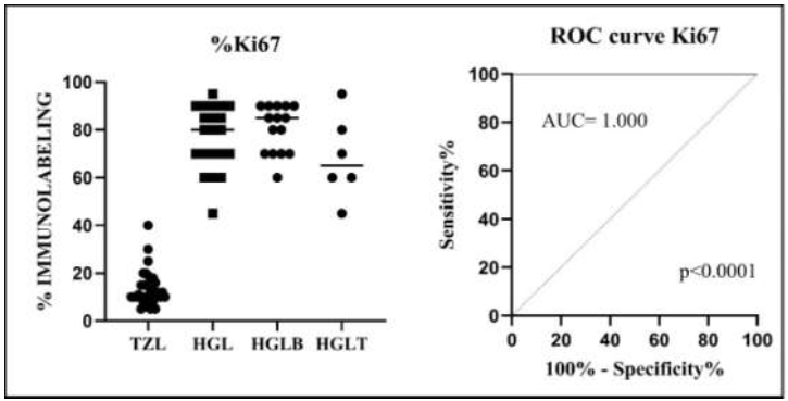
The Ki67 proliferation marker was assessed in 37 out of 39 cases of indolent T-zone lymphoma and in 21 cases of high-grade lymphomas. A significant difference was found in the mean Ki67 immunostaining between TZL and both high-grade B and T lymphomas (p<0.0001). The mean Ki67 labeling index for T-zone lymphomas was 13.32% (±7.06), while high-grade lymphomas showed a mean labeling index of 76.90% (±13.27) (Figure 1C and 1D; Table 4). No significant difference was noted between high-grade B and T lymphomas (p>0.05).
Statistical analysis of the Ki67 proliferation marker in T-zone lymphomas and high-grade B and T lymphomas. Different letters indicate statistical significance (p<0.05)
Concerning the Ki67 cutoff point for T-zone lymphomas, the area under the ROC curve (AUC) was 1.000 (CI 1.000 to 1.000) when compared to high-grade lymphomas (p<0.0001). The cutoff value was 42.5%, demonstrating 100% sensitivity and specificity in differentiating a T-zone lymphoma from a high-grade lymphoma (Fig. 2). When high-grade lymphomas were categorized into B and T subtypes and compared with T-zone lymphomas (TZL), sensitivity and specificity remained at 100%, with an AUC of 1.000 (CI 1.000 to 1.000) and cutoff values of 50% and 42.5%, respectively.

(A) Graphical representation of Ki67 immunostaining in TZL and high-grade lymphomas. (B) ROC curve for Ki67 immunostaining, comparing TZL with high-grade lymphomas.
Eight dogs died or were euthanized by the end of the study. Four dogs died, presumably due to disease progression. Three dogs passed away from causes unrelated to the disease, and one dog died of unknown causes. The mean overall survival of the eight deceased dogs was 256 days (ranging from 107 to 610 days), while the mean disease-specific survival-considering only dogs that died due to the disease-was 334 days (ranging from 107 to 610 days). The disease-free interval assessed at the end of the study was 565 days (ranging from 163 to 1012 days) (Table 5).
Statistical analysis of overall survival, disease-specific survival, and disease-free intervals in dogs diagnosed with T-zone lymphoma
DISCUSSION
This study described the epidemiological and immunophenotypic characteristics of multicentric indolent T-zone lymphoma in Brazilian dogs and compared the Ki67 proliferation index with high-grade B and T lymphomas. T-cell lymphoma encompasses a broad spectrum of diseases with variable biological behavior and distinct responses to treatment, including T-zone lymphoma. It is estimated that T-zone lymphoma is relatively common, representing between 3-12% of canine lymphomas and 60% of indolent lymphomas in dogs. However, to date, few studies have documented the clinical presentations of dogs with T-zone lymphoma (Flood-Knapik et al., 2013; Martini et al., 2016; Seelig et al., 2014; Sánchez-Solé et al., 2021).
The mean age of dogs at diagnosis was 8.8 years, consistent with that observed in other T-cell lymphomas and confirming previous data indicating that the mean age of dogs with this disease was 8 and 8.9 years, respectively. Furthermore, there appears to be no gender predilection for T-zone lymphoma (Valli et al. 2006; Mizutani et al. 2016a; Pinello et al., 2023).
The present study demonstrated that the Golden Retriever (n=17, 44%) was the most affected breed among dogs diagnosed with T-zone lymphoma. This data supports previous findings in other reports where the Golden Retriever breed was the most prevalent in this diagnosis, comprising 45%, 56%, 33%, 44%, and 40% respectively (Seelig et al., 2014; Mizutani et al., 2016a; Harris et al., 2018; Jark et al., 2020; Sánchez-Solé et al., 2021).
Considering this information together, one can infer that a genetic factor may increase the risk of T-zone lymphoma in this breed. This fact is observed in dogs bred in various countries, including Brazilian, North American, and Japanese Golden Retrievers, which may share the same genetic lineage. However, no cases of T-zone lymphoma were diagnosed in Golden Retrievers during a study conducted in Italy (Martini et al., 2015).
The second most frequently affected breed was the Shih Tzu, with a total of 5 animals (13%). Similar to other studies that identified the prevalence of this breed in cases of T-zone lymphoma (Seelig et al., 2014; Flood-Kanpik et al., 2013; Martini et al., 2015; Mizutani et al., 2016a; Harris et al., 2018), the results suggest that this breed warrants greater attention concerning lymphoma diagnosis, as they are often affected by indolent lymphomas.
In the initial clinical approach, the most frequently reported alteration based on responses from the Google Forms® questionnaire was generalized lymphadenomegaly (n=12, 52%), followed by regional (n=7, 31%) and nodal (n=4, 17%) lymphadenomegaly. Valli et al. (2006) also noted lymphadenomegaly in all 10 (100%) cases of T-zone lymphoma, with 4 (40%) being generalized, 4 (40%) involving only one lymph node, and 2 (20%) showing involvement of two lymph nodes. Mizutani et al. (2016a) similarly reported that generalized lymphadenomegaly was observed in all cases of T-zone lymphoma (n=16, 100%) included in their study. Likewise, Harris et al. (2018) found that 8 dogs (75%) diagnosed with T-zone lymphoma in the tongue exhibited lymphadenomegaly (one or more lymph nodes) at the time of the initial clinical evaluation. Therefore, lymphadenomegaly is a common clinical alteration in this type of lymphoma.
Lymphocytosis at the time of diagnosis of T-zone lymphoma was not common in this study; it was a laboratory finding noted in seven (30%) of the 23 samples obtained. However, Flood-Knapik et al. (2013) observed lymphocytosis in 47.5% of dogs diagnosed with T-zone lymphoma, while Seelig et al. (2014) reported it in 53%. This suggests that lymphocytosis, along with lymphadenomegaly, are consistent findings in T-zone lymphomas, indicating a more advanced stage of the disease, although it does not appear to point to a more aggressive progression.
In the present study, 39 lymph node samples affected by lymphoma were tested for the markers anti-CD3 and anti-CD79a, with all samples showing positivity for anti-CD3 and negativity for anti-CD79a. Previously, Valli et al. (2006) documented reactivity in flow cytometry using the same marker in 90% (n=9) of T-zone lymphoma samples. In the study at hand, it was found that 28 (100%) of the samples submitted for immunohistochemical examination of the anti-CD45 marker tested negative, corroborating studies that reported similar results, although they used flow cytometry for this marker (Seelig et al., 2014; Mizutani et al., 2016b; Hughes et al., 2018). Nevertheless, in a recent study, Stein et al. (2021) evaluated 27 samples of T-zone lymphoma and observed positive CD45 staining in 2 of 27 cases, thereby highlighting the importance of evaluating other criteria to obtain a definitive diagnosis.
T-zone lymphoma appears to exhibit a distinct phenotype where neoplastic T lymphocytes lack expression of the pan-leukocyte antigen CD45, facilitating a reliable diagnosis through immunophenotyping. CD45 is a tyrosine phosphatase that plays a complex role in regulating signaling through T-cell receptors and in the activation of cytokine receptors. One possible mechanism connecting the absence of CD45 to T-cell lymphoma is the binding of CD45 by galectin on both immature (thymic) and activated T lymphocytes, which induces apoptosis. The loss of CD45 expression may enable T cells to escape deletion in the thymus or evade apoptosis, ultimately leading to neoplastic transformation (Seelig et al., 2014).
Although the loss of CD45 expression is characteristic of T-zone lymphomas, Parachini-Winter et al. (2018) recently published a case of high-grade peripheral T lymphoma in a dog, which showed a loss of CD45 in flow cytometry analysis. This highlights the necessity for a joint evaluation of clinical and morphological data, proliferation indices, and immunophenotyping findings to reach a diagnostic conclusion.
Although Ki67 is a well-established immunohistochemical marker in veterinary medicine, there is limited data on its application in lymphomas concerning cutoff values, particularly in indolent lymphomas. This study revealed a significant difference in Ki67 values between high-grade lymphoma (76.90%, ±13.27) and low-grade lymphoma (13.32%, ±7.49). These rates align with those observed by Flood-Knapik et al. (2013), who reported a mean Ki67 staining of 20.95% (±17.3) among 50 tested indolent lymphomas, including follicular lymphomas, marginal zone lymphomas, and T-zone lymphomas. Flood-Knapik et al. (2013) reported that the mean Ki67 staining in T-zone lymphomas was 17.6% (±12.1), lower than the means for mantle zone lymphomas and follicular lymphomas, showing mean staining of 20% (±9.6) and 59.9% (±28.5), respectively.
The proliferation index indicated by Ki67 reactivity in T-zone lymphoma in this study was low compared to high-grade lymphomas, supporting the likely mechanism of the disease, primarily linked to delayed apoptosis rather than increased cell proliferation (Seelig et al., 2014). The 42.5% Ki67 cutoff point demonstrated 100% sensitivity and specificity in differentiating T-zone lymphomas from high-grade. Consequently, our study suggests that Ki67 assessment reliably aids in distinguishing between indolent T-zone lymphomas and high-grade lymphomas. Establishing consistent references for the cutoff values of Ki67 staining percentages between indolent and high-grade lymphomas is crucial, especially for veterinary pathologists in routine practice. It is often not feasible to differentiate high-grade from low-grade lymphomas solely through morphological analysis of samples, and this information is essential for providing an accurate diagnosis, allowing the oncologist to formulate a better therapeutic plan and prognosis for the disease.
In this study, the overall survival was 8 months, and the specific survival was 10 months, suggesting a late diagnosis of the disease. T-zone lymphoma generally has a longer survival compared to other T-type lymphomas. Seelig et al. (2014) reported an average survival of 21 months, while Valli et al. (2006) and Flood-Knapik et al. (2013) recorded 22.5 and 33 months, respectively. Martini et al. (2016) observed an overall survival of 760 days and a specific survival of 180 days in 26 dogs with T-zone lymphoma. The survival times observed in our study may be shorter due to the limited number of animals that died. Only 8 of the 23 dogs died, with half due to disease (4/50%) and half due to other causes (4/50%). Thus, the overall and specific survival rates may not accurately reflect reality since most dogs (15/23) remained alive until the end of the study, indicating a long survival rate for dogs with T-zone lymphoma.
Understanding the epidemiological profile of dogs in Brazil and the immunohistochemical characterization can aid in diagnosing animals affected by the disease and help target patient treatment, given that this type of lymphoma is less severe and requires less aggressive therapies. One limitation of this study was the retrospective analysis based on responses to a questionnaire, whose low adherence among participants may have affected the robustness of the analysis. Although this practice is common in developed countries, it is less so in Brazil.
The results of these studies, given the methodological conditions employed, indicate that in the Brazilian sample population analyzed, the Golden Retriever was the dog breed most affected by T-zone lymphoma. Immunohistochemistry effectively demonstrated the loss of anti-CD45 antibody staining in T-zone lymphomas, proving to be a valuable diagnostic tool. The low Ki67 proliferation index, along with a cutoff point of 42.5%, assists in differentiating T-zone lymphomas from high-grade lymphomas. Furthermore, dogs with T-zone lymphomas exhibited a low mortality rate at the end of the study, suggesting a longer survival rate and confirming their indolent nature.
ACKNOWLEDGMENTS
The authors thank the VetPat Veterinary Pathology Laboratory for their assistance with the project.
REFERENCES
- AMORIM, R.L.; NETO R.T.; KUIPEL, M. Imuno-histoquímica no diagnóstico oncológico. In.: DALECK, C.R.; NARDI, A.B. (Eds.) Oncologia em cães e gatos. 2.ed, Rio de Janeiro: Roca, 2016. p.217-237.
- ARESU, L.; MARTINI, V.; ROSSI, F. et al. Canine indolent and aggressive lymphoma: Clinical spectrum with histologic correlation. Veterinary and comparative oncology. v.13, p.348-362, 2015.
- BACCHI, C.E.; GOWN, A.M. Detection of cell proliferation in tissue sections. Braz. J. Med. Biol. Res., v.26, p.677-687, 1993.
- COMAZZI, S.; MARELLI, S.; COZZI, M. et al. Breed-associated risks for developing canine lymphoma differ among countries: an European canine lymphoma network study. BMC Vet. Res., v.14, p.1-7, 2018.
- DAHLKE, M.H.; LARSEN, S.R.; RASKO, J.E.J. et al. The biology of CD45 and its use as a therapeutic target. Leuk. Lymphoma, v.45, p.229-236, 2004.
- FLOOD‐KNAPIK, K.E.; DURHAM, A.C.; GREGOR, T.P. et al. Clinical, histopathological and immunohistochemical characterization of canine indolent lymphoma. Vet. Comp. Oncol., v.11, p.272-286, 2013.
- FOURNEL-FLEURY, C.; MAGNOL, J.P.; CHABANNE, L. et al. Growth fractions in canine non-Hodgkin's lymphomas as determined in situ by the expression of the Ki-67 antigen. J. Comp. Pathol., v.117, p.61-72, 1997.
- HARRIS, L.J.; ROUT, E.D.; HUGHES, K.L. et al. Clinicopathologic features of lingual canine T‐zone lymphoma. Vet. Comp. Oncol., v.16, p.131-139, 2018.
- HUGHES, K.L.; LABADIE, J.D.; YOSHIMOTO, J.A. et al. Increased frequency of CD45 negative T cells (T zone cells) in older Golden retriever dogs. Vet. Comp. Oncol., v.16, p.E109-E116, 2018.
- JARK, P.C.; FRACACIO, C.P.; ANAI, L.A. et al. Histopathological and immunophenotypical characterization of canine multicentric lymphoma in Brazil: a study of 203 cases. Arq. Bras. Med. Vet. Zootec., v.72, p.787-793, 2020.
- KIUPEL, M.; TESKE, E.; BOSTOCK, D. Prognostic factors for treated canine malignant lymphoma. Vet. Pathol., v.36, p.292-300, 1999.
- KOJIMA, K.; CHAMBERS, J.K.; MIZUNO, T. et al. Nodal T-zone lymphoma and T-zone hyperplasia in dogs. Vet. Pathol., v.59, p.733-739, 2022.
- MARTINI, V.; MARCONATO, L.; POGGI, A. et al. Canine small clear cell/T‐zone lymphoma: clinical presentation and outcome in a retrospective case series. Vet. Comp. Oncol., v.14, p.117-126, 2016.
- MARTINI, V.; POGGI, A.; RIONDATO, F. et al. Flow-cytometry detection of phenotypic aberrancies in canine small clear cell lymphoma. Vet. Comp. Oncol., v.13, p.281-287, 2015.
- MIZUTANI, N.; KOSHINO, Y.G.; TAKAHASHI, M. et al. Clinical and histopathological evaluation of 16 dogs with T-zone lymphoma. J. Vet. Med. Sci., v.78, p.1237-1244, 2016a.
- MIZUTANI, N.; KOSHINO, Y.G.; TSUBOI, M. et al. Evaluation of CD25-positive cells in relation to the subtypes and prognoses in various lymphoid tumours in dogs. Vet. Immunol. Immunopathol., v.173, p.39-43, 2016b.
- PARACHINI‐WINTER, C.; CURRAN, K.M.; RUSSELL, D.S. et al. A case of canine high‐grade T‐cell lymphoma immunophenotypically consistent with T‐zone lymphoma. Vet. Clin. Pathol., v.47, p.643-648, 2018.
- PINELLO, K.; SANTOS, M.; PEREIRA, P.D. et al. A retrospective histopathological survey on canine lymphomas subtypes of Porto District. Open Vet. J., v.13, p.443-450, 2023.
- ROSENBAUM, C.S.; SEELIG, D.M.; BURTON, E.N. et al. Monitoring of large B-cell lymphoma and T-zone lymphoma in a dog via flow cytometry. J. Vet. Diag. Invest., v.33, p.1008-1012, 2021.
- SÁNCHEZ-SOLÉ, R.; MOSQUILLO, F.; JARK, P. et al. Hematological and biochemical profiles of canine CD45− T lymphomas are different from other immunophenotypes. Open Vet. J., v.11, p.734-746, 2021.
- SEELIG, D.M.; AVERY, P.; WEBB, T. et al. Canine T-zone lymphoma: unique immunophenotypic features, outcome, and population characteristics. J. Vet. Intern. Med., v.28, p.878-886, 2014.
- STEIN, L.; BACMEISTER, C.; KIUPEL, M. Immunophenotypic characterization of canine nodal T-zone lymphoma. Vet. Pathol., v.58, p.288-292, 2021.
- VALLI, V.E.; VERNAU,W.; LORIMIER, L.P. et al. Canine indolent nodular lymphoma. Vet. Pathol., v.43, p.241-256, 2006.
- VALLI, V.E.; SAN MYINT, M.; BARTHEL, A. et. al. Classification of canine malignant lymphoma according to the World Health Organization criteria. Vet. Pathol., v.48, p198-211, 2011.
-
FUNDING
This work was supported by the Coordenação de Aperfeiçoamento de Pessoa de Nível Superior - Brazil (CAPES) - Funding Code 001.88882.330339/2019-01.
-
DATA AVAILABILITY STATEMENT
The research data supporting this publication are openly available in the institutional repository at https://repositorio.unesp.br
The research data supporting this publication are openly available in the institutional repository at https://repositorio.unesp.br