
 Femoral nerve block by pre-iliac approach and sciatic nerve block by parassacral approach in a dog undergoing unilateral colocephalectomy - case report
ABSTRACT
Multimodal anesthesia, which combines locoregional blocks with systemic analgesia, is widely used in veterinary orthopedic procedures to provide effective pain control. This case report describes the use of femoral nerve block via a pre-iliac approach and sciatic nerve block via a parassacral approach in a dog undergoing unilateral femoral head and neck excision. The techniques were performed with the aid of a nerve stimulator, using 0.25% bupivacaine (1.5mg/kg). The anesthetic protocol also included the administration of gabapentin (5mg/kg), dipyrone (25mg/kg), meloxicam (0.1mg/kg), ketamine (0.6mg/kg/h), methadone (0.3mg/kg), and fentanyl (5µg/kg/h), forming a multimodal protocol. The effectiveness was assessed based on physiological parameters, sensory-motor block of the limb, and pain scores, demonstrating effective pain control and hemodynamic stability during the perioperative period. In conclusion, the femoral and sciatic nerve blocks were efficient in providing analgesia for the coxofemoral joint, making the procedure safe and comfortable for the patient.
Keywords:
multimodal anesthesia; peripheral nerve block; osteotomy
RESUMO
A anestesia multimodal, que combina bloqueios locorregionais com analgesia sistêmica, é amplamente utilizada em procedimentos ortopédicos veterinários para proporcionar controle eficaz da dor. Neste relato, descreve-se o uso do bloqueio do nervo femoral por abordagem pré-ilíaca e do nervo isquiático por abordagem parassacral em um cão submetido à colocefalectomia unilateral. As técnicas foram realizadas com auxílio de neurolocalizador, utilizando-se bupivacaína a 0,25% (1,5mg/kg). O protocolo anestésico incluiu também a administração de gabapentina (5mg/kg), dipirona (25mg/kg), meloxicam (0,1mg/kg), cetamina (0,6mg/kg/h), metadona (0,3mg/kg) e fentanil (5µg/kg/h), compondo um protocolo multimodal. A avaliação da eficácia foi realizada com base nos parâmetros fisiológicos, no bloqueio sensitivo-motor do membro e no escore de dor, o que demonstrou controle eficaz da dor e estabilidade hemodinâmica durante o trans e o pós-operatório. Diante do exposto, conclui-se que o bloqueio dos nervos femoral e isquiático mostrou-se eficiente na analgesia da articulação coxofemoral, tornando o procedimento seguro e confortável para o paciente.
Palavras-chave:
anestesia multimodal; bloqueio de nervos periféricos; osteotomia
INTRODUCTION
Coxofemoral dysplasia is a highly prevalent orthopedic condition in veterinary medicine, particularly in dogs. It is characterized by an incongruence between the femoral head and the pelvic acetabulum. This misalignment affects the joint capsule, leading to pain, crepitus, and severe locomotor impairment (Fossum, 2014). The most common clinical signs include lameness, intense pain, and apathy, directly impacting the animal's quality of life (King, 2017).
The management of this condition primarily aims at pain relief and functional improvement. Treatment may include the use of analgesic and anti-inflammatory drugs, joint supplements, and integrative therapies such as physiotherapy and acupuncture to enhance mobility (Ferreira, 2014; Magalhães et al., 2024). In more advanced cases, femoral head and neck excision arthroplasty (FHNEA) is indicated to create a functional pseudoarthrosis, alleviating symptoms and restoring mobility (Fossum, 2014).
One critical aspect of this surgical procedure is effective pain control, both intraoperatively and postoperatively. In this context, multimodal anesthesia, which combines locoregional blocks and systemic analgesia, is widely recommended. This approach reduces the need for general anesthetics, minimizes adverse effects, and enhances both the safety and efficacy of the procedure (Grimm et al., 2017).
Several regional block techniques have been described for effective analgesia of the pelvic limbs. Epidural anesthesia is commonly used, providing potent analgesia for both pelvic limbs and facilitating pain management for procedures involving the limbs and abdominal region (Pereira, 2018).
However, targeted peripheral nerve blocks, such as femoral and sciatic nerve blocks, offer an effective alternative by providing localized analgesia and reducing the risks associated with epidural blocks, such as hypotension and infection (Otero and Portela, 2018).
Given the above, this report describes the use of a pre-iliac approach for femoral nerve block and a parasacral approach for sciatic nerve block in a dog undergoing unilateral femoral head and neck excision arthroplasty, aiming to ensure adequate analgesia during the surgical procedure and the postoperative period.
ETHICAL ASPECTS
This research has not been submitted to the Ethics Committee on the Use of Animals.
CASE REPORT
A 3-year-old male Shih Tzu dog weighing 7.4kg was presented to the Sylvio Barbosa Cardoso Veterinary Hospital with a complaint of left pelvic limb lameness. Radiographic examination confirmed coxofemoral luxation. Initial treatment included tramadol (3mg/kg, PO, BID for 5 days) and gabapentin (5mg/kg, PO, BID for 30 days) for pain management. Following orthopedic evaluation, unilateral femoral head and neck excision arthroplasty was indicated.
Preoperative tests, including complete blood count, biochemistry panel, echocardiography, and blood pressure measurement, were within reference ranges, except for left atrial overload on electrocardiography. The patient was classified as ASA II by the American Society of Anesthesiology, indicating a mild surgical risk, and was deemed fit for the procedure.
The preoperative anesthetic protocol included premedication with acepromazine (0.02 mg/kg), ketamine (1mg/kg), and methadone (0.3mg/kg), administered intramuscularly. After stabilization and trichotomy, venipuncture was performed using a 22G catheter, and anesthetic induction was achieved with propofol (3mg/kg, IV), followed by endotracheal intubation with a 5.5-mm cuffed tube. Anesthesia was maintained with isoflurane in a universal vaporizer using a Baraka system.
Femoral and sciatic nerve blocks were performed using a nerve stimulator (Delta Life® DL 250) set at a frequency of 1 Hz, a current range of 0.2-2 mA, and a pulse duration of 100 µs, with 0.25% bupivacaine (1.5mg/kg). Intraoperative monitoring included oxygen saturation, heart rate, respiratory rate, blood pressure, and temperature using a multiparameter monitor.
During surgery, the patient received continuous infusions of fentanyl (5µg/kg/h, IV) and ketamine (0.6mg/kg/h, IV), as well as dipyrone (25mg/kg), meloxicam (0.1mg/kg), and ceftriaxone (30mg/kg, IV) for analgesia and prophylaxis. The procedure lasted 15 minutes without complications, and vital parameters remained stable, with no need for rescue analgesia.
In the immediate postoperative period, pain was assessed using a modified Glasgow pain scale. The patient remained stable, showing no significant signs of pain or discomfort. Sensory and motor blockade were confirmed, ensuring effective analgesia in the operated limb.
For the lateral pre-iliac approach to the femoral nerve block, the spinous process of the sixth lumbar vertebra (L6) and the iliac crest were first identified (Figure 1A). The technique involves inserting the nerve stimulator needle at the intersection of a perpendicular line to the longitudinal axis of the spine and a parallel line drawn from the L6 spinous process and the iliac crest. The needle was inserted caudomedially at a 45° angle through the iliocostal lumbar muscle, advancing toward the greater trochanter of the femur (Figure 1B).
The nerve stimulator current was initially set at 2mA, then progressively reduced to 0.2mA, at which point the motor response (knee extension) ceased, indicating proper nerve localization. After confirming correct positioning, 0.25% bupivacaine (1.5mg/kg) was administered at a total volume of 2.2mL.
Regarding the sciatic nerve block using the parasacral approach, the anatomical landmarks used were the iliac crest and the left ischial tuberosity. A line was drawn between these two structures, and the needle was inserted perpendicularly (90° angle) at the end of the first third of this line (Fig. 2).
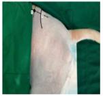
Locoregional femoral nerve block using a lateral pre-iliac approach with the aid of a nerve stimulator in a dog undergoing left unilateral femoral head and neck excision arthroplasty. In (1), the spinous process of the sixth lumbar vertebra (L6) is identified. In (2), the iliac crest is observed. The intersection of the lines drawn perpendicularly and parallel to the longitudinal axis of the spine represents the needle insertion site (●). At this point, the needle was positioned at a 45° angle, directed caudomedially through the iliocostal lumbar muscle toward the greater trochanter of the femur.

Sciatic nerve block using the parasacral approach in a dog undergoing left unilateral femoral head and neck excision arthroplasty. The iliac crest “(” and ischial tuberosity “3” were identified. A line was drawn between these two structures and divided into three equal segments to guide needle insertion at the first third of the drawn line. At point 1, the needle was positioned perpendicular to the skin, with the aid of a nerve stimulator to confirm proper placement before the administration of bupivacaine.
The nerve stimulator was initially set to a current of 2mA, which was gradually reduced to 0.2mA. At this point, the muscle response, characterized by contraction of the tarsal musculature, ceased, indicating proper localization of the sciatic nerve. After confirmation, bupivacaine (1.5mg/kg), diluted in saline to a final concentration of 0.25%, was administered in a total volume of 2.2mL
RESULTS AND DISCUSSION
The neuroanatomy of the pelvic limb comprises the branches of the lumbosacral plexus. The primary nerves involved in the innervation of the pelvic joint capsule include the femoral, sciatic, obturator, and cranial gluteal nerves, along with branches of the lumbar nerves L4 to L7 and the sacral nerves S1 and S2. These nerves ensure both sensory and motor innervation of the pelvic limbs (Klaumann and Otero, 2013; King, 2017).
The femoral nerve block via the paravertebral approach to the lumbar plexus, which targets the L4, L5, and L6 nerve roots, provides effective analgesia for procedures involving the femur, knee, tibia, and tarsus. However, complete desensitization of the pelvic limb requires its association with the sciatic nerve block. This method also affects the obturator and cranial gluteal nerves, ensuring analgesia of the pelvic joint capsule, making it particularly suitable for procedures such as femoral head and neck excision arthroplasty (Klaumann and Otero, 2013).
The choice of peripheral nerve blocks over neuroaxial anesthesia, such as epidural or spinal anesthesia, has proven to be effective and advantageous. Peripheral nerve blocks provide precise unilateral desensitization, minimizing the risk of complications associated with epidural anesthesia, such as hypotension, epidural abscess formation, and contamination (Portela et al., 2013). This method offers a safe and effective alternative with a lower risk of systemic complications.
The effectiveness of the nerve block was monitored through physiological parameters recorded every 5 minutes. In this patient, no rescue analgesia was required, as the values remained within the baseline threshold (Fig. 3).
According to Muir (2008), an increase of more than 20% in baseline vital signs (heart rate, respiratory rate, and blood pressure) would indicate pain, due to catecholamine release, leading to tachypnea, tachycardia, and hypertension.

Intraoperative monitoring of physiological parameters in a dog undergoing left unilateral femoral head and neck excision arthroplasty. Heart rate (HR), respiratory rate (RR), systolic arterial pressure (SAP), body temperature, and oxygen saturation (SpO₂) values were recorded at five time points: M0 - moment of anesthetic block execution; M1 - start of surgery; M2 and M3 - intermediate surgical time points; M4 - end of surgery.
Regarding the anesthetic protocol used, continuous infusion of fentanyl (5µg/kg/h, IV) and ketamine (0.6mg/kg/h, IV) was chosen. According to Grimm et al. (2017), the combination of a full μ-opioid agonist with ketamine at an analgesic dose ensured effective pain control for a period of 30 minutes to 2 hours. At the end of surgery, the infusion was discontinued, and after 30 minutes, the patient's parameters were reassessed. Indeed, no alterations exceeding 20% were observed, with a heart rate of 114 bpm, respiratory rate of 28rpm, and systolic blood pressure of 135 mmHg, confirming the efficacy of the block.
The femoral nerve block primarily desensitizes the dermatomes of the medial thigh, knee, and tarsus, whereas the sciatic nerve block covers the craniolateral and caudomedial regions of the pelvic limb (Otero and Portela, 2018; Hermanson et al., 2020). Thus, the surgical incision site was adequately desensitized by the blocks, reinforcing the effectiveness of multimodal anesthesia, which was complemented by the administration of methadone, fentanyl, and ketamine.
Postoperatively, the animal recovered quickly and without complications. The assessed parameters (heart rate, respiratory rate, blood pressure, oxygen saturation, and temperature) remained within baseline values. Pain levels, evaluated using the Glasgow Pain Scale, were recorded as 1 (indicating the absence of pain), and the animal showed no signs of discomfort upon palpation. These findings support the efficacy of the anesthetic protocol, aligning with the findings of Corrêa and Grados (2021), which demonstrate the benefits of locoregional blocks in surgical procedures.
Additionally, the effectiveness of both sensory and motor block of the limb was confirmed. The anesthetic used, bupivacaine (1.5mg/kg at 0.25%), initially produced a sensory block with an onset time of approximately 20 minutes, followed by complete motor blockade. Although the latency period was not fully respected before the start of surgery, desensitization was confirmed throughout the intraoperative and postoperative periods, supporting the choice of anesthetic (Klaumann and Otero, 2013; Grimm et al., 2017).
Neuroaxial anesthesia techniques, such as epidural or spinal anesthesia, are commonly used in veterinary orthopedic procedures, but peripheral nerve blocks stand out for their precision and lower risk of postoperative complications. Risks associated with epidural anesthesia include apnea, hematomas, epidural abscesses, dural sac laceration, and hypotension due to sympathetic blockade (Portela et al., 2013; Gurney and Leece, 2014; Sanches and Henriques, 2024).
However, the success of femoral and sciatic nerve blocks depends on the anesthetist's anatomical knowledge and experience, particularly in using a nerve stimulator or ultrasound. Potential complications include iatrogenic nerve injury, inadvertent injection of anesthetic into an incorrect site, allergic reactions, and systemic adverse effects (Klaumann and Otero, 2013).
In conclusion, the femoral nerve block using the lateral pre-iliac approach, combined with the sciatic nerve block via the parasacral approach, provides effective analgesia for unilateral femoral head and neck excision arthroplasty. Although some specific regions were not directly desensitized by the blocks, the association with systemic analgesics ensured adequate pain control and patient comfort. Thus, multimodal anesthesia is crucial for the appropriate pain management of patients undergoing orthopedic surgeries.
CONCLUSION
The femoral nerve block via the pre-iliac approach, combined with the sciatic nerve block via the parasacral approach, has proven to be an effective technique for pain management in a dog undergoing unilateral femoral head and neck excision arthroplasty, within a multimodal anesthesia protocol. This approach provided adequate analgesia, hemodynamic stability, and minimized complications associated with epidural anesthesia. The success of peripheral nerve blocks reinforces their utility in orthopedic procedures, requiring technical proficiency from the veterinarian. Despite their advantages, these techniques carry potential risks that necessitate rigorous monitoring. This case highlights the importance of individualized protocols for pain management in orthopedic surgeries and suggests that future studies explore the application of these techniques in different contexts.
REFERENCES
- CORRÊA, L.M.; GRADOS, F.M. Retrospective assessment of the relationship between intraoperative nociception and postoperative analgesia requirements in dogs undergoing thoracic surgery. Vet. Rec., v.190, p.1-7, 2021.
- FERREIRA, P.C. Efeitos da acupuntura na marcha de cães Labradores com displasia coxofemoral - estudo preliminar. 2014. 121f. Dissertação (Mestrado em Medicina Tradicional Chinesa) - Instituto de Ciências Biomédicas Abel Salazar da Universidade do Porto. Porto.
- FOSSUM, T.W. Cirurgia de pequenos animais. 4.ed. Rio de Janeiro: Elsevier, 2014. 1640p.
- GURNEY, M.A.; LEECE, E.A. Analgesia for pelvic limb surgery. A review of peripheral nerve blocks and the extradural technique. Vet. Anaesth. Analg., v.41, p.445-458, 2014.
- HERMANSON, J.W.; DELAHUNTA, A.; EVANS, H.E. et al. Miller and Evans' anatomy of the dog. 5.ed. St. Louis: Elsevier, 2020. 981p.
- GRIMM, K.A. et al. Lumb & Jones: Anestesiologia e analgesia em veterinária. 5.ed. Rio de Janeiro: Roca, 2017. 1056p.
- KING, M.D. Etiopathogenesis of canine hip dysplasia, prevalence, and genetics. Vet. Clin. North Am. Small Anim. Pract., v.47, p.753-767, 2017.
- KLAUMANN, P.R.; OTERO, P.E. Anestesia locorregional em pequenos animais. São Paulo: Roca, 2013. 288p.
- MAGALHÃES, L.A.; SOUZA, J.A.; RIBEIRO, L.F. Displasia coxofemoral em cães. GETEC., v.16, p.1-15, 2024.
- MUIR, W.W. Pain and stress. In: GAYNOR, J.; MUIR W.W. Handbook of veterinary pain management. 2.ed. Missouri: Mosby, 2008. p.42-55.
- OTERO, P.E.; PORTELA, D.A. Manual de anestesia regional em animais de estimação: anatomia para bloqueios guiados por ultrassonografia e neuroestimulação. 1.ed. São Paulo: Medvet, 2018. 452p.
- PEREIRA, F.G. Epidural anesthesia and analgesia in small animal practice: An update. Vet. J., v.242, p.24-32, 2018.
- PORTELA, D.A.; OTERO, P.E.; BIONDI, M. et al. Peripheral nerve stimulation under ultrasonographic control to determine the needle‐to‐nerve relationship. Vet. Anaesth. Analg., v.40, p.e91-e99, 2013.
- SANCHES, F.C.M.; HENRIQUES, D.P. Epidural block for bilateral perineal herniorrhaphy in canine patient. Pubvet, v.18, p.1-10, 2024.
The research data are available upon request.


