Biodegradable Chitosan Sutures Enhanced with N-Acetyl-D-Glucosamine: Comparative Study with Catgut Sutures
Abstract
This study aimed to develop and evaluate chitosan (CS) and chitosan with N-Acetyl-D-glucosamine (CS/GlcNAc) sutures, comparing them to commercial Catgut sutures. Medium molecular weight chitosan was processed into sutures using the wet-spinning method, both with and without the addition of GlcNAc. The mechanical properties, surface morphology, biocompatibility, biodegradability and in vivo response of the sutures were evaluated. Scanning electron microscopy (SEM) and enzymatic degradation tests in phosphate-buffered saline (PBS) with lysozyme were conducted. Cytotoxicity was assessed using an agar diffusion assay on L929 cells. In vivo studies involved suturing rat skin, with monitoring at 5, 15 and 25 days post-surgery. Both macroscopic and microscopic analyses were performed to evaluate wound healing, tissue response, and complications. The chitosan-based sutures exhibited tensile strength within the standards for surgical sutures under dry and hydrated conditions. Particularly, the CS/GlcNAc sutures exhibited superior biodegradability, and biocompatibility compared to Catgut sutures. These sutures promoted tissue healing, reduced adverse tissue reactions and infections, and displayed non-toxic to L929 cells and favorable microbiological characteristics. Thus, CS and CS/GlcNAc sutures present promising alternatives to traditional Catgut sutures, contributing valuable advancements to the field of biodegradable surgical materials.
Keywords:
Chitosan; Suture; N-Acetyl-D-glucosamine; Catgut; Threads
1. Introduction
The selection of materials for biodegradable surgical sutures involves critical considerations regarding biocompatibility, biodegradability, mechanical strength, flexibility, healing capacity, compatibility with sterilization methods, and cost-effectiveness1-4. The surgical sutures must not induce adverse reactions, degrade naturally, exhibit adequate mechanical properties, flexibility, and promote effective wound healing5-10.
Chitosan (CS), derived from chitin found in crustacean exoskeletons and fungi, possesses a chemical structure comparable to cellulose but with amino groups in each glucose unit, imparting unique properties including biocompatibility, non-toxicity, biodegradability, and hydrophilicity. Chitosan also exhibits hemostatic and wound healing capabilities, antimicrobial and antifungal activities, anti-tumor properties, analgesic effects, and potential as an anthelmintic agent11-22.
The versatility of chitosan allows it to be processed into various forms films, gels, granules, and fibers, rendering it suitable for a wide range of biomedical applications. As a biomaterial for surgical sutures, the biodegradability, hemostaticity, and antimicrobial activity of chitosan are desirable characteristics. These qualities make it a safe and effective option for wound healing following surgical procedures. It is well tolerated by the human body and naturally decomposes over time, eliminating the need for removal and reducing the risk of long-term foreign body reactions5,7,23-25.
Despite these advantages, comparative studies between chitosan sutures and conventional absorbable sutures such as Catgut remain limited in the literature. Previous studies have explored various aspects of chitosan-based sutures, including their mechanical properties, biocompatibility, and performance in wound healing5,7,26-28. However, comprehensive evaluations comparing these sutures to established commercial products like Catgut are sparse.
The objective of this study was to develop chitosan (CS) sutures and chitosan sutures incorporating N-acetyl-D-glucosamine (GlcNAc), an amino monosaccharide derived from glucose essential for cartilage cell formation and biological processes such as wound healing and anti-inflammatory activity29,30. These sutures were fabricated using the wet-spinning method with medium molecular weight chitosan and were compared to commercial Catgut sutures. The research aimed to assess the biological, cytotoxicological, healing, microbiological, and biodegradation properties of the chitosan-based sutures, as well as their mechanical strength and in vivo performance, to determine their potential for future medical applications.
2. Materials and Methods
2.1. Materials
Medium molecular weight chitosan, suitable for medical applications, was extracted from the exoskeleton of the shrimp Liopenaeus vannamei at the Northeastern Biomaterials Evaluation and Development Laboratory - CERTBIO (Campina Grande, PB, Brazil). The molecular weight (Mv = 270 kDa) was determined by viscometry (PSL Rheotek, São Paulo, Brazil), and the degree of deacetylation (DD = 88%) was determined by infrared spectroscopy (Perkin Elmer, Beaconsfield, U.K.), both following the methods reported by Brugnerotto et al.31. Lactic acid was sourced from Vetec® (Duque de Caixas/Rio de Janeiro, Brazil), while sodium hydroxide (NaOH) and methanol (CH3OH) were purchased from Neon® (São Paulo, Brazil). N-Acetyl-D-glucosamine (>99%), phosphate-buffered saline (PBS), and lysozyme (hen egg-white - HEW) were purchased from Sigma-Aldrich® (Darmstadt, Germany). The Catgut thread used as a control was purchased from Shalon (Goiânia, Goiás, Brazil), adhering to the same USP standards. The needle was semi-circular, traumatic, and cylindrical.
2.2. Obtaining of chitosan with N-Acetyl-D-glucosamine (CS/GlcNAc) threads
A chitosan solution (4% w/v) was prepared by dissolving the polymer in an aqueous solution of lactic acid, with the latter being used stoichiometrically (0.21 mol/L) in relation to the amine groups of chitosan. The CS solution was continuously mechanically stirred at 600 rpm at room temperature (25 ± 1 °C) for 2 hours. Posteriorly, the GlcNAc (0.2 g) was then slowly added to the chitosan solution (100 mL), which was maintained for 30 minutes at 600 rpm7. The CS and CS/GlcNAc threads were prepared using the wet-spun method. The solutions was transferred to a syringe (20 mL capacity and 1 mm diameter outlet tip) and pumped into a coagulation bath (300 mL-70% aqueous solution of 1M NaOH and 30% methanol; pH 13±0.2) at a fixed flow rate (45 mL/h)7, using a syringe pump (Pump 11 Pico Plus Elite, Harvard Apparatus, Holliston, MA, USA). The threads were removed, washed with distilled water until the wash water reached a pH close to 7, and then underwent stretching (with a deformation, ϵ, of ~10%) the threads were oven-dried at 60 °C for a duration of 1.5 hours.
2.3. Surgical procedure and postoperative management
For the surgical intervention, the animals underwent a four-hour fasting period before the procedure. Subsequently, they received pre-anesthetic medication, which included an intraperitoneal injection of a 10% ketamine hydrochloride mixture (100mg/kg), 2% xylazine hydrochloride (10mg/kg), and 0.15mL of 0.9% sodium chloride, administered intramuscularly (IM).
The surgical procedure began once the animals reached the anesthetic plane, starting with the antisepsis of the surgical field, dressing of the surgeon, and securing of the field cloths. Trichotomy of the right and left dorsal regions of the animal was performed, followed by antisepsis of the surgical area with 2% chlorhexidine solution.
Two longitudinal incisions, each 1.5 cm in length, were made on the dorsal region of the animal, one on each side along the midline, using a number 15 scalpel blade. The subcutaneous tissue was then dissected with blunt-tipped scissors32. For the threads of Groups CS, CS/GlcNAc, and Catgut, the wound edges were approximated and sutured with three simple stitches, while in Group C, the incision was not sutured. In 18 animals, sutures were performed on the right side with chitosan with glucosamine thread (CS/GlcNAc) and on the left side with chitosan thread (CS). In the remaining 18 animals, the left side was sutured with commercial Catgut thread, and the right side was not sutured (Control group). All surgical procedures were performed by the same surgical team. Subcutaneous administration of dipyrone sodium 500mg/mL was given to the animals at 5, 15, or 25 postoperative days, according to the subgroup, for three consecutive days.
The clinical progression of the animals was monitored during this period, focusing on their return to food, defecation, and the identification of possible surgical complications (exudation, signs of pain on abdominal palpation, and apathy).
The animals were monitored daily, with a macroscopic assessment of the wound and photographic documentation.
2.4. Histomorphological
The animals in each subgroup were euthanized at the end of the postoperative period by administering an overdose of ketamine hydrochloride (100 mg/kg) and xylazine hydrochloride (10 mg/kg) intraperitoneal. The cervical dislocation technique was then performed according to the AVMA Guidelines for the Euthanasia of Animals: 2020 Edition33. Euthanasia was confirmed by the absence of eyelid reflex, thoracic movements, and heartbeats for three minutes. Subsequently, samples of the scar tissue surrounded by adjacent connective tissue, with a 5 mm safety margin from the edges of the healing area, were removed. The specimens were fixed in 10% buffered formalin solution for 24 hours and transferred to a 70% alcohol solution until processing and paraffin embedding.
The samples sectioned to a thickness of 6 μm, mounted on histological slides, and stained with hematoxylin and eosin for microscopic evaluation34. Histomorphological evaluation was performed in a double-blind manner, focusing on regions near the suture threads, considering variables such as the presence of inflammatory infiltrate, acute and chronic inflammatory infiltrates, necrotic focus, multinucleated giant cells, granulation reaction, and collagen fibers. The intensity of these variables (absent, scant, moderate, and intense) was scored using a scale from five (absent) to twenty (intense) (Table 1). Data were analyzed using GraphPad Prism version 5.0 (San Diego, CA, USA). Slides were photographed using an Axio Scope.A1 microscope with an Axio Cam MRc5 digital camera controlled by Zen. 3.2012.4 software.
2.5. Scanning Electron Microscopy (SEM)
The surface and cross-sectional morphology of the CS, CS/GlcNAc, and Catgut threads were examined using a scanning electron microscope (TESCAN, model VEGA 4, Brno – Kohoutovice, Czech Republic). With variable up to 30 kV, and high vacuum (7 to 150 Pa). The samples were mounted on double-sided carbon tape and coated with a thin layer of gold. The cross-sections of the threads were obtained by fracturing the filaments perpendicular to the filament axis under liquid nitrogen. The images were captured using an electron beam accelerating voltage of 15 kV, with a depth of focus of 1 mm and a resolution of 30 nm.
2.6. X-ray Diffractometry (XRD)
The drying conditions for both the stretched and unstretched CS, CS/GlcNAc, and Catgut filaments were assessed using XRD (X-ray Diffraction) analysis conducted with a Shimadzu XRD-7000 diffractometer (Shimadzu, Tokyo/Kyoto, Japan), equipped with Ni-filtered Cu-Kα radiation. XRD profiles were acquired within the scattering range of 5° < 2θ < 40°, employing a resolution of 0.02° and a scanning rate of 1°/min. The analysis was conducted with a voltage of 40 kV and a current of 30 mA. Samples were aligned in parallel and subsequently subjected to the assessment.
The samples were drawn and arranged in parallel, then submitted to the assay. To evaluate the change of crystallinity in the filaments of CS, CS/GlcNAc and Catgut, the crystallinity index (CI) was calculated. We performed the deconvolution of the X-ray diffraction patterns and the data and peak fitting. The peaks were fitted with Gaussian functions using nonlinear least squares fitting, with Origin Pro 9.0 software (OriginLab Corporation, Northampton, Massachusetts, MI, USA). The crystallinity index of the samples was calculated by the following equation:
where a Apeak is the area of the crystalline peaks and Asample is the area under the sample intensity curve.
2.7. Mechanical properties analysis
Uniaxial tensile testing was performed to assess the mechanical properties of CS, CS/GlcNAc, and Catgut threads. Experiments were conducted on a universal mechanical testing apparatus (Instron Model 6633) (Norwood, MA, USA), equipped with a load cell of 500 kN, under 24 ± 2 °C, and relative humidity of 60% ± 2%, a speed of 100 mm/min, and a claw distance of 100 mm. All threads, both non-knotted and knotted, were tested in dry conditions (CS, CS/GlcNAc, and Catgut). For the CS, CS/GlcNAc and Catgut threads under wet conditions, the threads were soaked for 5 minutes in PBS at 37 °C to simulate in vivo conditions. The threads were placed in the Instron machine with the knot approximately midway between the clamps. Data are expressed as mean ± standard deviation for ten determinations.
2.8. In Vitro enzymatic degradation
The in vitro degradation of CS, CS/GlcNAc, and Catgut threads was carried out with independent samples with 5.0 cm long (n= 6) for each biodegradation period. Was used 10 mL phosphate-buffered solution (PBS, pH 7.34) at a controlled temperature of 37 ± 0.5 °C and supplemented with 1.5 µg/mL lysozyme. The mass loss measurement was conducted following ASTM F1635-04. Therefore, the CS, CS/GlcNAc and Catgut filaments were accurately weighed () placed in PBS or PBS–lysozyme solution, and periodically retrieved at biodegradation intervals of 7, 14, 21, 28 and 35 days. After removal, the threads were rinsed with distilled water, gently dried with absorbent paper, subjected to another 6-hour drying cycle at 50°C and weighed again (Wt). The percentage of mass loss was calculated using Equation 2.
where W0, is the initial mass of the sample, and Wt is the mass of the samples degraded at time t. The FTIR spectra of CS, CS/GlcNAc, and Catgut threads after 21 days of biodegradation in PBS and PBS/Lysozyme solution, along with the tensile strength test (N), were conducted to confirm and evaluate any morphological changes resulting from degradation.
2.9. Cytotoxicity test
An agar diffusion assay was used to evaluate the cytotoxicity of CS, CS/GlcNAc and Catgut threads, according to ISO 10993-5 35. Extracts were prepared from the samples at a ratio of 0.2 g/mL of extractive solvent (ultrapure water), and autoclaved at 121 °C ± 2 °C for 1 h. Filter papers with an area of 100 mm2 (10 mm × 10 mm) were soaked with the extracts. For the cytotoxicity experiments, L929 cells, obtained from the Rio de Janeiro Cell Bank and preserved at the Northeastern Biomaterials Evaluation and Development Laboratory (CERTBIO; Campina Grande-PB, Brazil), were cultured in a six-well plate with a 35 mm diameter, in RPMI 1640 medium containing 10% fetal bovine serum (FBS), in humidified ovens at a constant temperature of 37 °C ± 1 °C in a 5% ± 1% CO2 atmosphere. Cell suspensions with concentrations of 1.1 to 1.3 × 10⁵ cells/mL, in a volume of 5 mL, were seeded for 24 h. After this period, the cultures that presented a uniform cell monolayer and confluency greater than 80% were used for the assay. Then, the medium was replaced with 1 mL of prepared agar medium containing 1.8% agar and 0.01% neutral red solution and MEM (Minimum Essential Medium) 2× concentrated. After the solidification of the agar (10 min), filter papers soaked with the extracts of the threads (CS, CS/GlcNAc and Catgut) were placed on the agar surface, as well as the positive (latex) and negative controls (filter paper Whatman n1), which were placed in the center of each plate, and the samples were made in duplicate. After 24 h of incubation, the decolorization index (halo formation) and lysis index were assessed using an optical microscope. Cell lysis was defined as a loss of cell membrane integrity, visible under a light inverted microscope (NIKON TS100 digital, (Tokyo, Japan) with the image magnification resource of NISElements software (Version 3.2, Melville, NY, USA). The assay was performed in duplicate. Cell lysis is scored as Table 2.
International Organization of Standardization (ISO) 10993-5:2009 criteria for reactivity scoring of the agar diffusion assay35.
2.10. Statistical evaluation
Data were expressed as mean ± standard deviation. The difference in mean values for the mechanical properties and in vitro biodegradation were compared using one-way analysis of variance (ANOVA), followed by t-test analysis. The level of significance was considered when p < 0.05.
The statistical method was chosen based on the distribution model and variance of the data evaluated by Kolmogorov-Smirnov and Levene tests, respectively. Groups were compared using the Kruskal-Wallis test, followed by the Dunn test to determine differences between groups (p < 0.05).
3. Results and Discussion
3.1. Filament morphology (SEM)
The SEM images (Figure 1) were obtained to assess the surface and internal morphology of CS and CS/GlcNAc sutures, comparing them with the commercial Catgut suture after cryogenic cutting and gold coating.

SEM micrographs of chitosan suture (CS) in longitudinal (a,b) and transversal sections (c,d), CS/GlcNAc suture in longitudinal (e,f) and transversal sections (g,h), and Catgut suture in longitudinal (i,j) and transversal sections (k,l).
In the micrographs of the superficial region of the CS Figure 1a, b and CS/GlcNAc, Figure 1e, f samples, a homogeneous and continuous aspect was observed, with a well-defined cylindrical shape, devoid of apparent pores and depressions. However, grooves were identified, originating from the wet spinning process and drying. During this process, chitosan macromolecules were oriented as the polymer solution passed through the profile, due to shear stresses, resulting in a suture with an oriented and ordered structure36.
Figure 1 corresponding to the micrographs of CS (b) and CS/GlcNAc (f) sutures also show the presence of residues, likely sodium lactate (white spots), resulting from the use of lactic acid in chitosan dissolution (Figure 2). This is a non-toxic byproduct, which may promote collagen deposition in the wound, improving the healing process37-39.

In the internal structure of chitosan samples (CS and CS/GlcNAc), the presence of pores resulting from the "skin-core" structure formed during the wet coagulation process used in suture production was observed. This process involves rapid surface coagulation at the initial stages, resulting in a dense outer layer. The neutralization reaction between NH3+ and OH- (NaOH) occurs instantaneously, and the diffusion of OH- (NaOH) through the precipitated chitosan controls the coagulation kinetics in the system (Figure 1g). However, coagulation speed is slower inside the suture, forming the structure known as “skin-core”40.
In the transverse section of chitosan suture samples (CS) with and without N-Acetyl-D-glucosamine (GlcNAc) Figure 1d and (h), an internal morphology with the presence of pores and dark marks was observed, which could be interpreted as pores or empty spaces, possibly resulting from processing flaws or bubbles. In the transverse cut of the suture with the additive (GlcNAc), the presence of insolubilized GlcNAc crystals is likely (Figure 1h).
The commercial Catgut suture presents monofilament characteristics with small surface irregularities, likely resulting from the manufacturing process. Pronounced grooves were not observed on the surface of this type of suture (Figure 1i).
Commercial sutures were observed to have small needle-shaped structures on their surface (Figure 1j), which appeared lighter in tone compared to the rest of the suture, as previously noted by Dalben et al.41. According to Maksymenko and Hryn42, these fibrous elements are organized fascicles of collagen fibers, while the darker streaks are composed of loose fibrous connective tissue. In transverse sections, these longitudinal structures were not visible, instead revealing a denser and more uniform region Figure 1k and 1l.
3.2. XRD analysis
The X-ray Diffraction (XRD) analysis was performed on the CS, CS/GlcNAc, and Catgut thread samples. These were studied to evaluate the degree of crystallinity of the threads.
Figure 3 shows the diffractograms of the suture samples. In the CS and CS/GlcNAc diffractogram, a similar profile can be observed, with peaks at 2θ = 10° and 2θ = 20°, characteristic of the crystalline phase of chitosan.

The degree of crystallinity (%) was calculated by comparing the peak areas with the total area of the sample, according to the methodology of Stern and Segerman43. In Figure 3 it was observed that the presence of the drug did not cause significant changes (p-value>0.05) in the crystallinity of the chitosan thread. Instead, it favored a more amorphous profile, with values of 34.4% for CS and 31.2% for CS/GlcNAc. However, characteristic peaks of N-Acetyl-D-glucosamine were not observed in the CS wire (p-value>0.05). This indicates that the presence of the drug in the CS thread only favored a more amorphous profile.
For Catgut suture threads, a semicrystalline morphological pattern is observed, as reported by Alirezaie Alavijeh et al.44. The diffractogram of the Catgut threads shows a peak at 2θ = 7.4°, corresponding to the diffraction patterns of collagen, and a broad band around 12-26°, indicative of amorphous dispersion resulting from disordered collagen components45. This morphological pattern is expected, as Catgut sutures are traditionally made from collagen, a protein that tends to have a semicrystalline and amorphous structure.
The manufacturing process of Catgut, which involves cleaning, twisting, and drying collagen fibers, can further alter the crystalline structure of the material. These modifications during the manufacturing process result in a structure that retains semicrystalline characteristics but also exhibits amorphous regions due to the misalignment and disorder of the collagen chains42,46-48.
3.3. Mechanical test
The mechanical properties of the sutures under study, with knot and without knot, under dry and wet conditions, were evaluated and are presented in Figure 4.

Mechanical properties of CS, CS/GlcNAc and Catgut sutures. a) Breaking Load (N), b) Elongation at Break (%), c) Young Modulus (MPa) and d) Stress at Break (MPa).
The mechanical properties of surgical sutures are crucial for ensuring wound closure effectiveness and promoting healing. This study compares the mechanical properties of biodegradable chitosan (CS) and chitosan with N-Acetyl-D-glucosamine (CS/GlcNAc) sutures with commercial Catgut suture, both in dry and wet conditions, with and without knots. The analyzed properties include breaking load, elongation at break, Young's modulus, and stress at break.
Breaking loadis one of the most important properties of surgical sutures, as it determines the thread's ability to withstand forces applied during wound closure and the healing process. Catgut sutures showed significantly higher tensile strength (9.8 ± 1.19 and 10.23 ± 1.98 N in dry and wet states, respectively) compared to chitosan (CS) and chitosan with N-Acetyl-D-glucosamine (CS/GlcNAc) sutures (Figure 4a).
This result is consistent in both dry and wet conditions and with and without knots. CS and CS/GlcNAc sutures exhibited lower tensile strength, particularly in wet conditions, which may be due to water absorption, leading to a reduction in the structural integrity of the chitosan polymer.
Elongation at break indicates the suture's ability to stretch before failure, being a measure of its ductility. Chitosan (CS) sutures showed higher elongation at break in wet conditions (27.83 ± 7.87%) compared to dry (9.66 ± 0.77%), (Figure 4b). This increase in elongation may be beneficial in allowing the suture to adjust to tissue changes during healing. However, this higher elongation is accompanied by a decrease in tensile strength, which may limit the applicability of these sutures in high-tension situations. In contrast, Catgut sutures maintained relatively high elongation both in dry (19.1 ± 2.23%) and wet (18.5 ± 1.47%) conditions, suggesting a good combination of flexibility and strength.
Young's modulus, or elasticity modulus, measures the material's stiffness, indicating its ability to return to its original shape after deformation. Catgut sutures exhibited a significantly higher Young's modulus (5490.66 ± 967.94 MPa dry and 4190.43 ± 337.82 MPa wet) compared to chitosan (CS) and chitosan with N-Acetyl-D-glucosamine (CS/GlcNAc) sutures (Figure 4c). This result suggests that Catgut sutures are stiffer and less prone to permanent deformation, a desirable characteristic for sutures that need to maintain tissue integrity over time. In contrast, CS and CS/GlcNAc sutures showed lower Young's moduli, indicating lower stiffness and therefore less ability to maintain shape under tension.
Stress at break is the maximum stress a material can withstand before failure. Catgut sutures demonstrated significantly higher rupture stress (838.01 ± 174.0 MPa dry and 579.11 ± 96.96 MPa wet) than chitosan (CS) and chitosan with N-Acetyl-D-glucosamine (CS/GlcNAc) sutures (Figure 4d). This result indicates that Catgut sutures can withstand higher loads before breaking, a critical feature for the safety and efficacy of surgical sutures. CS and CS/GlcNAc sutures showed lower rupture stresses, particularly in wet conditions, suggesting that moisture can significantly compromise the strength of these sutures.
Although catgut sutures exhibited better mechanical property values compared to chitosan-based sutures (CS and CS/GlcNAc), it is important to emphasize that the chitosan sutures achieved mechanical resistance values that are within the limits established by the U.S. Pharmacopeia for 6-0 sutures (filament diameter of 100–149 μm). This compliance with the standard demonstrates that, despite being less flexible than catgut, chitosan sutures still offer sufficient mechanical resistance for suture applications, which is reinforced by their composition and manufacturing process.
3.4. In vitro enzymatic biodegration
3.4.1. Mass loss analysis
The degradation behavior of CS, CS/GlcNAc, and Catgut threads was evaluated in PBS and PBS/Lysozyme solutions, considering their application in surgical sutures, which is of great importance. Figure 5 presents the biodegradation data of the threads in this solution.

Mass loss and Mass Gain of CS, CS/GlcNAc and Catgut suture in time of 21, 28, 35 and 42 days.
Initially, two stages were evidenced in the biodegradation process of chitosan threads the first stage is swelling and diffusion, and the second stage is degradation.
This phenomenon was also observed by Sabino et al.49, who pointed out that the hydrolysis process in semicrystalline polymers occurs in two stages. Initially, there is an attack on the amorphous regions, which are less dense, where the diffusion of the medium by hydrolysis is easier and faster than the attack on the crystalline regions. These differences in the kinetics of hydrolytic degradation generate these two stages in the degradation process.
Therefore, during the swelling and diffusion period (first stage), the degradation process is characterized by the loss of tensile strength, followed by the second stage, which occurs when the crystalline regions begin to be affected, resulting in the loss of polymeric mass due to degradation50.
As observed in Figure 5, for the sutures made solely of CS, the onset of mass loss began at the 35-day mark (for both sutures with and without the active ingredient). Within the initial 21 days of analysis, only the swelling process was evident. For the suture containing GlcNAc, mass loss was noticeable after 28 days. This can be explained by the fact that hydrolytic degradation initiates in the amorphous regions of the polymer chain, where water molecules diffuse more easily compared to the crystalline region, which has a densely ordered structure.
The mass gain observed in CS sutures up to day 28 and in catgut sutures up to day 35 can be attributed to swelling and water absorption. In the initial biodegradation phase, chitosan fibers absorb water, leading to increased mass due to the amorphous regions of the polymer allowing easier diffusion. Similarly, catgut sutures, as a natural material, absorb water and swell. This swelling continues until the material's absorption capacity is balanced by the onset of degradation, which occurs slower in catgut due to its composition and structure. The significant mass gain in catgut sutures (60% to 200%) is due to extensive swelling from water and other solution components absorbed into the suture structure.
The biodegradation over 28 days resulted in a mass loss of 2.9% ± 1.1 and 5.2% ± 3.4 for the 4% chitosan threads in PBS and PBS/Lysozyme solution, respectively. In contrast, the GlcNAc sutures experienced a mass loss of 45.3% ± 1.1 in the presence of lysozyme, while the suture in PBS alone showed a mass loss of 36.7% ± 1.4. These results indicate faster degradation in the presence of lysozyme. This outcome was statistically significant, with a p-value of 0.042 (p < 0.05) in the t-test, demonstrating that the difference between the degradation means of the samples in PBS and PBS/Lysozyme is statistically significant. These results revealed that the presence of lysozyme in phosphate-buffered saline (PBS) significantly accelerates the degradation of chitosan samples, affecting their susceptibility to enzymatic degradation51-55.
According to Lončarević et al.54, this occurs because the degradation of chitosan sutures in the absence of lysozyme results solely from the dissolution process of chitosan. In the presence of the enzyme, specific interactions between chitosan and lysozyme can occur, leading to the cleavage of chitosan's glycosidic bond and an acceleration of degradation.
The enzymatic degradation of chitosan typically results in the release of biocompatible monosaccharides that are non-toxic and do not cause significant inflammation or tissue damage. These monosaccharides can be incorporated into metabolic pathways or excreted subsequently56-61.
It is important to note that the biodegradation of chitosan/GlcNAc suture occurs more rapidly compared to chitosan threads without GlcNAc. This accelerated degradation may be due to the addition of GlcNAc, which acted as an accelerator of the degradation process. This thread could be used in an area requiring a shorter healing time for effective wound closure.This accelerated biodegradability is beneficial in sutures, as it reduces the time foreign materials spend in the patient's body, increasing the risk of infections and inflammation, especially in contaminated wounds62-64. In pediatric surgeries, these wires avoid complications associated with the patient's growth. Furthermore, plastic surgery, helps to minimize visible scars, while when closing superficial wounds, it prevents infections and irritation, providing faster healing.
This phenomenon is similar to the findings of Yang et al.65, who suggested in their study with chitosan threads that the amount of acetylated derivatives accelerates the degradation process, meaning that the degradation rate increases with an increase in acetylated derivatives in the system.
Additionally, according to Lim et al.66, chitosan is initially degraded by lysozyme into oligomers or low molecular weight species, which are then further degraded by N-Acetyl-D-Glucosaminidase into monomers.
The results of the commercial Catgut sutures revealed a significant increase in mass in PBS solution, reaching 65.20% at 28 days, followed by an even larger increase of 175.01% at 35 days. In contrast, in PBS/Lysozyme solution, the mass gain was 41.5% at 28 days and 109.82% at 35 days. These results indicate a differential water absorption and a possible influence of lysozyme on the material degradation.
After 42 days, a notable mass loss of 97.90% was observed in PBS/Lysozyme solution, evidencing significant degradation. Catgut is degraded and absorbed mainly by phagocytic proteolytic enzymes. The biodegradation process in this type of suture occurs through a dual mechanism. Initially, the bonds are broken down by the action of hydrolytic acid and the activity of collagenases. Subsequently, digestion and absorption occur through the action of proteolytic lysosomal enzymes from neutrophils and macrophages67.
Conversely, in PBS solution, there was a mass gain of 26.43% at 42 days, suggesting a complex interaction between lysozyme and the suture material.
The results suggest that the presence of lysozyme significantly influences the degradation of Catgut, resulting in a substantial mass loss compared to sutures immersed only in PBS. This effect can be attributed to the enzymatic activity of lysozyme, which can break down the polymeric bonds of Catgut more efficiently, leading to faster material degradation.
3.4.2. Fourier Transform Infrared Spectroscopy - (FTIR)
In Figure 6, the FTIR spectra of CS, CS/GlcNAc, and Catgut sutures after biodegradation in PBS and PBS/Lysozyme solutions are presented. These spectra were evaluated to confirm the biodegradation after the 35-day assay. The analysis was based on identifying the functional groups present in the sample molecules.

FTIR spectra of CS threads, before and after the biodegradation assay in PBS and PBS/lysozyme solution over a 35-day period.
Initially, the spectra of non-degraded CS threads revealed characteristic bands corresponding to functional groups such as NH, CH2, and amide groups, among others. After biodegradation, a notable reduction in the intensity of these bands was observed, indicating structural alterations due to the degradation process. The changes were more pronounced in the presence of lysozyme, suggesting that enzymatic degradation complemented hydrolytic degradation50,68,69.
For the CS/GlcNAc thread, the FTIR spectra showed typical bands of both chitosan and GlcNAc. The spectra indicated that the addition of GlcNAc introduced new acetylated groups, reflected in the increased intensity of specific peaks, such as those at 1426 cm−1 for CN stretching. Despite these modifications, the characteristic bands of chitosan remained prominent, indicating that the structural integrity of chitosan was largely preserved. This observation suggests that the amount of GlcNAc added was relatively small, and thus the main chitosan structure dominated the spectra70-73.
The spectrum of Catgut sutures exhibits structural features characteristic of collagen, with key bands corresponding to amides I, II, and III, which are indicative of collagen's protein structure. Upon degradation in PBS and PBS/Lysozyme, the intensity of these bands decreased, especially in the presence of lysozyme. This reduction pointed to the breakdown of peptide bonds and other structural components, signifying collagen degradation. The observed changes highlight the susceptibility of collagen in Catgut sutures to both hydrolytic and enzymatic degradation45,74-76.
In summary, the FTIR analysis revealed significant insights into the degradation behavior of CS, CS/GlcNAc, and Catgut sutures. The reduction in band intensities after exposure to degradation conditions indicates structural changes in these materials, with lysozyme enhancing the degradation process75-79.
3.4.3. Mechanical tensile test (MPa)
The tensile test was also conducted on the CS, CS/GlcNAc and Catgut sutures after 21 days of incubation in the PBS/lysozyme solution. Table 3 presents the tensile strength values for CS CS/GlcNAc, and Catgut sutures measured before and after 21 days of biodegradation.
Tensile strength of CS, CS/GlcNAc and Catgut sutures before and after 21 days of biodegradation.
The results show a significant decrease in tensile strength for all types of sutures. The test was conducted over 21 days, during which, as indicated by the mass loss test (Table 3), only the swelling process occurred. According Dart and Dart80, absorbable sutures lose tensile strength as they degrade and absorb. However, the loss of tensile strength is independent of absorption. This means that an absorbable suture can lose tensile strength quickly but be absorbed slowly.
The results indicate that both CS and CS/GlcNAc sutures experienced a significant reduction in tensile strength after 21 days of biodegradation. The loss of strength was more pronounced in CS/GlcNAc sutures (47.44%) compared to CS sutures (33.41%). The statistical significance of these reductions was confirmed by t-tests, with p-values < 0.05 for CS and < 0.01 for CS/GlcNAc. These results suggest that the incorporation of GlcNAc may increase the degradation rate, thereby affecting the mechanical strength of the sutures over time.
Catgut sutures, which are primarily composed of collagen, showed an initial tensile strength of 838.33 ± 67.58 MPa. After 21 days of biodegradation, the tensile strength drastically decreased to 126.61 ± 35.07 MPa, representing a loss of 84.90% (p < 0.04). This phenomenon was much more pronounced compared to the chitosan samples. These findings corroborate with those of Dart and Dart80, who described that simple Catgut sutures maintain their mechanical tensile strength (MPa) for only the initial 5 to 7 days.
These data suggest that although chitosan sutures (with and without active components) initially present lower tensile strength than commercial Catgut sutures, their degradation is more controlled and gradual compared to Catgut sutures.
3.5. In Vitro cytotoxicity
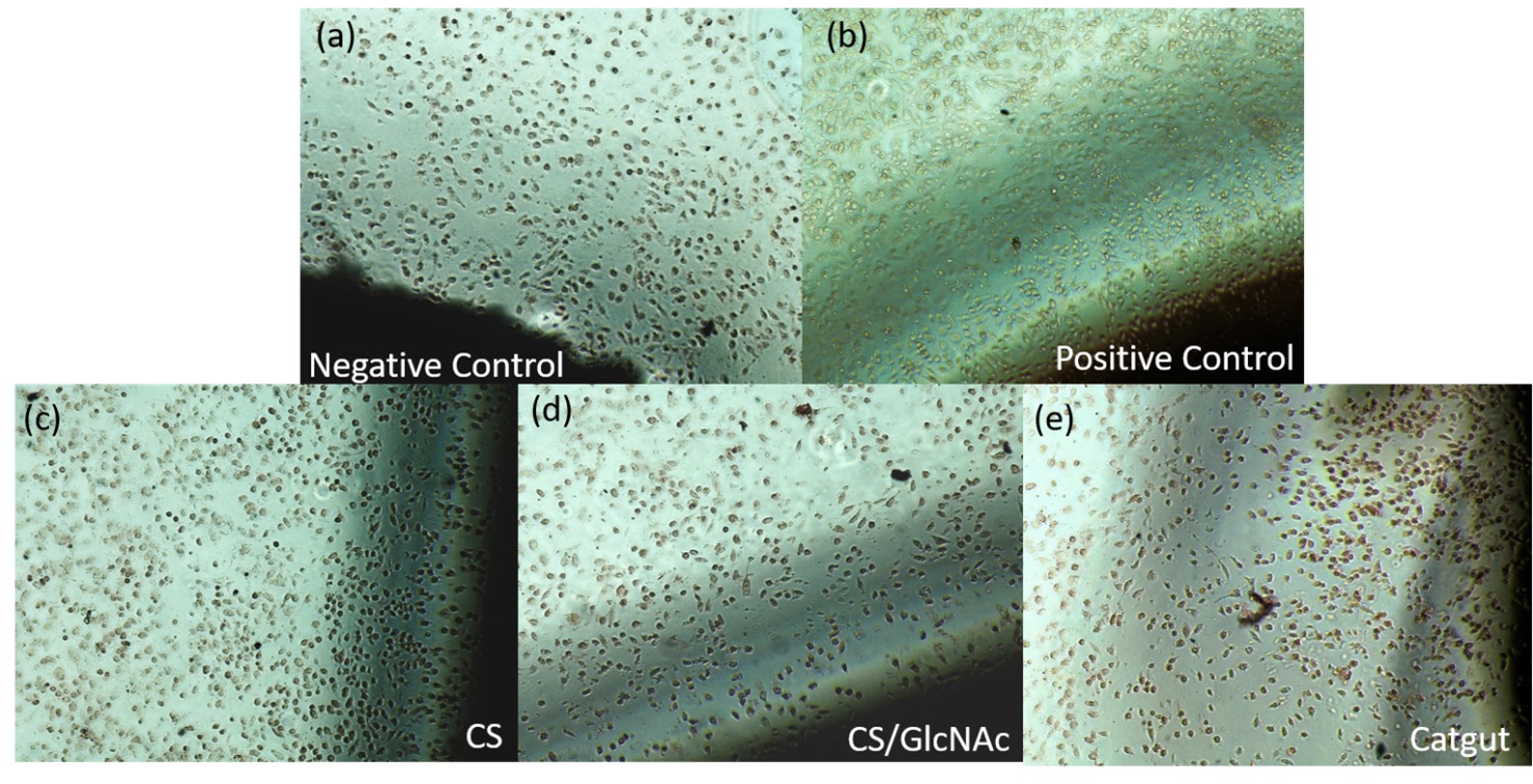
The in vitro cytotoxicity of CS, CS/GlcNAc, and Catgut sutures on L929 cells was assessed using the agar diffusion method. Cytotoxicity was determined by measuring the halo size around each material after 24 hours of incubation. Qualitative results for the positive and negative controls, as well as for the CS, CS/GlcNAc, and Catgut sutures, were obtained based on the halo analysis, with mean values recorded (Figure 7).

Phase contrast microscopy images of L929 cells in the agar diffusion cytotoxicity test: a) Negative control, b) positive control, c) CS suture, d) CS/GlcNAc suture, e) Catgut suture. All of the images were taken on the same magnification (100x).
In the positive control, a halo of 0.70 cm indicated cell death (lysis) and toxicity (Table 4). This result corresponds to severely cytotoxicity (grade 4) according to ISO 10993-5 standards, which define a halo size greater than 1.0 cm as indicative of severely cytotoxicity (Table 2).
The negative control demonstrated no cytotoxicity, with a halo size of 0.00 cm in the plate readings and a cytotoxicity grade of 0 (zero). The cytotoxicity tests for CS, CS/GlcNAc, and Catgut sutures revealed no halo formation around the filaments by the L929 cell line, and there were no observed changes in cell morphology, mirroring the negative controls (Figure 7).
These results suggest that CS, CS/GlcNAc, and Catgut sutures are non-toxic to L929 cells and can be safely used as absorbable surgical suture material. This conclusion aligns with previous studies that also found no cytotoxicity in chitosan-based materials81-84 and Catgut44,85.
3.6. Antibacterial in vitro Test
The antimicrobial effect of chitosan threads with and without N-Acetyl-D-glucosamine, as well as commercial Catgut thread, was evaluated against two representative microorganisms from the Gram-positive and Gram-negative groups: Staphylococcus aureus and Escherichia coli.
The samples consisted of threads approximately 0.5 mm thick. Fragments of the threads were applied directly onto Petri dishes containing Mueller-Hinton medium, previously inoculated with the tested microorganisms. The plates were incubated upside down for 24 hours at 35 ± 2 °C. After incubation, the formation of halos was observed and recorded in Table 5.
Sutures can serve as entry points for infections, hindering wound healing. Surgical threads made from natural fibers may increase the risk of developing infections because once colonized, local mechanisms to prevent infection become less effective. Additionally, some oral pathogens are resistant to antibiotics. Multifilament sutures, with their larger surface area for microorganism adherence, tend to exhibit a higher bacterial adhesion rate than monofilament sutures. Therefore, it is important to avoid leaving sutures in place for extended periods86.
The positive control exhibited significant inhibition zones, confirming the sensitivity of the microorganisms to the antimicrobial used. The negative control showed no inhibition zones, indicating no contamination or undesired antimicrobial activity (Table 5).
The results showed a very small inhibition of S. aureus and E. coli bacterial growth (0.5 mm) for the commercial Catgut sutures, suggesting low or no antimicrobial activity against these bacteria, as presented in Table 5.
Similar findings were reported by Saraí et al.87, who evaluated the antimicrobial activity of Catgut sutures with and without silver nanoparticle coatings, finding that sutures without silver nanoparticles showed no significant inhibition of Escherichia coli and Staphylococcus aureus growth. Chitosan sutures with N-Acetyl-D-glucosamine exhibited inhibition zones similar to the Catgut sutures (0.5 mm), suggesting that the addition of N-Acetyl-D-glucosamine negatively interfered with the antimicrobial activity of the chitosan sutures.
In contrast, the pure chitosan sutures showed larger inhibition zones, especially against S. aureus (4-5 mm), indicating moderate antimicrobial activity, primarily against Gram-positive bacteria like S. aureus. The greater efficacy of chitosan can be attributed to several mechanisms. For Gram-negative bacteria like E. coli, high molecular weight chitosan hinders nutrient exchange by binding to porins in the outer membrane, resulting in bacterial cell death88,89.
For Gram-positive bacteria like S. aureus, chitosan binds to teichoic acids and potentially extracts lipids from the membrane, leading to bacterial death90.
3.7. Analysis of healing in rat skin in Vivo
Initially, the development of wound healing after the use of CS, CS/GlcNAc, and Catgut sutures was evaluated in vivo. It was observed that the animals had a satisfactory clinical evolution in the postoperative period, with a return to feeding and excretion within 48 hours. In the macroscopic analysis (Figure 7), a satisfactory evolution of the surgical wound was observed, with no signs of bacterial infection or purulent secretion in all groups (CS, CS/GlcNAc, Catgut, and Control) at different time points (0,1,4,7, and 14 days).
As shown in Figure 8, wounds treated with CS and CS/GlcNAc sutures showed a seemingly smaller swelling effect and a moderate inflammatory response, which was less pronounced than the response exhibited by animals treated with Catgut sutures.

Representative images of wound treated with CS, CS/GlcNAc and Catgut for 0, 1, 4, 7 and 14 days.
Edema was observed in animals from the Catgut and Control groups until the 4th postoperative day. Animals from the CS and CS/GlcNAc groups had a lower inflammatory response, which allowed for faster healing of the skin wound (Figure 8).
On the 4th day after suturing, the absence of CS and CS/GlcNAc threads was confirmed, and it was observed that 100% of the wound was closed using the CS/GlcNAc suture, demonstrating the efficacy of the thread containing N-Acetyl-D-glucosamine. This was anticipated because GlcNAc, in conjunction with chitosan, not only exhibits hemostatic properties but also enhances the production of hyaluronic acid in the wound, facilitating a rapid healing process91,92.
On day 7, there was total closure of the wound treated with CS suture, while the wound treated with Catgut suture presented a mild inflammatory process. In this period, the formation of scar tissue in wounds healed with CS and CS/GlcNAc visually revealed a smooth appearance and clear aspect, without leaving a scar. By 14 days, more than 50% of the hair had already grown in animals treated with chitosan suture and those treated with CS/GlcNAc suture. While in the animal treated with commercial Catgut suture, hair growth was not observed yet, and the presence of the thread at the incision site was still evident.
During the healing process, the initial phase of tissue repair involves the release of several chemical mediators that induce vasodilation and increased vascular permeability, triggering signs of inflammation93,94. Edema following surgical procedures is a significant clinical concern that can impact patient recovery and rehabilitation. The development of edema is a complex process influenced by various factors, including the body's inflammatory response to surgical injury, which leads to vasodilation and increased vascular permeability. This allows fluid to leak into the surrounding tissues, which is a normal part of the healing process as it facilitates the transport of nutrients, oxygen, and immune cells to the site of injury. However, when edema becomes excessive, it can have detrimental effects on healing by impairing tissue nutrition and oxygenation and increasing the risk of infection95,96.
In group Control, the lack of wound stabilization due to the absence of approximation of its edges may have contributed to the formation of edema. In group Catgut, the suture thread may have caused a more intense tissue reaction.
Greenberg and Clark67, suggest that the intensity of inflammation depends on the nature and characteristics of the suture material used, which may explain the presence of inflammatory infiltrate and the type of chronic inflammation that predominated in the Catgut group, as well as the increased granulation tissue reaction, indicating that this suture acted as a foreign body. This is supported by the studies of Huaixan et al.97 and Ávila et al.27, which also found a mild inflammatory reaction inherent to chitosan sutures, as well as to chitosan-coated sutures85.
In the histological evaluation of the suture region (Table 6), the occurrences of necrosis, multinucleated giant cells, and type of inflammation did not show statistically significant differences between the experimental suture thread groups and the control group (P>0.05) throughout the experimental periods (7, 14, and 28 postoperative days). No focus of necrosis was presented in the histological examination in any of the groups.
Comparison of medians for the presence of inflammatory infiltrate, acute inflammatory infiltrate, chronic inflammatory infiltrate, and necrotic focus, obtained at different postoperative periods (7, 14, and 28 postoperative days).
A higher presence of inflammatory infiltrate and chronic inflammation was observed in the Catgut group throughout the experimental period. However, a statistically significant difference was only found compared to the control and CS/GlcNAc groups (P=0.011) at the 28-day mark. No events of acute inflammatory infiltrate or necrosis were demonstrated among the experimental groups during the evaluated periods (P>0.05) (Table 6). The Catgut group showed a higher chronic inflammatory infiltrate compared to the other groups, indicating a more prolonged inflammatory response associated with the use of Catgut suture threads
The intensity of multinucleated giant cells, granulation tissue proliferation, and collagen fiber evaluation in relation to different groups and postoperative evaluation times are shown in Table 7. The results indicate that there was no statistically significant difference between the sutures from the experimental groups (CS, CS/GlcNAc, and Catgut) and the control group regarding the number of multinucleated giant cells at all evaluation times (7, 14, and 28 days), as indicated by p-values above 0.05 for all periods evaluated.
Comparison of scores for histological parameters in groups CS, CS/GlcNAc, Catgut, and Control: giant cells, granulation tissue, and collagen fibers evaluated at different postoperative periods (7, 14, and 28 postoperative days).
For granulation tissue, no statistically significant differences were observed between the experimental groups and the control group at 7 and 14 days. However, at 28 days, a statistically significant difference was observed between the groups (p = 0.001), with the Catgut group showing a different score than the other groups evaluated (Table 7).
Regarding collagen fibers, there was no significant difference between the groups at 7 days. However, at 14 and 28 days, statistically significant differences were observed between the groups (p = 0.010 and p = 0.014, respectively). These differences indicate that the Catgut and CS/GlcNAc groups showed differences in collagen fiber scores compared to the CS and Control groups.
The faster healing and reduced signs of inflammation in CS and CS/GlcNAc groups are supported by studies by Le et al.98, which investigated wound healing in Sprague Dawley rats using chitosan-based hydrocolloid adhesives. These studies found a significant increase in wound healing rate, accelerating the inflammatory stage by suppressing the activity of pro-inflammatory cytokines (e.g., TNF-α, IL-6, MCP-1, and IL-1β).
Anushree et al.99, in your study found that chitosan accelerated the healing of dermal wounds in diabetic Wistar rats. Additionally, the product was effective in promoting skin regeneration, as demonstrated by an increase in the number of fibroblasts through specific biomarkers. This can be explained by the fact that chitosan participates in all phases of wound repair, potentially increasing extracellular matrix formation and promoting cell growth due to cells adhering strongly to the polymer and proliferating rapidly. In addition to its ability to induce blood coagulation, antimicrobial activity3,22,85,100-107
4. Conclusions
This study successfully developed chitosan (CS) and chitosan with N-Acetyl-D-glucosamine (CS/GlcNAc) sutures using the wet-spinning method with medium molecular weight chitosan and compared them to commercial Catgut sutures. The chitosan sutures demonstrated promising biological, cytotoxicological, healing, microbiological, and biodegradation properties, meeting the standards for surgical suture threads. Although Catgut sutures exhibited superior mechanical performance, essential for effective wound closure and healing, the incorporation of GlcNAc into chitosan sutures accelerated their degradation rate, making them suitable for applications requiring faster degradation. Additionally, the chitosan-based sutures exhibited tensile strength within the standards for surgical sutures (U.S. Pharmacopeia (1.7 N) for number 6-0 sutures). In vivo observations indicated that both chitosan and chitosan/GlcNAc sutures elicited a lower inflammatory response and facilitated faster wound healing compared to animals treated with commercial Catgut sutures, underscoring their potential for future medical applications. While further improvements in the mechanical properties of chitosan sutures are necessary to compete effectively with Catgut sutures across a wide range of surgical applications, their potential benefits in terms of biocompatibility and accelerated healing make them a promising alternative worth exploring. The unique structural features and performance characteristics of chitosan fibers obtained through the wet coagulation process affirm their suitability for biomedical applications.
5. References
- 1 Sneha KR, Steny PS, Sailaja GS. Intrinsically radiopaque and antimicrobial cellulose based surgical sutures from mechanically powerful Agave sisalana plant leaf fibers. Biomater Sci. 2021;9(23):7944-61.
- 2 Li Y, Meng Q, Chen S, Ling P, Kuss MA, Duan B, et al. Advances, challenges, and prospects for surgical suture materials. Acta Biomater. 2023;168:78-112.
- 3 Rakhmatullayeva D, Ospanova A, Bekissanova Z, Jumagaziyeva A, Savdenbekova B, Seidulayeva A, et al. Development and characterization of antibacterial coatings on surgical sutures based on sodium carboxymethyl cellulose/chitosan/chlorhexidine. Int J Biol Macromol. 2023;236:124024.
- 4 Perez AR, Coladonato C, Muchintala R, Christopher N, Matthews J, Tjoumakaris FP, et al. A systematic review of the biomechanical properties of suture materials used in orthopaedics. J Orthop. 2024;51:122-9.
- 5 Silva MCD, Leal RDCA, Silva HND, Fook MVL. Biodegradable suture threads as controlled drug delivery systems. Mater Res Innov. 2020;24(3):161-5.
- 6 Mohammadi H, Alihosseini F, Hosseini SA. Improving physical and biological properties of nylon monofilament as suture by Chitosan/Hyaluronic acid. Int J Biol Macromol. 2020;164:3394-402.
- 7 Silva MC, Silva HN, Alves Leal Cruz RC, Sagoe Amoah SK, Lima Silva SM, Lia Fook MV. N-Acetyl-D-glucosamine-loaded chitosan filaments biodegradable and biocompatible for use as absorbable surgical suture materials. Materials. 2019;12(11):1807.
- 8 Perrin N, Mohammadkhani G, Homayouni Moghadam F, Delattre C, Zamani A. Biocompatible fibers from fungal and shrimp chitosans for suture application. Curr Res Biotechnol. 2022;4:530-6.
- 9 Shah R, Taylor L, Saeinasab M, Zhang X, Zhang W, Nair K, et al. Bioactive sutures: advances in surgical suture functionalization. In: Thomas S, Coates P, Whiteside B, Joseph B, Nair K, editors. Advanced technologies and polymer materials for surgical sutures. Cambridge: Woodhead Publishing; 2023. p. 149-69. Chapter 7.
- 10 Zhang Q, Zou Y, Tang L, Liu X, Hu M, Han H, et al. Stage-controlled antibacterial surgical sutures based on curcumin@ZIF-8 functional coating for improved wound healing. Prog Org Coat. 2023;184:107829.
- 11 Mostafa NA, Hamdi SAH, Fol MF. Potential anthelmintic effect of chitosan on Syphacia muris infecting Wistar rats: biochemical, immunological, and histopathological studies. Sci Rep. 2024;14(1):2825.
- 12 Lee JE, Park S, Park M, Kim MH, Park CG, Lee SH, et al. Surgical suture assembled with polymeric drug-delivery sheet for sustained, local pain relief. Acta Biomater. 2013;9(9):8318-27.
- 13 Shen Z, Kamdem DP. Development and characterization of biodegradable chitosan films containing two essential oils. Int J Biol Macromol. 2015;74:289-96.
- 14 Gu BK, Park SJ, Kim MS, Lee YJ, Kim J-I, Kim C-H. Gelatin blending and sonication of chitosan nanofiber mats produce synergistic effects on hemostatic functions. Int J Biol Macromol. 2016;82:89-96.
- 15 Mohammadi A, Hashemi M, Masoud Hosseini S. Effect of chitosan molecular weight as micro and nanoparticles on antibacterial activity against some soft rot pathogenic bacteria. Lebensm Wiss Technol. 2016;71:347-55.
- 16 Wang J, Wang L, Yu H, Zain-ul-Abdin, Chen Y, Chen Q, et al. Recent progress on synthesis, property and application of modified chitosan: an overview. Int J Biol Macromol. 2016;88:333-44.
- 17 Pereira dos Santos E, Nicácio PH, Coêlho Barbosa F, Nunes da Silva H, Andrade AL, Lia Fook MV, et al. Chitosan/essential oils formulations for potential use as wound dressing: physical and antimicrobial properties. Materials. 2019;12(14):2223.
- 18 Silva HN, Tissiani MP, Souza MF, Barreto MEV, Barbosa RC, Lia Fook MV. Evaluation of crosslinking conditions on the properties of chitosan woven meshes. Mater Res. 2022;25:e20210472.
- 19 Ning S, Wang C, Zhao L, Yang J, Shi X, Zheng Y. Lecithin/chitosan nanoparticle drug carrier improves anti-tumor efficacy of Monascus pigment rubropunctatin. Int J Biol Macromol. 2023;242(Pt 3):125058.
- 20 Shi L, Xu S, Zhu Q, Wei Y. Chitosan-coated miconazole as an effective anti-inflammatory agent for the treatment of postoperative infections in obstetrics and vaginal yeast infection control on in vitro evaluations. Microb Pathog. 2023;184:106312.
- 21 Jagdale S, Agarwal B, Dixit A, Gaware S. Chitosan as excellent bio-macromolecule with myriad of anti-activities in biomedical applications: a review. Int J Biol Macromol. 2024;257(Pt 2):128697.
- 22 Okamoto Y, Kawakami K, Miyatake K, Morimoto M, Shigemasa Y, Minami S. Analgesic effects of chitin and chitosan. Carbohydr Polym. 2002;49(3):249-52.
- 23 Liao J, Li X, Fan Y. Prevention strategies of postoperative adhesion in soft tissues by applying biomaterials: based on the mechanisms of occurrence and development of adhesions. Bioact Mater. 2023;26:387-412.
- 24 Yin X, Du M, Sun Z, Zhu H, Xu P, Wang H. Solid-waste-based keratin/chitosan hydrogel for controlling drug release in vitro. Eur Polym J. 2023;199:112451.
- 25 Polez RT, Ajiboye MA, Österberg M, Horn MM. Chitosan hydrogels enriched with bioactive phloroglucinol for controlled drug diffusion and potential wound healing. Int J Biol Macromol. 2024;265(Pt 1):130808.
- 26 Tan Y, Rajoka MS, Ke Z, Mehwish HM, Deng W, Li J, et al. Effect of squid cartilage chitosan molecular structure on the properties of its monofilament as an absorbable surgical suture. Polymers. 2022;14(7):1306.
- 27 Ávila SH Fo, Moura V, Queiroz PJB, Faleiro MBR, Lima BSG, Freitas SLR, et al. Chitosan thread in the healing of the cecal wall of rabbits (Oryctolagus cuniculus) submitted to cecorrhaphy. J Biomed Mater Res B Appl Biomater. 2024;112(1):e35314.
- 28 Prabha S, Sowndarya J, Ram P, Rubini D, Hari BNV, Aruni W, et al. Chitosan-coated surgical sutures prevent adherence and biofilms of mixed microbial communities. Curr Microbiol. 2021;78(2):502-12.
- 29 Marchetti M, De Berardis B, Bigioni I, Mariano A, Superti F, Scotto d’Abusco A. In vitro antiviral and anti-inflammatory activities of n-acetylglucosamine: development of an alternative and safe approach to fight viral respiratory infections. Int J Mol Sci. 2023;24(6):5129.
- 30 Jain T, Kumar H, Dutta PK. D-glucosamine and N-acetyl D-glucosamine: their potential use as regenerative medicine. In: Dutta PK, editor. Chitin and chitosan for regenerative medicine. New Delhi: Springer India; 2016. p. 279-95.
- 31 Brugnerotto J, Lizardi J, Goycoolea FM, Argüelles-Monal W, Desbrières J, Rinaudo M. An infrared investigation in relation with chitin and chitosan characterization. Polymer. 2001;42(8):3569-80.
- 32 Saito Y, Liang G, Egger G, Friedman JM, Chuang JC, Coetzee GA, et al. Specific activation of microRNA-127 with downregulation of the proto-oncogene BCL6 by chromatin-modifying drugs in human cancer cells. Cancer Cell. 2006;9(6):435-43.
- 33 Leary S, Underwood W, Anthony R, Cartner S. Guidelines for the euthanasia of animals. Schaumburg, IL: American Veterinary Medical Association; 2020.
- 34 Okamoto T, Yabushita K, Nakama HH, Okamoto R. Processo de reparação cutânea após incisão e sutura com fios de poliglactina 910 e poliglecaprone 25: estudo microscópico comparativo em ratos. Rev Odontol Araçatuba. 2003;24(2):62-7.
- 35 Casalini T, Masi M, Perale G. Drug eluting sutures: a model for in vivo estimations. Int J Pharm. 2012;429(1-2):148-57.
- 36 Dresvyanina EN, Dobrovol’skaya IP, Popryadukhin PV, Yudin VE, Ivan’kova EM, Elokhovskii VY, et al. Influence of spinning conditions on properties of chitosan fibers. Fibre Chem. 2013;44(5):280-3.
- 37 Ruan G-X, Kazlauskas A. Lactate engages receptor tyrosine kinases Axl, Tie2, and vascular endothelial growth factor receptor 2 to activate phosphoinositide 3-kinase/Akt and promote angiogenesis. J Biol Chem. 2013;288(29):21161-72.
- 38 Hunt TK, Aslam RS, Beckert S, Wagner S, Ghani QP, Hussain MZ, et al. Aerobically derived lactate stimulates revascularization and tissue repair via redox mechanisms. Antioxid Redox Signal. 2007;9(8):1115-24.
- 39 Porporato PE, Payen VL, De Saedeleer CJ, Préat V, Thissen J-P, Feron O, et al. Lactate stimulates angiogenesis and accelerates the healing of superficial and ischemic wounds in mice. Angiogenesis. 2012;15(4):581-92.
- 40 Silva HN, Silva MC, Santos FSF, Silva JAC Jr, Barbosa RC, Fook MVL. Chitosan woven meshes: influence of threads configuration on mechanical, morphological, and physiological properties. Polymers. 2020;13(1):47.
- 41 Dalben GS, Francischone LA, Prieto-Oliveira P, Consolaro A. Evaluation of sutures after immersion in nonalcoholic benzydamine hydrochloride mouthrinse by scanning electron microscopy. Clin Oral Investig. 2008;12(3):287-90.
- 42 Maksymenko O, Hryn V. Methods and results of studying the structure and tinctorial properties of catgut thread. Med Ecol Problems. 2023;27(1-2):13-6.
- 43 Stern PG, Segerman E. On the structure of polypropylene fibres. Polymer. 1968;9:471-7.
- 44 Alirezaie Alavijeh A, Rajabi M, Barati F, Javdani M, Karimi I, Barati M, et al. Catgut enriched with CuSO4 nanoparticles as a surgical suture: morphology, antibacterial activity, cytotoxicity and tissue reaction. Nanomed Res J. 2020;5(3):256-64.
- 45 Bak SY, Lee SW, Choi CH, Kim HW. Assessment of the influence of acetic acid residue on type I collagen during isolation and characterization. Materials. 2018;11(12):2518.
- 46 Sumner-Smith G, Dingwall JS. A new absorbable suture. Can Vet J. 1972;13(2):53.
- 47 Carroll RE. Surgical catgut: the myth of allergy. J Hand Surg Br. 1989;14(2):218-20.
- 48 Meyer M. Processing of collagen based biomaterials and the resulting materials properties. Biomed Eng Online. 2019;18(1):24.
- 49 Sabino MA, Albuerne J, Müller AJ, Brisson J, Prud’homme RE. Influence of in vitro hydrolytic degradation on the morphology and crystallization behavior of poly(p-dioxanone). Biomacromolecules. 2004;5(2):358-70.
- 50 Sun L, Wanasekara N, Chalivendra V, Calvert P. Nano-mechanical studies on polyglactin sutures subjected to in vitro hydrolytic and enzymatic degradation. J Nanosci Nanotechnol. 2015;15(1):93-9.
- 51 Nordtveit RJ, Vårum KM, Smidsrød O. Degradation of fully water-soluble, partially N-acetylated chitosans with lysozyme. Carbohydr Polym. 1994;23(4):253-60.
- 52 Hong Y, Song H, Gong Y, Mao Z, Gao C, Shen J. Covalently crosslinked chitosan hydrogel: properties of in vitro degradation and chondrocyte encapsulation. Acta Biomater. 2007;3(1):23-31.
- 53 Lu G, Sheng B, Wang G, Wei Y, Gong Y, Zhang X, et al. Controlling the degradation of covalently cross-linked carboxymethyl chitosan utilizing bimodal molecular weight distribution. J Biomater Appl. 2009;23(5):435-51.
- 54 Lončarević A, Ivanković M, Rogina A. Lysozyme-induced degradation of chitosan: the characterisation of degraded chitosan scaffolds. J Tissue Repair Regen. 2017;1(1):12-22.
- 55 Islam N, Wang H, Maqbool F, Ferro V. In vitro enzymatic digestibility of glutaraldehyde-crosslinked chitosan nanoparticles in lysozyme solution and their applicability in pulmonary drug delivery. J Biomater Appl. 2019;24(7):1271.
- 56 Pangburn SH, Trescony PV, Heller J. Lysozyme degradation of partially deacetylated chitin, its films and hydrogels. Biomaterials. 1982;3(2):105-8.
- 57 Li Q, Dunn ET, Grandmaison EW, Goosen MFA. Applications and properties of chitosan. J Bioact Compat Polym. 1992;7(4):370-97.
- 58 Ravi Kumar MNV. A review of chitin and chitosan applications. React Funct Polym. 2000;46(1):1-27.
- 59 Wang X, Yan Y, Zhang R. A comparison of chitosan and collagen sponges as hemostatic dressings. J Bioact Compat Polym. 2006;21(1):39-54.
- 60 Kean T, Thanou M. Biodegradation, biodistribution and toxicity of chitosan. Adv Drug Deliv Rev. 2010;62(1):3-11.
- 61 Lizardi-Mendoza J, Argüelles Monal WM, Goycoolea Valencia FM. Chemical characteristics and functional properties of chitosan. In: Bautista-Baños S, Romanazzi G, Jiménez-Aparicio A, editors. Chitosan in the preservation of agricultural commodities. San Diego: Academic Press; 2016. p. 3-31. Chapter 1.
- 62 Zhu S, Dou W, Zeng X, Chen X, Gao Y, Liu H, et al. Recent advances in the degradability and applications of tissue adhesives based on biodegradable polymers. Int J Mol Sci. 2024;25(10):5249.
- 63 Seitz JM, Durisin M, Goldman J, Drelich JW. Recent advances in biodegradable metals for medical sutures: a critical review. Adv Healthc Mater. 2015;4(13):1915-36.
- 64 He H, Li X, Wu J, Xiao J, Zhang H. Bioactive degradable surgical suture and preparation method thereof. Australian patent AU 2019210543 B1. 2020, Feb 6.
- 65 Yang YM, Hu W, Wang XD, Gu XS. The controlling biodegradation of chitosan fibers by N-acetylation in vitro and in vivo. J Mater Sci Mater Med. 2007;18(11):2117-21.
- 66 Lim SM, Song DK, Oh SH, Lee-Yoon DS, Bae EH, Lee JH. In vitro and in vivo degradation behavior of acetylated chitosan porous beads. J Biomater Sci Polym Ed. 2008;19(4):453-66.
- 67 Greenberg JA, Clark RM. Advances in suture material for obstetric and gynecologic surgery. Rev Obstet Gynecol. 2009;2(3):146-58.
- 68 Mucha M, Pawlak A. Complex study on chitosan degradability. Polimery. 2002;47(7):509-16.
- 69 Jin Q, Yu H, Wang X, Li K, Li P. Effect of the molecular weight of water-soluble chitosan on its fat-/cholesterol-binding capacities and inhibitory activities to pancreatic lipase. PeerJ. 2017;5:e3279.
- 70 Takeda M, Kondo K, Sanda S, Kan D, Borges IK, Suzuki I, et al. Enzymatic degradation of β-1,4-linked N-acetylglucosaminoglucan prepared from Thiothrix nivea Int J Biol Macromol. 2018;109:323-8.
- 71 Alhosseini SN, Moztarzadeh F, Mozafari M, Asgari S, Dodel M, Samadikuchaksaraei A, et al. Synthesis and characterization of electrospun polyvinyl alcohol nanofibrous scaffolds modified by blending with chitosan for neural tissue engineering. Int J Nanomedicine. 2012;7:25-34.
- 72 Luo H, Li W, Ji D, Zuo G, Xiong G, Zhu Y, et al. One-step exfoliation and surface modification of lamellar hydroxyapatite by intercalation of glucosamine. Mater Chem Phys. 2016;173:262-7.
- 73 Hao XP, Chen SG, Wang WH, Yang ZQ, Yue LF, Sun HY, et al. AgNP-coordinated glucosamine-grafted carbon nanotubes with enhanced antibacterial properties. New J Chem. 2017;41(15):7045-51.
- 74 Riaz T, Zeeshan R, Zarif F, Ilyas K, Muhammad N, Safi SZ, et al. FTIR analysis of natural and synthetic collagen. Appl Spectrosc Rev. 2018;53(9):703-46.
- 75 Wang S-S, Yu Y, Sun Y, Liu N, Zhou D-Q. Comparison of physicochemical characteristics and fibril formation ability of collagens extracted from the skin of farmed river puffer (Takifugu obscurus) and tiger puffer (Takifugu rubripes). Mar Drugs. 2019;17(8):462.
- 76 Nashchekina YA, Starostina AA, Trusova NA, Sirotkina MY, Lihachev AI, Nashchekin AV. Molecular and fibrillar structure collagen analysis by FTIR spectroscopy. J Phys Conf Ser. 2020;1697(1):012053.
- 77 Gonzalez L, Wess T. Use of attenuated total reflection-Fourier transform infrared spectroscopy to measure collagen degradation in historical parchments. Appl Spectrosc. 2008;62(10):1108-14.
- 78 Belbachir K, Noreen R, Gouspillou G, Petibois C. Collagen types analysis and differentiation by FTIR spectroscopy. Anal Bioanal Chem. 2009;395(3):829-37.
- 79 Kristoffersen KA, Måge I, Wubshet SG, Böcker U, Riiser Dankel K, Lislelid A, et al. FTIR-based prediction of collagen content in hydrolyzed protein samples. Spectrochim Acta A Mol Biomol Spectrosc. 2023;301:122919.
- 80 Dart AJ, Dart CM. Suture material: conventional and stimuli responsive. Comp Biomaterials. 2011;6:573-87.
- 81 Rejinold NS, Muthunarayanan M, Muthuchelian K, Chennazhi KP, Nair SV, Jayakumar R. Saponin-loaded chitosan nanoparticles and their cytotoxicity to cancer cell lines in vitro. Carbohydr Polym. 2011;84(1):407-16.
- 82 Vulcani VAS, Bizarria MTM, d’Ávila MA, Mei LHI, Bernal C, Perussi JR. Cytotoxicity tests for nanostructured chitosan/PEO membranes using the agar diffusion method. Mater Res. 2012;15:213-7.
- 83 Zhao R, Li X, Sun B, Zhang Y, Zhang D, Tang Z, et al. Electrospun chitosan/sericin composite nanofibers with antibacterial property as potential wound dressings. Int J Biol Macromol. 2014;68:92-7.
- 84 Zoe LH, David SR, Rajabalaya R. Chitosan nanoparticle toxicity: a comprehensive literature review of in vivo and in vitro assessments for medical applications. Toxicol Rep. 2023;11:83-106.
- 85 Chen Y-G, Li C-X, Zhang Y, Qi Y-D, Feng J, Zhang X-Z. Antibacterial sutures coated with smooth chitosan layer by gradient deposition. Chin J Polym Sci. 2022;40(9):1050-61.
- 86 Mahesh L, Kumar VR, Jain A, Shukla S, Aragoneses JM, Martínez González JM, et al. Bacterial adherence around sutures of different material at grafted site: a microbiological analysis. Materials. 2019;12(18):2848.
- 87 Saraí CG-R, Rogelio JS-V, Raúl AM-L, Víctor S-M, Rafael López C. Antimicrobial effect of silk and catgut suture threads coated with biogenic silver nanoparticles. In: Khan M, editor. Silver nanoparticles. Rijeka: IntechOpen; 2018. Chapter 13.
- 88 Li X, Feng X, Yang S, Fu G, Wang T, Su Z. Chitosan kills Escherichia coli through damage to be of cell membrane mechanism. Carbohydr Polym. 2010;79(3):493-9.
- 89 Yan D, Li Y, Liu Y, Li N, Zhang X, Yan C. Antimicrobial properties of chitosan and chitosan derivatives in the treatment of enteric infections. Molecules. 2021;26(23):7136.
- 90 Raafat D, von Bargen K, Haas A, Sahl H-G. Insights into the mode of action of chitosan as an antibacterial compound. Appl Environ Microbiol. 2008;74(12):3764-73.
- 91 Güneş S, Tıhmınlıoğlu F. Hypericum perforatum incorporated chitosan films as potential bioactive wound dressing material. Int J Biol Macromol. 2017;102:933-43.
- 92 Ouyang QQ, Hu Z, Lin ZP, Quan WY, Deng YF, Li SD, et al. Chitosan hydrogel in combination with marine peptides from tilapia for burns healing. Int J Biol Macromol. 2018;112:1191-8.
- 93 Tiruppathi C, Minshall RD, Paria BC, Vogel SM, Malik AB. Role of Ca2+ signaling in the regulation of endothelial permeability. Vascul Pharmacol. 2002;39(4-5):173-85.
- 94 Udell IJ, Abelson MB. Chemical mediators of inflammation. Int Ophthalmol Clin. 1983;23(1):15-26.
- 95 Lehnert B, Jhala G. The use of foam as a postoperative compression dressing. J Foot Ankle Surg. 2005;44(1):68-9.
- 96 Slaa AT, Dolmans DEJGJ, Ho GH, Moll FL, van der Laan L. Pathophysiology and treatment of edema following femoropopliteal bypass surgery. Vascular. 2012;20(6):350-9.
- 97 Huaixan LN, Arruda SS, Leonardo AS, Viana JC, Barreto-Vianna AR, Ximenes FH, et al. Macroscopic, histochemical, and immunohistochemical comparison of hysterorrhaphy using catgut and chitosan suture wires. J Biomed Mater Res B Appl Biomater. 2016;104(1):50-7.
- 98 Le LTT, Giang NN, Chien PN, Trinh XT, Long NV, Van Anh LT, et al. Enhancement of wound healing efficacy by chitosan-based hydrocolloid on sprague dawley rats. In Vivo. 2023;37(3):1052-64.
- 99 Anushree U, Punj P, Vasumathi, Bharati S. Phosphorylated chitosan accelerates dermal wound healing in diabetic wistar rats. Glycoconj J. 2023;40(1):19-31.
- 100 Stricker-Krongrad AH, Alikhassy Z, Matsangos N, Sebastian R, Marti G, Lay F, et al. Efficacy of chitosan-based dressing for control of bleeding in excisional wounds. Eplasty. 2018;18:e14.
- 101 Notario-Pérez F, Martín-Illana A, Cazorla-Luna R, Ruiz-Caro R, Veiga MD. Applications of chitosan in surgical and post-surgical materials. Mar Drugs. 2022;20(6):396.
- 102 Gheorghiță D, Moldovan H, Robu A, Bița AI, Grosu E, Antoniac A, et al. Chitosan-based biomaterials for hemostatic applications: a review of recent advances. Int J Mol Sci. 2023;24(13):10540.
- 103 Mititelu Tartau L, Bogdan M, Buca BR, Pauna AM, Tartau CG, Dijmarescu LA, et al. Evaluation of antinociceptive effects of chitosan-coated liposomes entrapping the selective kappa opioid receptor agonist U50,488 in mice. Medicina. 2021;57(2):138.
- 104 Huang S, Han B, Shao K, Yu M, Liu W. Analgesis and wound healing effect of chitosan and carboxymethyl chitosan on scalded rats. J Ocean Univ China. 2014;13(5):837-41.
- 105 Ueno H, Mori T, Fujinaga T. Topical formulations and wound healing applications of chitosan. Adv Drug Deliv Rev. 2001;52(2):105-15.
- 106 Guarnieri A, Triunfo M, Scieuzo C, Ianniciello D, Tafi E, Hahn T, et al. Antimicrobial properties of chitosan from different developmental stages of the bioconverter insect Hermetia illucens. Sci Rep. 2022;12(1):8084.
- 107 Matica MA, Aachmann FL, Tøndervik A, Sletta H, Ostafe V. Chitosan as a wound dressing starting material: antimicrobial properties and mode of action. Int J Mol Sci. 2019;20(23):5889.