Floral Morphoanatomy and Pollen Viability in Miconia polystachya (naudin) r. Goldenb., (Miconieae, Melastomataceae)
Abstract
Many Melastomataceae are distributed in highland rocky fields (Campos rupestres) landscapes with their intrinsic environmental factors. These factors influence the expression of morphoanatomical characteristics and determine which adaptations are necessary for survival, including reproductive processes. The genus Miconia belongs to the Neotropical tribe Miconieae, the largest in Melastomataceae. Species of Miconieae are known to engage in apomixis, which results in much of the tribe possibly carrying out autonomous or pseudogamous reproduction. Thus, this study seeks to describe the morphoanatomy of the flowers of Miconia polystachya (Naudin) R. Goldenb. and contribute to the understanding of their reproduction. For this, flowers were collected at different stages of development in different seasons (autumn and spring) for morphoanatomical study. The flowers were fixed and cross and longitudinal sections were carried out with a rotating microtome. Pollen viability tests were also performed. The individuals of Miconia polystachya studied in highland rocky fields of the state of Paraná have a high percentage of unviable pollen, verified by the structure of pollen grains showing different degrees of degeneration. However, individuals with mature fruits were observed, corroborating the association between reproduction of this species with apomixis. The described morphological characteristics are similar to those found in previous studies for the family and genus. However, our study provides novel ecological information about the flowering and floral anatomy of M. polystachya, emphasizing the anatomy of hypanthium, fertile verticils and embryonic development of the species.
Keywords:
apomixis; agamospermy; highland rocky fields; parthenogenesis; reproduction
HIGHLIGHTS
Low frequency of viable pollen grains was confirmed by staining tests and records of spore development phases.
The low frequency is related to low pollen viability and fruit set observed with occurrence of apomixis.
General floral morphological and anatomical features of the species are similar to the described for the genus and family.
Floral anatomy features of the species are described for the first time.
INTRODUCTION
Miconia polystachya (Naudin) R. Goldenb. belongs to the tribe Miconieae in family Melastomataceae, one of the plant families with greatest species diversity in Neotropical biomes [1, 2]. In Brazil, 1,436 of the 5,750 species described in the family worldwide are found [3, 4]. Furthermore, 929 of these species are endemic to the country [5]. Tribe Miconieae is the largest tribe in the family and the richest in the Neotropics with 1,900 species distributed in 16-19 genera [6]. Recent phylogenetic studies have found that Miconieae is polyphyletic and supported changes in its circumscription, both at genus and species level. Among those species is M. polystachya which, according to Michelangeli and coauthors (2019) [6], should be transferred from Leandra to Miconia. However, Flora and Funga from Brazil (2022) [1] still recognizes M. polystachia as a synonym of Leandra polystachya (Naudin) Cogn. We chose to follow the wider circumscription of Miconia, as species of Leandra have emerged in 10 different clades of Miconieae [7] in phylogenetic studies.
Miconia has 560 species recorded in Brazil [8], and in the state of Paraná, 32 species are found, belonging to seven sections that can be recognized by features like mostly terminal inflorescences, without leafy bracts, non-contrite hypanthium at the apex, reduced external laciniae, petals with a rounded or emarginated apex and bacaceous fruits. [9].
Miconia polystachya has a wide distribution in the country, with records in the states of Minas Gerais, São Paulo, Paraná, Santa Catarina, and Rio Grande do Sul, and a corresponding wide morphological variation. It can be identified by oval leaves with basal veins, short petiole and leaf blade glabrous or with sparse trichomes [10]. Its habit can vary according to the environment where it is found, but it is usually seen as subshrubs or shrubs that can reach more than one meter in height [11]. In Paraná, the species is found in the Guartelá State Park, which protects a rich and abundant native flora [11, 12]. This protected area contains a vegetation mosaic formed by highland rocky fields, Cerrado and Mixed Ombrophilous Forest, for example [13]. This diversity makes up the so-called High-Altitude Complex, which has a high rate of plant endemism and is rich in Melastomataceae [14].
The flowers of most species of Melastomataceae have sickle-shaped tubular anthers with apical pores, through which the pollen is released [15]. The anther morphology and this type of dehiscence are intertwined and related to pollination, which in most cases is carried out through vibration from bees [16, 17]. However, biotic pollination does not seem to occur in M. polystachya, with authors reporting a low frequency of pollinator visits to individuals [18]. It is likely that biotic pollination of the species is negatively affected by the low rate of viability of pollen grains (2 to 4%), given that pollen is the main floral resource in Melastomataceae and unviable pollen grains may have reduced nutritional value [12, 19, 20].
The unviability of pollen grains in M. polystachya may be related to agamospermy or apomixis, which is significant in Miconieae, with approximately 63% of the species having some degree of apomixis in their reproductive processes [21]. Apomixis, the process in which new generations are guaranteed through the production of embryos originating from integument cells, nucellus or ovule, without the need for fertilization, occurs in many species [22]. There are types of apomixis in which embryos originating from embryonic sacs fertilized together with apomictic embryos can be formed, resulting in seeds with more than one embryo. Furthermore, fertilization may be important in pseudogamous type apomixis, where it acts in the formation of the endosperm [23].
With that, the existing relationships between reproductive strategies adopted by plants with the environment where they live and pollinating agents need to be better understood. The increase in global average temperatures can interfere with phenological cycles, leading plants to start their reproductive periods earlier, thus altering the ecological dynamics between flowering, pollination, fruiting, and seed dispersal [24]. Although studies have been carried out with the species in highland rocky fields [11, 12], the description of the anatomical characteristics of floral whorls has not been addressed. Thus, this study seeks to collect this information, as it can contribute to the understanding of reproduction in the species and subsequently help preserve highland rocky fields, a hotspot of biodiversity [25].
MATERIAL AND METHODS
Individuals of the studied species were collected in Guartelá State Park (24º34'07.7”S/50º15'40.7”W), a protected area with an extension of approximately 789.97 acres, where the soil is mainly formed by sediments from the Paleozoic period and sandstone outcrops from the Devonian Escarpment [25]. Its implementation took place in 1997, due to the need to preserve the vegetation and archaeological heritage of the region [26]. Grassland, Cerrado and Mixed Ombrophilous Forest occur naturally in the park, forming a mosaic of vegetation formations with ecotone regions [27]. Thus, its vegetation is defined as Grassy-Woody Steppe, Humid Field, and Highland Rocky Fields [13].
Collections were carried out in March and October 2022 and in March 2023. Flowers at different stages of development were collected, fixed in 50% FAA (37% formaldehyde, glacial acetic acid and 50% ethanol in a 1: 1: 18 ratio) [28] for 24 hours and preserved in 70% ethanol. The identification of the collected material was carried out with the aid of specific literature [10, 11, 29, 30] and the resulting vouchers were deposited in the UNOP herbarium (UNOP 11464) (Thiers, continuously updated).
The description of the morphological characteristics was carried out from photographs of the species taken in their habitat and from the fixed material, and this material was measured using graph paper to measure the sizes of the floral pieces. For light microscopy analysis, buds (or fragments) and flowers in complete anthesis fixed were dehydrated in a regular butyl series [31] and embedded in historesin (Leica Historesin Embedding Kit, Nussloch, Germany). Afterwards, cross and longitudinal sections were made with rotating microtome with a thickness of 3 to 5 μm, stained with 0.05% 0.1 M toluidine blue in sodium phosphate buffer at pH 6.8 [31] and mounted in Entellan (Merck, Darmstadt, Germany). Histochemical tests were carried out to identify phenolic compounds with ferric chloride [28], mucilage with methylene blue, lipids with sudan III [32] and starch with lugol [33]. Photomicrographs were obtained with the aid of a DP041 digital camera coupled to an Olympus Bx70 photomicroscope using the DP Controller program.
Calculation of pollen viability was carried out from the analysis of pollen grains from pre-anthesis buds. Pollen particles obtained by crushing the anthers with glass mixture [34] were treated in two ways: (1) prepared according to the method above (stained with Toluidine Blue) and (2) from slides stained with 1% basic fuchsine. A total of 900 pollen grains, 300 each from three individuals, were analyzed by staining. Pollen grains that did not present stained cytoplasm or in which the staining was not intense were considered non-viable. The average values obtained with each stain were compared with a Tukey's test at 5% significance using R (R Core Team 2021) [35]. Formula 1 (below) was used to calculate the average for each stain.
(1) 'VP' being the percentage of viable pollen, 'Nºnpg
' the number of pollen grains considered viable and 'Nºcpg' the number of counted pollen grains.
RESULTS
Morphology
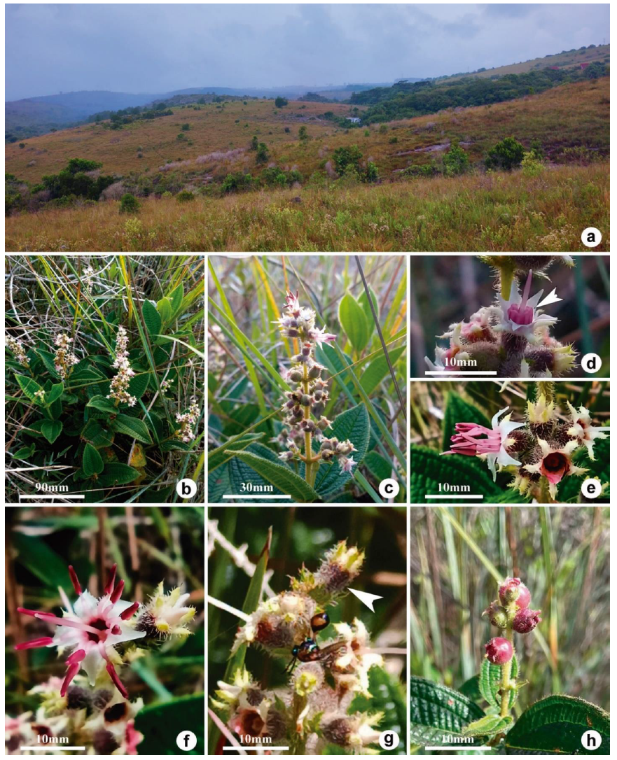
The species was found in abundance and flowering in all collection expeditions (March and October); both subshrubs and shrubs with semi-woody stems were observed. The phytophysiognomy of the collection localities was grassland vegetation (Figure 1a, b). Some individuals were collected in transition areas between grassland and forest vegetation (Mixed Ombrophilous Forest), but they did not show differences in size when compared to the others. Most individuals collected belonged to subpopulations found in the vegetation that makes up the edge of the park's main walking trail, although we also explored regions deeper into the vegetation.
The flowers of Miconia polystachya are bisexual, radial, dichlamydeous, and pentamerous, occurring in varying numbers in each glomerulus, which in turn make up the glomerulus-shaped panicle of the species (Figure 1b, c). Regarding the panicle, bud maturation occurs from the base to the apex, thus, reproductive organs in several stages of development are found in the same inflorescence, from developing buds to fruits. Flowers in different development stages are also observed in each glomerulus (Figure 1c, d).
The hypanthium is perigynous, tubular (4.5 × 2−3 mm) and campanulate (Figure 1f; Figure 2a). The calyx is formed by five laciniae; the outer laciniae are linear (2 × 1 mm) and the inner (2 × 0.5 mm) are triangular to oblong.

Miconia polystachya. A - collection localit, B - habit, C - glomerulus-shaped panicle, D - arrow shows anthers bent over the calyx, E - stamens in anthesis with apices facing the style, F- flower in complete anthesis with introrse stamens arranged radially around the style, arrow indicates the hypanthium, G - insect scraping the hypanthium epidermis covered by trichomes, H- fruits at different stages of development.
The laciniae remain until fruit maturation. The corolla is composed of five free petals (4-5 × 2-2.5 mm), white to pink and lanceolate-acuminate (Figure 1d, e, f). The androecium is composed of two whorls of five stamens each, which are curved in early development (Figure 1d), with the anthers (3 × 1 mm) close to the base of the hypanthium (Figure 1e, f). When full anthesis is reached, the stamens are positioned radially around the style and become introrse (Figure 1f).
The anthers are dorsifixed, tubular, subulate, pink and with prolonged connective (Figure 1f). During the collection and observation period, we did not record occurrence of biotic pollination, as we did not observe insects or other possible pollinators performing pollinator behavior with the fertile parts of the flowers. Nectaries were not identified in the flower structures. However, we observed insects (possibly from order Hymenoptera) interacting with the hypanthium, performing a scrapping behavior on the surface (Figure 1g). The fruits are fleshy and purple when mature (Figure 1h).
Anatomy
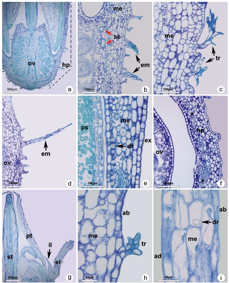
The hypanthium has uniseriate outer and inner epidermis with a thin cuticle (Figure 2b, e). In the outer epidermis, thin non-glandular emergences are visible (Figure 2b, d) and stellate-furfuraceous dendritic trichomes are present (Figure 2c). The inner epidermis consists of a layer of longitudinally elongated cells with a thin wall (Figure 2e). Just below the first cell layer, crystalline druse idioblasts are found in large numbers and present throughout the entire extension of the inner subepidermal region (Figure 2e). The mesophyll is homogeneous and made up of six to eight layers of parenchyma cells (Figure 2b, d, e), with sclereids (Figure 2b red arrows) and idioblasts with scattered druses. The vascular bundles are arranged concentrically, varying between large and small caliber (Figure 2f).
The laciniae epidermis and mesophyll (Figure 2g, i) have similar emergences and trichomes to the ones in the hypanthium (Figure 2h) but idioblasts containing druses are rare and restricted to the mesophyll when present. The petals have uniseriate epidermis composed of isodiametric cells with thin walls and a thin cuticle (Figure 2g). The occurrence of homogeneous mesophyll with parenchyma cells can be observed internally to the epidermis, where idioblasts containing druses can be found (Figure 2i).
The stamens have filaments with simple epidermis covered by a thin cuticle and without trichomes. The connective tissue is a little prolonged, and the mesophyll is homogeneous (Figure 3a). The anthers are sickle shaped and tetrasporangiate, with an epidermis formed by elongated and thin rectangular cells (Figure 3a). Crystalline idioblasts of the druse type are found in all anther tissues (Figure 3c, e, h). The developmental pattern of the anthers follows the dicotyledonous type, where the middle layer is single layered. The tapetum is uninucleate glandular (Figure 3b - d, h). Both structures degenerate as the anthers mature (Figure 3e, f). In mature anthers, the endothecium becomes thin and heterogeneously arranged in the pollen sac, being absent in the apical region close to the pore (Figure 3e - g).
During another development, the mother cells of the microspores (sporogenic cells) (Figure 3b) complete their development and form the microspore tetrahedral tetrads (Figure 3d), but cytoplasmic degradation is observed during the formation of most microgametophytes (pollen grains) (Figure 3e, h). Only some of the formed pollen grains have cellular constitution that indicates viability (dense cytoplasm and intact nuclei), most are empty, showing only the cell wall with deposited callose and exine (Figure 3e - i). Few pollen grains with cytoplasmic content are found in mature anthers (Figure 3f - i).
The gynoecium has a single, terminal style (Figure 2g) formed by an uniseriate epidermis composed of isodiametric cubic cells with thin walls and thin cuticle. The mesophyll shows elongated parenchyma cells in which prismatic crystals and druses are found (Figure 4a). The stigma has papillae that store phenolic compounds (Figure 4b). No pollen grains were observed deposited and/or germinating on the stigma and along the style.
The ovary is adnate to the hypanthium up to the upper two thirds (Figure 2a; Figure 4c). The placentation is axillary, and the ovules are arranged in three locules (Figure 4d). The ovary septum has an epidermis of cubic cells and homogeneous parenchymal mesophyll (Figure 4d). Four regions of vascular bundles are observed in the central region of the septum, three of them irrigating the locules and the central region vascularizing the style (Figure 4d). The ovules (seminal rudiment) are anatropous (Figure 4e), characterized as crassinucellate, due to the thickness of the nucellus, and the funiculus is simple, with one vascular bundle, that extends to the funiculus region, irrigating the ovules. (Figure 4e). The ovules are 2-integumented, and the external and internal integuments have three and two layers, respectively (Figure 4f). The chalaza has a cluster of cells with dense cytoplasm, defining a hypostasis region (Figure 4e, f). The micropyle is zigzag (Figure 4g). In the seminal rudiment, the archesporial cell differentiates into a megaspore mother cell during megasporogenesis (Figure 4h). We did not follow the development of the megaspore mother cell, but seminal rudiments can be seen in the megaspore tetrad phase and possible mature embryo sacs in the senescent flower (Figure 4i).

Sections of Miconia polystachya bud (a, c, e, g) and mature flower (b, d, f, h, i). A. overview of the flower bud in pre-anthesis, dashed line indicates the extent of the hypanthium; B. sclereids (red arrows) in the mesophyll and emergences in the outer epidermis of the hypanthium; C. dendritic trichomes; D. detail of non-glandular emergence (arrow); E. detail of subepidermal druse in the hypanthium (arrow); F. hypanthium mesophyll, vascular bundles of different calibers (asterisks); G. laciniae and petals in bud; H. lacinia mesophyll and adaxial epidermis; I. petal mesophyll and epidermis, arrow shows druse-type crystal. ab - abaxial epidermis, ad - adaxial epidermis, dr - druse, em - emergence, el - outer lacinia, ex - outer epidermis, hp - hypanthium, il - inner lacinia, me- mesophyll, ov - ovary, pt -petal, ps - pollen sac, st - style, tr - trichome. A, B, C, D, E, G, H and I - longitudinal section; F - cross section.ovary, pt -petal, ps - pollen sac, st - style, tr - trichome. A, B, C, D, E, G, H and I - longitudinal section; F - cross section.

Androecium of Miconia polystachya. A. overview of the anther; B. sporogenic tissue in immature anther; C. parietal layers in the immature anther; D. detail of microsporogenesis in the spore tetrad stage; E. overview and detailed view of the anther in the pre-anthesis stage; F detail of the parietal layers of the dehiscent anther; G. detail of apical portion of mature anther; H. pollen grains in anther in pre-anthesis bud; I. viable and non-viable pollen grains in mature anther. c - connective, ep - epidermis, en - endothecium, id - idioblast, mi - microsporangium, ml - middle layers, mmc - microspore mother cell, np - non-viable pollen grain, ps - pollen sac, s - sporangial septum, t - theca, ta - tapetum, te - microspore tetrahedral tetrads, vb - vascular bundle, vp - viable pollen grain. B, D, G, H, I- longitudinal sections; A, C, E, F - cross sections.

Anatomical sections of the gynoecium of M. polystachya. A. druses and prismatic crystals in the basal portion of the style; B. style apex, detail of stigma papillae; C. overview of the perigynous ovary, dashed line indicates the portion of the ovary adnate to the hypanthium; D. vascularization of ovarian septum, bundles indicated by asterisks; E. detail of the anatropous ovule; F. detail of ovule integuments and hypostasis region; G. zigzag micropyle; H. immature ovule at megaspore mother cell stage; I. mature ovule with complete gametophyte. at - antipodes, c - chalaza, dr - druse, ei - external integument, f - funiculus, gm - gametophyte, h - hypostasis, it -internal integument, m - micropyle, mmc - megaspore mother cell, n - nucellus, o - oosphere, ol - ovary locule, os - ovarian septum, pc - prismatic crystal, pn - polar nuclei, pp - papilla, si - synergids, vb - vascular bundle. A, B, C, E - longitudinal sections; D, F, G, H, I - cross sections.
Pollen viability
The calculation of pollen viability through a staining test with 1% basic fuchsin and 0.05% toluidine blue, revealed average of 3.27% and 2.39%, respectively, with the staining treatments not being statistically different (Table 1). Pollen grains with intense staining of the cytoplasmic content were considered viable (Figure 5).

Pollen viability test in Miconia polystachya. A, B. viable pollen grains; C, D. non-viable pollen grains. A, C - staining with toluidine blue, B, D - staining with basic fuchsin.
DISCUSSION
The pollen analyses carried out with individuals of Miconia polystachya confirmed the low viability of this species’ pollen, previously identified in other study [12]. The pollen viability average, 3.27% with 1% basic fuchsin and 2.39% with 0.05% toluidine blue, are close to the percentage of 2-4% described by Maia and coauthors (2016) [12], who analyzed pollen of individuals collected in the same study site. The authors attributed the small amount of viable pollen to apomixis, which could explain the observed fruiting.
Apomixis is common in the tribe Miconieae, with several reproductive biology studies reporting its occurrence [12, 18, 21, 36]. Controlled pollination experiments with species of Miconia and Leandra from higher altitude and Cerrado regions, environments that share features with the highland rocky fields from our study, showed a strong association between low pollen viability and apomixis [18, 21]. Furthermore, self-compatibility was shown to be more frequent than self-incompatibility in species of Melastomataceae from highland rocky fields [20], which might favor the production of autogamous seeds. Although we have not seen it in this study, polyembryony is also commonly associated with apomictic species of Melastomataceae from the Cerrado [36].
Most of the pollen grains observed in mature anthers of M. polystachya have an irregular shape with little or absent cytoplasm, being visualized as empty gametophytes with deposition of exine and callose. This condition is observed from pre-anthesis buds onwards, indicating that most of the pollen is unviable before anthesis. This observation can be explained by the relationship of low pollen viability with apomixis, which is the consensus in Melastomataceae studies [22, 37]. In apomictic individuals, the pollen grain formation process is marked by meiotic irregularities, such as chromosomal bridges in anaphases I and II, which can cause lagging chromosomes, originating tetrads with micronuclei, in addition to symmetric mitoses [21, 38]. These genetic disorders culminate in production of pollen grains that do not complete their development or are not viable when mature.
Apomixis in Miconieae is also related to polyploidy, as there seems to be a close relationship between polyploidy, unviable pollen and apomixis [21, 39]. Miconieae has a polyploid origin, where the polyploidy of Neotropical Miconieae has its origins in aneuploidy of a chromosomal base numbers of Paleotropical polyploid species [40]. Studies on the reproduction of polyploid Melastomataceae species indicate that low pollen viability is less frequently related to pseudogamous apomixis than autonomous apomixis [20]. These differ, respectively, in the occurrence or not of fertilization of the central cell for endosperm formation [23]. However, floral ontogeny studies, focusing on observing the development of embryo sacs and embryos, is necessary to determine the type of apomixis that occurs in M. polystachya.
Interestingly, in opposition to the expected in an apomictic species, where pollination has reduced importance and high rates of unviable pollen are common, M. polystachya presents herkogamy in the floral development. In herkogamy processes, there is a spatial separation between the female and male phases of the flower [41]. In this species, the stigma is exposed at the apex of the bud before anthesis, making it available for pollination with pollen from other flowers. Herkogamy is a common strategy to limit self-fertilization, keeping more ovules available to be fertilized by gametes with different genetic make-up, increasing cross-pollination and genetic variability of embryos [42-44].
A possible explanation for this phenomenon is that herkogamy is common in Melastomataceae, having been recorded since the first reproduction biology work in the family [2, 44, 45], and has been retained in M. polystachya. However, manual pollination of exposed stigmas and anatomical follow-up of subsequent phases are necessary to confirm the hypothesis that the rare pollen grains produced by M. polystachya are insufficient for effective fertilization, whether autogamous or allogamous.
The anatomical characteristics of the ovules, i.e., anatropous, crassinucellate, with hypostasis and integuments with more than one cell layer, are similar to those described for Miconia pepericarpa DC. and M. fallax DC., differing only from the hemi-anatropous ovules of M. pepericarpa [46]. In Miconieae, the ovule integuments may be arranged in more than one layer, which can be an useful character state for taxonomy, since it may be related to the formation of fleshy fruits in the tribe [ 47]. M. albicans has ovules with similar characteristics, including the crystalline idioblasts of the druse type concentrated in the subepidermal region of the inner epidermis of the hypanthium and sclereids dispersed in the mesophyll [48].
In almost all analyzed tissues, we observed the presence of large amounts of parenchyma and subepidermal cells differentiated into crystalliferous idioblasts containing druses and prismatic crystals. These crystals are synthesized endogenously, mainly composed of calcium oxalate and their existence is relatively common in plant tissues [49]. Druses can be observed as multifaceted clumps of calcium oxalate forming a crystal, which can be isolated or clustered in plant cells [50]. The deposition of crystals in plant tissues has several causes, such as concentration of metals and minerals in the environment, and assume different functions, such as protection against herbivory, improvement of mechanical resistance of structures or deposition of excess minerals [51]. Considering the information collected in this study, the druses could contribute to decrease fruit palatability, thus reducing predation. During development, the anthers are nested in the internal region of the hypanthium, possibly exerting pressure on its internal wall, which suggests a role of the subepidermal druses in increasing wall resistance.
Based on our results and previous studies, we classify the species as having annual flowering as flowering and fruiting individuals were found in autumn and spring [30, 52]. The berry-like fruits turn purple to reddish when mature. The partial predation observed in fruits in the final stages of maturation suggests occurrence of biotic seed dispersal, with the fruits included in the diet of the local fauna, probably birds, as they are most often reported preying on fleshy fruits of Miconieae [2, 53].
The interaction recorded between Hymenoptera and the flower hypanthium may be associated with the dense trichome layer and non-glandular emergences present. In Melastomataceae, the presence of tector and glandular trichomes on the stem, leaves and hypanthium is common, and their morphology is used for species identification [10, 48, 54]. The occurrence of emergences in the epidermis is also not uncommon [55], however the emergences we found in Miconia polystachya are not glandular, as seen in Microlepis oleaefolia (DC.) Triana [56]. These epidermal appendages are sometimes associated to the production and exudation of phenolic compounds, suggesting that this insect-plant interaction might be related to obtaining these compounds. Nevertheless, this interaction needs further investigation, as studies have shown that pollution caused by human action can negatively affect production of phenolic substances in plants [57].
CONCLUSION
This study contributes to the understanding of reproduction in Miconieae, as the high rate of unviable pollen grains found in Miconia polystachya, together with their incomplete development, support the interpretation of the species as apomictic. Although we have reported a small number of viable pollen grains, their germination needs to be studied in regards to pollen tube and viable gamete production. Likewise, the ontogeny of embryonic development of micro and megametophytes needs to be better studied to observe and confirm irregularities in cell divisions and the type of apomixis that occurs. The large number of idioblasts containing druses found in the studied individuals may be associated with structural functions and resistance to herbivory.
Acknowledgments
The State University of West Paraná and the federal scientific promotion agencies, for providing research resources. I would also like to thank the team at the Laboratory of Anatomy and Morphology of Plants (LAMP) who helped collecting and preparing the material.
REFERENCES
-
1 Goldenberg R, Baumgratz JFA, Michelangeli FA, Guimarães PJF, Romero R, Versiane AFA, et al. Melastomataceae A. Juss [Internet]. [place unknown: publisher unknown]; [cited 2023 Nov]. Available from: http://floradobrasil.jbrj.gov.br/.
» http://floradobrasil.jbrj.gov.br -
2 Renner SS. A Survey of Reproductive Biology in Neotropical Melastomataceae and Memecylaceae. Ann Missouri Bot Gard [Internet]. 1989 [cited 2023 Nov];76:496. Available from: https://doi.org/10.2307/2399497
» https://doi.org/10.2307/2399497 -
3 Michelangeli FA, Almeida F, Goldenberg R, Penneys D. A Guide to Curating New World Melastomataceae Collections with a Linear Generic Sequence to World-Wide Melastomataceae. Preprints (Basel) [Internet]. 2020 [cited 2023 Nov]. Available from: https://doi.org/10.20944/preprints202010.0203.v2
» https://doi.org/10.20944/preprints202010.0203.v2 -
4 Gomes‐da‐Silva J, Filardi FLR, Barbosa MR V, Baumgratz JFA, Bicudo CEM, Cavalcanti TB, et al. Brazilian Flora 2020: Leveraging the power of a collaborative scientific network. Taxon [Internet]. 2022 [cited 2023 Nov]; 71:178-98. Available from: https://doi.org/10.1002/tax.12640
» https://doi.org/10.1002/tax.12640 -
5 Goldenberg R, Baumgratz JFA, Souza MLDR. [Taxonomy of Melastomataceae in Brazil: retrospective, perspectives and identification key for the genera]. Rodriguésia [Internet]. 2012 [cited 2023 Nov];63:145-61. Available from: https://doi.org/10.1590/S2175-78602012000100011
» https://doi.org/10.1590/S2175-78602012000100011 -
6 Michelangeli FA, Goldenberg R, Almeda F, Judd WS, Bécquer ER, Ocampo G, et al. Nomenclatural novelties in Miconia (Melastomataceae: Miconieae). Brittonia [Internet]. 2019 [cited 2023 Nov];71:82-121. Available from: https://doi.org/10.1007/s12228-018-9546-0
» https://doi.org/10.1007/s12228-018-9546-0 -
7 Martin CV, Little DP, Goldenberg R, Michelangeli FA. A phylogenetic evaluation of Leandra (Miconieae, Melastomataceae): a polyphyletic genus where the seeds tell the story, not the petals. Cladistics [Internet]. 2008 [cited 2023 Nov];24:315-27. Available from: https://doi.org/10.1111/j.1096-0031.2007.00185.x
» https://doi.org/10.1111/j.1096-0031.2007.00185.x -
8 Caddah MK, Meirelles J, Nery EK, Lima DF, Nicolas AN, Michelangeli FA, et al. Beneath a hairy problem: Phylogeny, morphology, and biogeography circumscribe the new Miconia supersection Discolores (Melastomataceae: Miconieae). Mol Phylogenet Evol [Internet]. 2022 [cited 2023 Nov];171:107461. Available from: https://doi.org/10.1016/j.ympev.2022.107461
» https://doi.org/10.1016/j.ympev.2022.107461 -
9 Goldenberg R. [The genus Miconia (Melastomataceae) in the State of Paraná, Brazil]. Acta bot bras [Internet]. 2004 [cited 2023 Nov];18:927-47. Available from: https://doi.org/10.1590/S0102-33062004000400024
» https://doi.org/10.1590/S0102-33062004000400024 - 10 Camargo EA de, Goldenberg R. [Leandra section Leandraria (Melastomataceae) in the State of Paraná, Brazil]. Iheringia Sér Bot 2007;62:103-11.
-
11 Maia FR, Goldenberg R. Melastomataceae from the “Parque Estadual do Guartelá”, Tibagi, Paraná, Brazil: Species list and field guide. Check List [Internet]. 2014 [cited 2023 Nov];10:1316-23. Available from: https://doi.org/10.15560/10.6.1316
» https://doi.org/10.15560/10.6.1316 -
12 Maia FR, Varassin IG, Goldenberg R. Apomixis does not affect visitation to flowers of Melastomataceae, but pollen sterility does. Plant Biol [Internet]. 2016 [cited 2023 Nov];18:132-8. Available from: https://doi.org/10.1111/plb.12364
» https://doi.org/10.1111/plb.12364 -
13 Benites V de M, Caiafa AN, Mendonça E de S, Schaefer CE, Ker JC. [Soils and vegetation in the Mantiqueira and Espinhaço Rocky Complexes]. Flora e Amb [Internet]. 2003;10:76-85. Available from: https://www.floram.org/article/588e2202e710ab87018b45f3
» https://www.floram.org/article/588e2202e710ab87018b45f3 -
14 Manjula RR, Kumar PR. A Review on Species of Melastomataceae. Int. J. Pharm. Sci. Res. [Internet]. 2021 [cited 2023 Nov];13. Available from: https://doi.org/10.31838/ijpr/2021.13.02.367
» https://doi.org/10.31838/ijpr/2021.13.02.367 -
15 Da Paz JRL, Pigozzo CM. [Aspects of pollination of two endemic species of Melastomataceae (tribe Melastomeae) in a Restinga in Bahia, Northeast Brazil]. Nat On [Internet]. 2017;15:009-18. Available from: https://naturezaonline.com.br/revista/article/view/34
» https://naturezaonline.com.br/revista/article/view/34 -
16 Montoro GR, Santos ML. [Phenology and Reproductive Biology of Tibouchina papyrus (Pohl) Toledo (Melastomataceae) in Serra dos Pireneus State Park, Goiás]. Rev. Biol. Neotrop. [Internet]. 2008 [cited 2023 Nov];4:21-9. Available from: https://doi.org/10.5216/rbn.v4i1.4654
» https://doi.org/10.5216/rbn.v4i1.4654 -
17 Goldenberg R, Varassin IG. [Reproductive systems of Melastomataceae species from Serra do Japi, Jundiaí, São Paulo, Brazil]. Rev Bras Bot [Internet]. 2001 [cited 2023 Nov];24:283-8. Available from: https://doi.org/10.1590/S0100-84042001000300006
» https://doi.org/10.1590/S0100-84042001000300006 - 18 Rech AR, Agostini K, Oliveira PE, Machado IC. [Pollination Biology]. 1st ed. Rio de Janeiro: Projeto Cultural; 2014.
-
19 Dos Santos APM, Fracasso CM, Luciene dos Santos M, Romero R, Sazima M, Oliveira PE. Reproductive biology and species geographical distribution in the Melastomataceae: a survey based on New World taxa. Ann Bot [Internet]. 2012 [cited 2023 Nov];110:667-79. Available from: https://doi.org/10.1093/aob/mcs125
» https://doi.org/10.1093/aob/mcs125 -
20 Goldenberg R, Shepherd GJ. Studies on the reproductive biology of Melastomataceae in “cerrado” vegetation. Pl Syst Evol. 1998;211:13-29. Available from: https://doi.org/10.1007/BF00984909
» https://doi.org/10.1007/BF00984909 - 21 Asker SE, Jerling L. Apomixis in Plants. 1o. Boca Raton e Londres: CRC Press; 1992.
-
22 Koltunow AM, Grossniklaus U. Apomixis: A Developmental Perspective. Annu Rev Plant Biol [Internet]. 2003 [cited 2023 Nov];54:547-74. Available from: https://doi.org/10.1146/annurev.arplant.54.110901.160842
» https://doi.org/10.1146/annurev.arplant.54.110901.160842 -
23 Morellato LPC, Alberton B, Alvarado ST, Borges B, Buisson E, Camargo MGG, et al. Linking plant phenology to conservation biology. Biol Conserv [Internet]. 2016 [cited 2023 Nov];195:60-72. Available from: https://doi.org/10.1016/j.biocon.2015.12.033
» https://doi.org/10.1016/j.biocon.2015.12.033 -
24 De Queiroz RT, de Souza BI, Souza JJLL, de Medeiros JR, de Fonseca CF, Cardoso ECM. Extending the Limits of Campos Rupestres in Brazil. RCG [Internet]. 2021 [cited 2023 Nov];22:115-37. Available from: https://doi.org/10.14393/RCG228255806
» https://doi.org/10.14393/RCG228255806 - 25 Maack R. [Physical geography of the state of Paraná]. Curitiba: Editora UEPG; 2017.
-
26 Instituto Água e Terra. [Environmental Institute of Paraná. Guartelá State Park Management Plan] [Internet]. Curitiba: IAP; 2002 Dec [cited 2023 Nov]. Available from: https://www.iat.pr.gov.br/Pagina/Plano-de-Manejo-Parque-Estadual-do-Guartela
» https://www.iat.pr.gov.br/Pagina/Plano-de-Manejo-Parque-Estadual-do-Guartela - 27 Veloso HP, Rangel-Filho ALR, Lima JCA. [Classification of Brazilian vegetation, adapted to a universal system]. vol. 1. CCDI. Rio de Janeiro: IBGE, Departamento de Recursos Naturais e Estudos Ambientais; 1991.
-
28 Do Carmo MRB, Andrade ALP, dos Santos GA da SD, Assis MA. [Structural analysis of Cerrado relics in Guartelá State Park, municipality of Tibagi, State of Paraná, Brazil]. Ciência Florestal [Internet]. 2012 [cited 2023 Nov];22:505-17. Available from: https://doi.org/10.5902/198050986618
» https://doi.org/10.5902/198050986618 - 29 Johansen DA. Plant Micro-Technique. New York: McGraw Hill Text; 1940.
- 30 Souza M, Baumgratz J. Taxonomic notes in Leandra raddi (Melastomataceae). Insula 2004;33:89-99.
-
31 De Camargo EA, de Souza CMF, Caddah MK, Goldenberg R. [The genus Leandra, sections Carassanae, Chaetodon, Niangae, Oxymeris and Secundiflorae (Melastomataceae) in the state of Paraná]. Rodriguésia [Internet]. 2009 [cited 2023 Nov];60:595-631. Available from: https://doi.org/10.1590/2175-7860200960308
» https://doi.org/10.1590/2175-7860200960308 -
32 Feder N, O’Brien TP. Plant Microtechnique: Some Principles and New Methods. Am J Bot 1968;55:123. Available from: https://doi.org/10.2307/2440500
» https://doi.org/10.2307/2440500 - 33 Sass JE. Botanical microtechnique. 2nd ed. Ames: Iowa State College Press; 1951.
- 34 Berlyn GP, Miksche JP. Botanical microtechnique and cytochemistry. Ames: The Iowa State University Press; 1976.
- 35 Guerra M, Souza MJ de. [How to observe chromosomes: a guide to techniques in plant, animal and human cytogenetics]. Ribeirão Preto: FUNPEC; 2002.
-
36 Mendes-Rodrigues C, Oliveira PE. Polyembryony in Melastomataceae from Brazilian Cerrado: multiple embryos in a small world. Plant Biol [Internet]. 2012 [cited 2023 Nov];14:845-53. Available from: https://doi.org/10.1111/j.1438-8677.2011.00551.x
» https://doi.org/10.1111/j.1438-8677.2011.00551.x -
37 Melo-de-Pinna GFA, Machado IC, Luceño M. [Reproduction of three species of Clidemia (Melastomataceae) in Brazil]. Rev Biol Trop [Internet]. 2018 [cited 2023 Nov];47:359-63. Available from: https://doi.org/10.15517/rbt.v47i3.33116
» https://doi.org/10.15517/rbt.v47i3.33116 -
38 Cortez PA, Carmello-Guerreiro SM, Teixeira SP. Understanding male sterility in Miconia species (Melastomataceae): a morphological approach. Aust J Bot [Internet]. 2012 [cited 2023 Nov];60:506. Available from: https://doi.org/10.1071/BT12076
» https://doi.org/10.1071/BT12076 -
39 Caetano APS, Teixeira SP, Forni-Martins ER, Carmello-Guerreiro SM. Pollen insights into apomictic and sexual Miconia (Miconieae, Melastomataceae). Int J Pl Sci [Internet]. 2013 [cited 2023 Nov];174:760-8. Available from: https://doi.org/10.1086/669927
» https://doi.org/10.1086/669927 - 40 Almeda F. Chromosome numbers and their evolutionary significance in some neotropical and paleotropical Melastomataceae. BioLlania 1997:167-90.
-
41 Lloyd DG, Webb CJ. The avoidance of interference between the presentation of pollen and stigmas in angiosperms I. Dichogamy. N Z J Bot 1986;24:135-62. Available from: https://doi.org/10.1080/0028825X.1986.10409725
» https://doi.org/10.1080/0028825X.1986.10409725 -
42 Takebayashi N, Morrell PL. Is self-fertilization an evolutionary dead end? Revisiting an old hypothesis with genetic theories and a macroevolutionary approach. Am J Bot [Internet]. 2001 [cited 2023 Nov];88:1143-50. Available from: https://doi.org/10.2307/3558325
» https://doi.org/10.2307/3558325 -
43 Armbruster WS, Hansen TF, Pelabon C, Perez-Barrales R, Maad J. The adaptive accuracy of flowers: measurement and microevolutionary patterns. Ann Bot [Internet]. 2009 [cited 2023 Nov];103:1529-45. Available from: https://doi.org/10.1093/aob/mcp095
» https://doi.org/10.1093/aob/mcp095 -
44 Renner SS. How common is heterodichogamy? Trends Ecol Evol [Internet]. 2001 [cited 2023 Nov];16:595-7. Available from: https://doi.org/10.1016/S0169-5347(01)02280-7
» https://doi.org/10.1016/S0169-5347(01)02280-7 -
45 Clausing G, Renner SS. Molecular phylogenetics of Melastomataceae and Memecylaceae: implications for character evolution. Am J Bot [Internet]. 2001 Mar [cited 2023 Nov] ;88:486-98. Available from: https://doi.org/10.2307/2657114
» https://doi.org/10.2307/2657114 -
46 Caetano APS, Cortez PA, Teixeira SP, Oliveira PE, Carmello-Guerreiro SM. Unusual diversity of apomictic mechanisms in a species of Miconia, Melastomataceae. Plant Syst Evol [Internet]. 2018 [cited 2023 Nov];304:343-55. Available from: https://doi.org/10.1007/s00606-017-1480-1
» https://doi.org/10.1007/s00606-017-1480-1 -
47 Caetano APS, Basso-Alves JP, Cortez PA, Brito VLG De, Michelangeli FA, Reginato M, et al. Evolution of the outer ovule integument and its systematic significance in Melastomataceae. Bot J Linn Soc [Internet]. 2018 [cited 2023 Nov];186:224-46. Available from: https://doi.org/10.1093/botlinnean/box093
» https://doi.org/10.1093/botlinnean/box093 -
48 Cortez PA, Carmello-Guerreiro SM. Ontogeny and structure of the pericarp and the seed coat of Miconia albicans (Sw.) Triana (Melastomataceae) from “ cerrado”, Brazil. Rev Bras Bot [Internet]. 2008 [cited 2023 Nov];31:71-9. Available from: https://doi.org/10.1590/S0100-84042008000100008
» https://doi.org/10.1590/S0100-84042008000100008 -
49 Nakata PA. Advances in our understanding of calcium oxalate crystal formation and function in plants. Plant Science [Internet]. 2003 [cited 2023 Nov];164:901-9. Available from: https://doi.org/10.1016/S0168-9452(03)00120-1
» https://doi.org/10.1016/S0168-9452(03)00120-1 -
50 Franceschi VR, Nakata PA. Calcium Oxalate In Plant: Formation and Function. Annu Rev Plant Biol [Internet]. 2005 [cited 2023 Nov];56:41-71. Available from: https://doi.org/10.1146/annurev.arplant.56.032604.144106
» https://doi.org/10.1146/annurev.arplant.56.032604.144106 -
51 Duarte MR, Lopes JF. Leaf and stem morphoanatomy of Petiveria alliacea. Fitoterapia [Internet]. 2005 [cited 2023 Nov];76:599-607. Available from: https://doi.org/10.1016/j.fitote.2005.05.004
» https://doi.org/10.1016/j.fitote.2005.05.004 -
52 Kinoshita LS, Martins AB, Bernardo KFR. [The Melastomataceae from the municipality of Poços de Caldas, Minas Gerais, Brazil]. Hoehnea [Internet]. 2007 [cited 2023 Nov];34:447-80. Available from: https://doi.org/10.1590/S2236-89062007000400003
» https://doi.org/10.1590/S2236-89062007000400003 -
53 Michelangeli FA, Judd WS, Penneys DS, Skean JD, Bécquer-Granados ER, Goldenberg R, et al. Multiple Events of Dispersal and Radiation of the Tribe Miconieae (Melastomataceae) in the Caribbean. Bot Ver [Internet]. 2008 [cited 2023 Nov];74:53-77. Available from: https://doi.org/10.1007/s12229-008-9004-x
» https://doi.org/10.1007/s12229-008-9004-x -
54 Cassiano DSA, Branco A, Silva TR dos S, Santos AKA dos. [Morphoanatomical characterization of leaves and stems of Microlicia hatschbachii Wurdack, Melastomataceae]. Rev Bras Farmacogn [Internet]. 2010 [cited 2023 Nov];20:529-35. Available from: https://doi.org/10.1590/S0102-695X2010000400011
» https://doi.org/10.1590/S0102-695X2010000400011 -
55 Somavilla NS, Graciano-Ribeiro D. [Comparative analysis of the leaf anatomy of Melastomataceae in vereda and cerrado sensu stricto environments]. Acta Bot Bras [Internet]. 2011 [cited 2023 Nov];25:764-75. Available from: https://doi.org/10.1590/S0102-33062011000400004
» https://doi.org/10.1590/S0102-33062011000400004 -
56 Milanez CRD, Machado SR. Leaf emergences in Microlepis oleaefolia (DC.) Triana (Melastomataceae) and their probable function: An anatomical and ultrastructural study. Micron [Internet]. 2008 [cited 2023 Nov];39:884-90. Available from: https://doi.org/10.1016/j.micron.2007.11.009
» https://doi.org/10.1016/j.micron.2007.11.009 -
57 Furlan CM, Salatino A, Domingos M. Leaf contents of nitrogen and phenolic compounds and their bearing with the herbivore damage to Tibouchina pulchra Cogn. (Melastomataceae), under the influence of air pollutants from industries of Cubatão, São Paulo. Rev Bras Bot [Internet].1999 [cited 2023 Nov];22:317-23. Available from: https://doi.org/10.1590/S0100-84041999000500014
» https://doi.org/10.1590/S0100-84041999000500014