Designing and Functionalization of dsRNA Targeted to DOPAL Synthase Gene of Aedes aegypti with Nanoparticles from Different Molecular Weight Chitosan
Abstract
RNA interference (RNAi) technology is an effective tool for silencing specific genes in living organisms, with a wide range of applications. However, its administration through ingestion in insects faces significant challenges, such as degradation by ribonucleases and chemical hydrolysis in natural environments. Functionalizing double-stranded RNA (dsRNA) with nanoparticles (NPs) constitutes an effective strategy to preserve and stabilize these RNAs, allowing their application in controlling populations of Aedes aegypti, a vector of serious diseases such as dengue, malaria, zika, and chikungunya. In this study, a segment of the 3,4-dihydroxyphenylacetaldehyde (DOPAL) synthase gene from wild specimens of A. aegypti originating from the northwest of Peru was amplified and sequenced. Sequencing revealed that the amplified segments contain two single nucleotide polymorphisms (SNPs), both A/G substitutions that do not lead to changes in the amino acid sequence. These segments were used to generate dsRNA through in vitro transcription and cloning in Escherichia coli HT115 (D3). Finally, the obtained dsRNA-DOPAL synthase was functionalized with chitosan NPs from different molecular weight ranges chitosan types (50-190, 190-310, and 310-375 kDa). The interaction between dsRNA-DOPAL synthase and chitosan NPs was confirmed by electrophoretic mobility shift assays (EMSA) and Fourier transformed infrared spectroscopy (FTIR-ATR) analysis. Functionalizing dsRNA-DOPAL synthase with chitosan NPs is a first step to develop an effective strategy for controlling A. aegypti populations. However, further in vivo tests with mosquito larvae in natural habitats (water containers or water bodies for example) are necessary to confirm its effectiveness.
Keywords:
Transformed E; coli; RNA interference; chitosan nanoparticles; DOPAL synthase gene; mosquito.
HIGHLIGHTS
Amplicons of the DOPAL synthase gene from Aedes aegypti from endemic areas in northwest Peru were isolated.
Two single nucleotide polymorphisms were detected in DOPAL synthase gene, both A/G replacement.
DOPAL synthase-dsRNA was designed and constructed by two methods, in vitro transcription and cloning.
DOPAL-dsRNA was functionalized with NPs from different molecular weight Chitosan
Insert a highlight no longer than 85 characters.
INTRODUCTION
RNA interference (RNAi) is a specific and efficient molecular mechanism to silence gene expression (1). RNAi includes the use of a dsRNA targeted to a gene of interest via homologous binding. This mechanism has been used to down-regulate the expression of key genes involved in insect development (2-4). However, to be effective, the dsRNA must access the cellular cytoplasm to participate in the gene silencing mechanism. It is well known that dsRNA is a very unstable molecule that it is easily degraded by environmental parameters such as pH of the insect’ digestive system, presence of nucleases or the repulsion by the negatively charged cell membranes (1). This drawback limits its application in real conditions such as in the case of insect control.
Dengue is a challenge for public health in the world. The region of the Americas has been one of the most affected by dengue and its most serious form, dengue hemorrhagic fever. In addition, Zika, Chikungunya and yellow fever diseases are added to this problem, which have in common the transmission by the same vector, A. aegypti [2, 5]. International efforts to prevent these epidemics revolve around epidemiological and virological surveillance and vector control. The ecological, social and environmental conditions presented by the Northern Region of Peru favor the reproduction, increasing and dispersion of A. aegypti. Although, A. aegypti is a human health problem northwest Peru, there are not characterization studies of key genes suitable to be used in the vector control via RNAi.
The use of the RNAi technique has already been successfully studied for the control of A. aegypti, targeting different genes [2, 3, 5-10] with or without the use of nanoparticles as a carrier. Among the genes on which RNA interference studies have focused in A. aegypti, it has recently been successfully studied that the silencing of the gene that produces 3,4-dihydroxyphenylacetaldehyde (DOPAL) synthase, an enzyme that is involved in the formation of the cuticle in insects and allows effective control of the vector using chitosan NPs with specific dsRNA (5).
On the other hand, chitosan is a biocompatible material derived from chitin obtained by deacetylation that exhibits important antimicrobial activity against different gram positive and negative bacteria and fungi [11]. The mechanisms of NPs to attach to biomolecules are very varied. Nucleic acids (RNA and DNA), being anionic molecules, need cationic nanoparticles such as chitosan to form complexes via electrostatic attraction interactions [12]. Different mechanisms of functionalization of chitosan with dsRNA have been reported, however, the most used method is ionic gelation [5, 7-10].
In this framework, the present research aimed to evaluate the functionalization of dsRNA of the A. aegypti DOPAL synthase gene with NPs from chitosan with different molecular weight. The study included isolation and characterization of DOPAL synthase gene for endemic mosquito zones in Piura, Peru, designing and producing dsRNA targeting the DOPAL synthase gene using cloning in E. coli HT115(D3) and in vitro transcription. Then, producing NPs from different molecular weight chitosan and their functionalization with the dsRNA in a single step.
MATERIAL AND METHODS
Biological sample
A total of 100 adults of A. aegypti mosquitoes were randomly sampled from Sullana-Mallares and Tambogrande-Locuto in Piura, a region of Peru. The mosquitoes were transported at room temperature in styrofoam containers. After reception, specimens were stored at -80°C until analysis was carried out.
The nucleic acid extraction process (RNA and genomic DNA) from adult mosquitoes was optimized using commercial kits (Direct-zol RNA Miniprep Plus Kit Zymo Research and ZymoBIOMICS DNA Miniprep Kit, respectively). In both cases, mosquitoes were selected based on the absence of visible blood ingestion. Selection was conducted randomly, without considering the sex of the mosquitoes.
The isolated RNA was used to generate cDNA. Both nucleic acids were quantitatively and qualitatively evaluated using a nanodrop (NanoDrop Implen NP80). The isolated genomic DNA and cDNA were used for PCR amplification using the DOPAL synthase F/DOPAL synthase R primers (Table 1). The amplicons obtained were sent for sequencing to detect polymorphisms as well as the presence or absence of introns.
Additionally, DOPAL synthase amplicon was amplified from mosquito’s cDNA by Touchdown-PCR with Restriction-Dopal-F / Restriction-Dopal-R primers, while the GUS insert was generated by PCR from cloning vector pBI121 using the Restriction-Gus-F/Restriction-Gus-R primers (see Table 1). Both PCR reactions were performed with Phusion High-Fidelity DNA Polymerase and evaluated by electrophoresis on a 1.5% agarose gel. Both primer sets, containing restriction sites, were incorporated into the primers to facilitate directional cloning into the corresponding vector.
Bioinformatic analysis of the DOPAL synthase gene from the mosquito A. aegypti
Amplicon sequences obtained from Sanger sequencing were curated using BioEdit software. The curated sequences were aligned with the nucleotide sequence reference “alpha-methyldopa hypersensitive protein” (DOPAL synthase) reported with accession number XM_001661007 (GenBank, NCBI), using MEGA XI and ClustalW software. The sequences obtained in this study were translated in silico using MegaX and compared with the published protein sequence (XP_001661057.2).
Production of interferent RNA
Interfering RNAs, also known as dsRNA, from DOPAL synthase and GUS were obtained through in vitro transcription and cloning in bacteria.
To obtain the insert, the DOPAL synthase amplicon was digested with PstI and BamHI, generating a 429 bp fragment. Although the amplicon contained a restriction site for MluI, BamHI was used instead due to its higher digestion efficiency. As a result, the final insert was shorter than originally expected (503 bp). In parallel, the GUS amplicon was processed with PstI and BmtI, yielding a 407 bp insert. The restriction enzymes used were carefully chosen to avoid unexpecting cuts in the cloned inserts. The cloning vector pL4440 was transformed into E. coli DH5α and extracted using the PureYield™ Plasmid Maxiprep kit (Promega). Then DOPAL synthase and GUS inserts were ligated into the pL4440 plasmid, a vector possessing convergent T7 promoters. The ligation step was carried out using T4 ligase enzyme at 16°C for 20h. The recombinant plasmids were transformed by heat shock into competent cells of HT115 (DE3), an RNase-III-deficient E. coli strain. The cells were grown in SOC medium at 37°C for 2h, and an aliquot was spread on LB agar plates with 100 µg/mL ampicillin and 12.5 µg/mL tetracycline, then incubated at 37°C for 20h. Plasmid extraction was performed using the PureLink Quick Plasmid Miniprep Kit (Invitrogen). The ligated plasmids, named constructs pL4440-DOPAL synthase and pL4440-GUS, along with linearized pL4440 plasmid, DOPAL synthase, and GUS inserts, were evaluated by 1.5% agarose gel electrophoresis.
The transformed strains were grown in 250 mL of LB broth with 100 µg/mL ampicillin and 12.5 µg/mL tetracycline at 37°C for approximately 4h until reaching an OD600= 0.5. IPTG was added to a final concentration of 0.9 mM and incubated at 30°C for 4h to induce T7 polymerase activity.
RNA extraction was carried out using the Direct-zol RNA Miniprep Plus Kit (Zymo Research), which includes DNase treatment. Total RNA was digested with the RNase T1 enzyme, that cleaves only single-stranded RNA and purified using Monarch® RNA Cleanup Kit (NEB). The quantification was performed using a Qubit (Invitrogen) fluorometer.
In vitro transcription assays were performed to obtain segments of dsRNA. PCR amplification was carried out using the primer set (Table 1), which includes the T7 promoter sequence as a recognition site for T7 RNA polymerase. 100 µL of DOPAL synthase and GUS amplicons were purified using AMPure XP beads (Beckman Coulter). The purified amplicons were utilized as templates for in vitro transcription with HiScribe™ T7 Quick High Yield RNA Synthesis kit (NEB). The resulting dsRNA was treated with Turbo DNase (Invitrogen) and subsequently purified using the Monarch® RNA Cleanup kit (NEB). Finally, the dsRNA was quantified using a Qubit (Invitrogen) fluorometer.
Validation by RT-qPCR
The presence of dsRNAs, DOPAL synthase and GUS, was validated by RT-qPCR amplification. The amplifications were performed using primers DOPAL synthase (qPCR) forward/DOPAL synthase (qPCR) reverse and GUS-qPCR-F/GUS-qPCR-R (Table 1). In both cases, the SYBR Green Master MIX kit (Thermo Scientific) was utilized. The amplification process was conducted in the Mic qPCR Cycler following a reverse transcription step at 50°C for 10 min, a predenaturation at 95°C for 3 min, followed by 45 cycles of denaturation at 95°C, annealing at 58°C, and extension at 72°C for 15 sec each. The fluorescent data were collected at 72°C, activating the SYBR Green channel. The threshold value was set, and the Cq values of the samples were recorded. Each replicate consisted of two technical readings. A Student's t-test for independent samples was used to compare the Cq values of two replicates of in vitro transcription (IVT) RNA and bacteria-produced RNA for both genes. The significance level was set at a p-value of 0.05 to evaluate significant differences.
Chitosan NPs preparation
For this case, 3 types of commercial chitosan samples were used, low, medium and high molecular weight (from Sigma-Aldrich). The characteristics of the chitosan samples used were taken from the information in the specification sheets, the viscosities were 20-300, 200-800 and 800-2000 cP for low, medium and high molecular weight chitosan, respectively, while the molecular weights were 50-190, ~190-310 and 310-375 kDa, respectively. The degree of deacetylation was between 75-85% for the low and medium molecular weight chitosan samples and >75 % for the high molecular weight chitosan. For the production of chitosan NPs, the ionic gelation method was used, 0.2 g of chitosan of different molecular weights (individually) was dissolved in 100 mL of a 1 % acetic acid solution (v/v), with constant stirring at room temperature for 60 min. Separately, a solution of sodium tripolyphosphate (TPP) (0.3%) was prepared, 0.3 g of TPP was weighed and dissolved in 100 mL of ultrapure water and then homogenized until obtaining a translucent solution. Then, the chitosan solution (0.2 %) was placed in a magnetic stirrer at maximum power (10 000 rpm) and titrated with the TPP solution (0.3%) drop by drop. Subsequently, it was centrifuged at 10 000 rpm and the supernatant was collected. The solutions were lyophilized to obtain a light powder in a Thermo Scientific lyophilizer (powerdry LL1500). Samples were codified as ChNPL, ChNPM and ChNPH for NPs obtained from low, medium, and high molecular weight chitosan.
The produced chitosan NPs were characterized by FTIR and X-ray diffraction (XRD). FTIR was conducted with an equipment Shimadzu Tracer100 with attenuated total reflectance (ATR) and between wavenumbers between 4000 - 400 cm-1. The peak analysis was done using Origin Pro 2021b software. Meanwhile, the XRD analysis was conducted by a Bruker (D8 Advance) diffractometer. The diffractometer used operated with a Cu kα1 radiation source (λ=0.15406 nm) at 40 kV and 40 mA. The measurement range was made from 10° to 80° (2θ) with a step of 0.02 degrees and a time of 0.03 s per step.
DOPAL synthase dsRNA and Chitosan NPs functionalization
For the functionalization of chitosan NPs with DOPAL synthase dsRNA, 20 µg of chitosan NPs (from low, medium and high molecular weight chitosan samples) was dissolved in 100 µL of acetate buffer (0.2 M - pH 5). Then 5 µL of the dsRNA was added. The methodology used was based on the work carried out by Kolge, Kadam [13]. The mixture was left inside a laminar flow chamber for 1.5 h at room temperature.
Detection of dsRNA and Chitosan NPs functionalization
To check the interaction and successful functionalization of the dsRNA and the chitosan NPs, the FTIR was carried out to the naked dsRNA and the functionalized material. FTIR was conducted on a Shimadzu Tracer100 equipment with ATR and between wavenumbers of 4000 - 400 cm-1. The peak analysis was done using Origin Pro 2021b software.
Additionally, for further checking of the functionalization, EMSA was conducted. DOPAL synthase -dsRNA functionalized with NPs from chitosan from the three MW were used as they were obtained, i.e. without any dilution. Naked DOPAL synthase dsRNA obtained from the two methods (cloning and IVT) were used in ultrapure sterile water (1/10 proportion). EMSA was carried out in 2.5% agarose gel, 70 V for 45 min using a SYBRTM Safe DNA Gel Stain (Invitrogen) and for comparison a GeneRuler 100 bp DNA ladder (Thermo Scientific).
RESULTS AND DISCUSSION
Structure of the DOPAL synthase and genetic stability
RNA was isolated from 5 adult A. aegypti mosquitoes collected in the town of Tambogrande-Locuto (TL), with a final concentration of 120.56 ng/µL. DNA was extracted from 10 A. aegypti mosquitoes collected from the towns of Sullana-Mallares (M) and TL, with concentrations of 174.25 ng/µL and 124.90 ng/µL, respectively. All these samples were used for sequencing the DOPAL synthase fragment of interest.
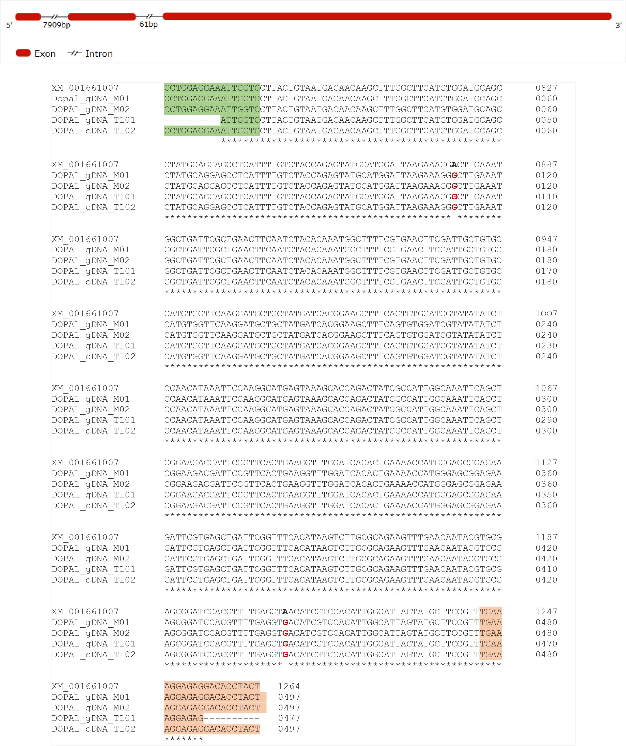
The DOPAL synthase gene, with a size of 9 851 bp, is located at position 406 432 747 - 406 422 897 on the negative strand of chromosome 2 of A. aegypti (NCBI Reference Sequence: NC_035108.1). The complete mRNA sequence is also reported in NCBI and has a size of 1 881 bp with a CDS of 1 566 bp (accession XM_001661007). This gene contains three exons (89, 235 and 1 557 bp) and two introns (7 909 and 61 bp). The 497 bp amplified fragments in this research correspond to a segment of the largest exon.
Sequences obtained using the Sanger method from amplicons derived from the genomic DNA of three specimens were aligned with the complementary DNA of a fourth specimen, revealing a 100% identity. These findings indicate a high level of genetic stability between the Sullana-Mallares and Tambogrande-Locuto mosquito populations in the Piura region. However, when comparing all sequences obtained in this study with the reference sequence of the DOPAL synthase gene mRNA of A. aegypti (accession XM_001661007), two single nucleotide polymorphisms (SNPs) were identified, both corresponding to an A/G substitution (highlighted in red in Figure 1).
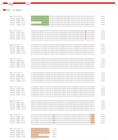
Upper: DOPAL synthase gene structure. This gene contains three exons (1 557 bp, 235 bp and 89 bp) and two introns (7 909 bp and 61 bp) in minus strand. Lower: Alignment of amplified DOPAL synthase gene fragments. Three genomic DNA sequences (gDNA) and one complementary DNA sequence (cDNA) corresponding to exon 1 were aligned with the reference sequence. Nucleotides highlighted in red represent polymorphisms identified in this study. M01-M02: Sullana-Mallares mosquitoes. TL01-TL02: Tambogande-Locuto mosquitoes. Note: DOPAL_gDNA_M01 and DOPAL_gDNA_M02 reads were amplified using different primer sets (DOPAL synthase F/DOPAL synthase R and Restriction-DOPAL-F/Restriction-DOPAL-R) from the same DNA sample from Sullana-Mallares. In addition, DOPAL_gDNA_TL01 reads were amplified from a DNA sample, while DOPAL_cDNA_TL02 reads were amplified from an RNA sample, both from Tambogrande-Locuto.
As these are synonymous mutations that do not alter the amino acid sequence, they further support the fact that this gene is highly conserved, a pattern consistent with its critical function in mosquito development. DOPAL synthase catalyzes the conversion of L-dopa to DOPAL, a highly reactive aldehyde that crosslinks cuticular proteins, contributing to the formation of a flexible and protective cuticle, a process essential for mosquito survival [14]. This process likely imposes strong selective pressure to maintain the integrity of this gene across populations.
Knockout studies of DOPAL synthase in A. aegypti and Anopheles gambiae have demonstrated its essential role in embryonic development, as embryos lacking functional DOPAL synthase fail to hatch [14], highlighting its crucial role in mosquito biology and reinforcing the need for further research on its potential as a target for vector control strategies.
Cloning DOPAL synthase and GUS inserts in E. coli HT115
The E. coli strain HT115 harbor in its chromosome the defective RNase III gene and an isopropyl-β-D-1-thiogalactopyranoside (IPTG)-inducible T7 polymerase gene [14]. RNase III enzymes are Mg2+-dependent dsRNA-specific endonucleases that enable bacteria to identify and degrade dsRNAs within their cells [15, 16]. T7 RNA polymerases are single-subunit enzymes known for their high and specific processivity; synthesizing RNA at a rate several times faster than E. coli RNA polymerase and transcribing entire genes without the need for additional proteins. Additionally, they exhibit high selectivity for initiation at their own promoter sequences [17, 18]. Therefore, E. coli HT115 strains are specifically engineered to produce large quantities of specific dsRNA segments [14]. Thanks to these characteristics, the HT115 strain is a key tool in RNAi-based control strategies. The effectiveness of this approach is evidenced by previous studies showing that administering dsRNA to mosquito larvae targeting testis-specific genes can successfully induce male sterility without compromising mating competitiveness, while targeting the female sex determination gene double sex skews the population sex ratio toward males [19].
In this research, strain E. coli HT115 was transformed with a pL4440 plasmid vectors that enabled the IPTG-inducible production of dsRNAs against DOPAL synthase of A. aegypti. The plasmid pL4440 vector contains two convergent T7 polymerase promoters in opposite orientation separated by a multicloning site to generate dsRNA from the cloned insert [14, 20]. Transforming E. coli HT115 with pL4440 plasmids enables large-scale production of specific dsRNAs, reinforcing RNAi-based approaches as species-specific and environmentally safe strategies for suppressing mosquito populations and other insects [19].
PCR was used to confirm the successful transformation of E. coli HT115 strains. Following transformation, 10 randomly selected clones per gene were screened. Among the DOPAL synthase gene clones, 1 out of 10 (pL4440-DOPAL-HT115) tested positive, whereas for the GUS gene, 8 out of 10 (pL4440-GUS-HT115) were positive. A positive clone per gene was then selected for further analysis.
The higher transformation efficiency observed for GUS (407 bp insert) compared to DOPAL synthase (429 bp insert) may be related to differences in sequence composition rather than insert size. Variations in GC content or the presence of secondary structures could influence ligation efficiency. However, since these factors were not experimentally evaluated in this study, their impact remains uncertain.
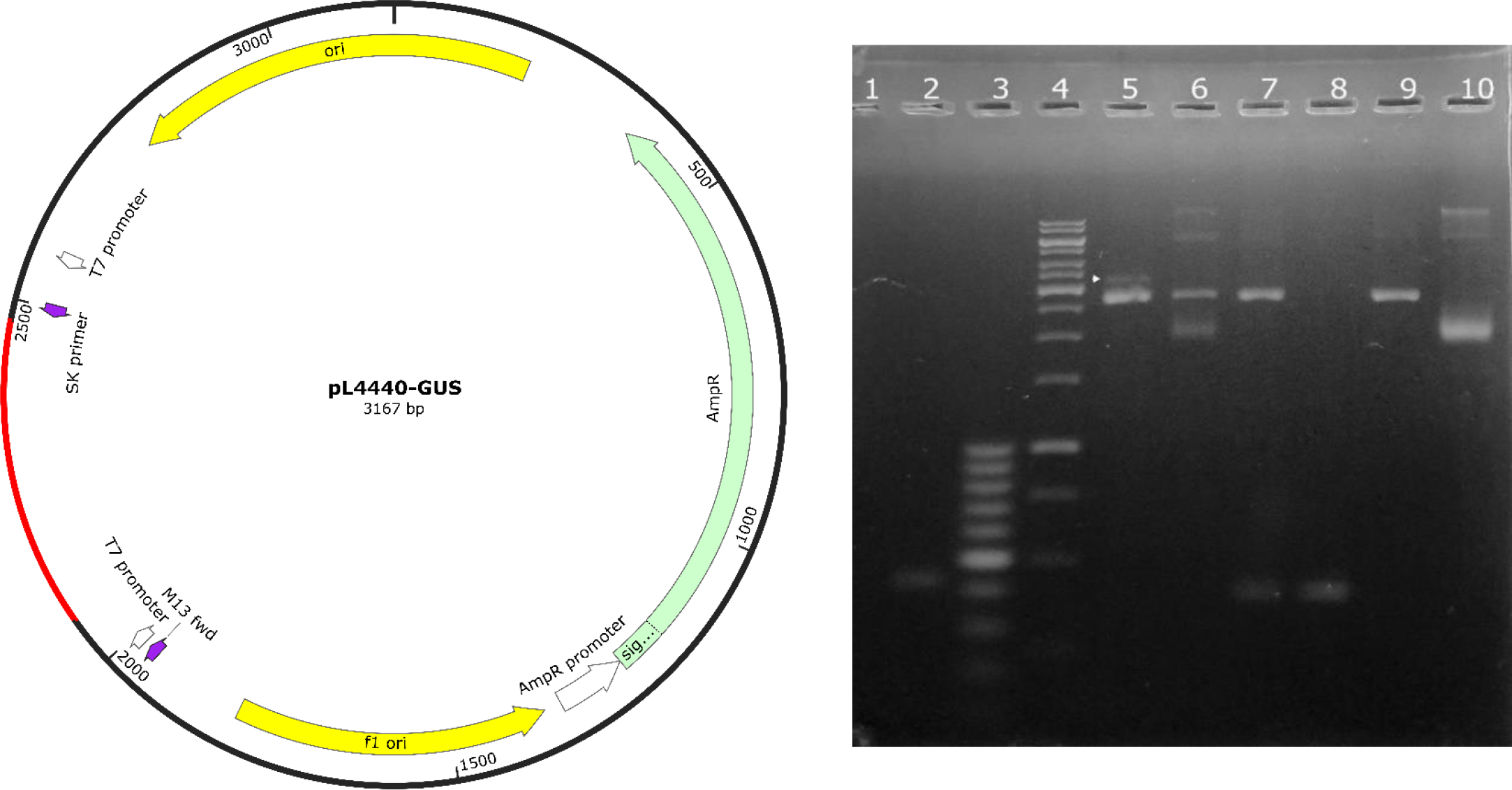
The evaluation of the recombinant plasmids was carried out using PCR amplification and enzymatic digestion. For GUS, the agarose gel showed that the linearization of the pL4440-GUS plasmid with the restriction enzyme BmtI resulted in a band that was 407 bp longer than the band produced by linearized pL4440 (Line 5, Figure 2). Enzymatic digestion with the restriction enzymes PstI and BmtI generated two bands of 2760 bp and 407 bp, corresponding to the pL4440 cloning vector and the GUS gene insert, respectively (Line 7, Figure 2). Furthermore, enzymatic digestion of the insert, amplified with primers containing restriction sites, produced a 407 bp band (Line 8, Figure 2).
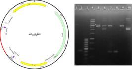
Cloning of GUS into pL4440 plasmid and transformation into E. coli HT115. Left. Schematic diagram of the recombinant plasmid pL4440-GUS for dsRNA expression in E. coli HT115. Right. Amplification and enzymatic digestion of pL4440-GUS plasmid. Lane 1: Non-template control (NTC). Lane 2: GUS amplicon (425 bp) from pL4440-GUS. Lane 3: 100 bp DNA Ladder. Lane 4: 1 kb DNA Ladder. Lane 5: Simultaneous migration of pL4440-GUS and pL4440 plasmids linearized by BmtI, resulting in a 3167 bp linearized fragment (indicated by the arrow) and 2760 bp, respectively. Lane 6: Digestion of pL4440 with PstI yielded a 2790 bp fragment. Lane 7: Digestion of pL4440-GUS with PstI and BmtI resulted in bands of 2760 bp (pL4440) and 407 bp (GUS). Lane 8: Digestion with PstI and BmtI of GUS amplicon containing restriction sites, produced a 407 bp fragment. Lane 9: Digestion of pL4440 with PstI and BmtI produced a 2760 bp fragment. Lane 10: Undigested pL4440.
These results confirm the successful insertion of the insert of the GUS gene into the pL4440 vector. The consistent detection of the expected fragment sizes across different digestion patterns demonstrates the reliability of PCR and enzymatic digestion as effective tools for verifying plasmid recombination. These findings support the structural integrity of the recombinant pL4440-GUS plasmid, validating its suitability for downstream applications, such as dsRNA production.
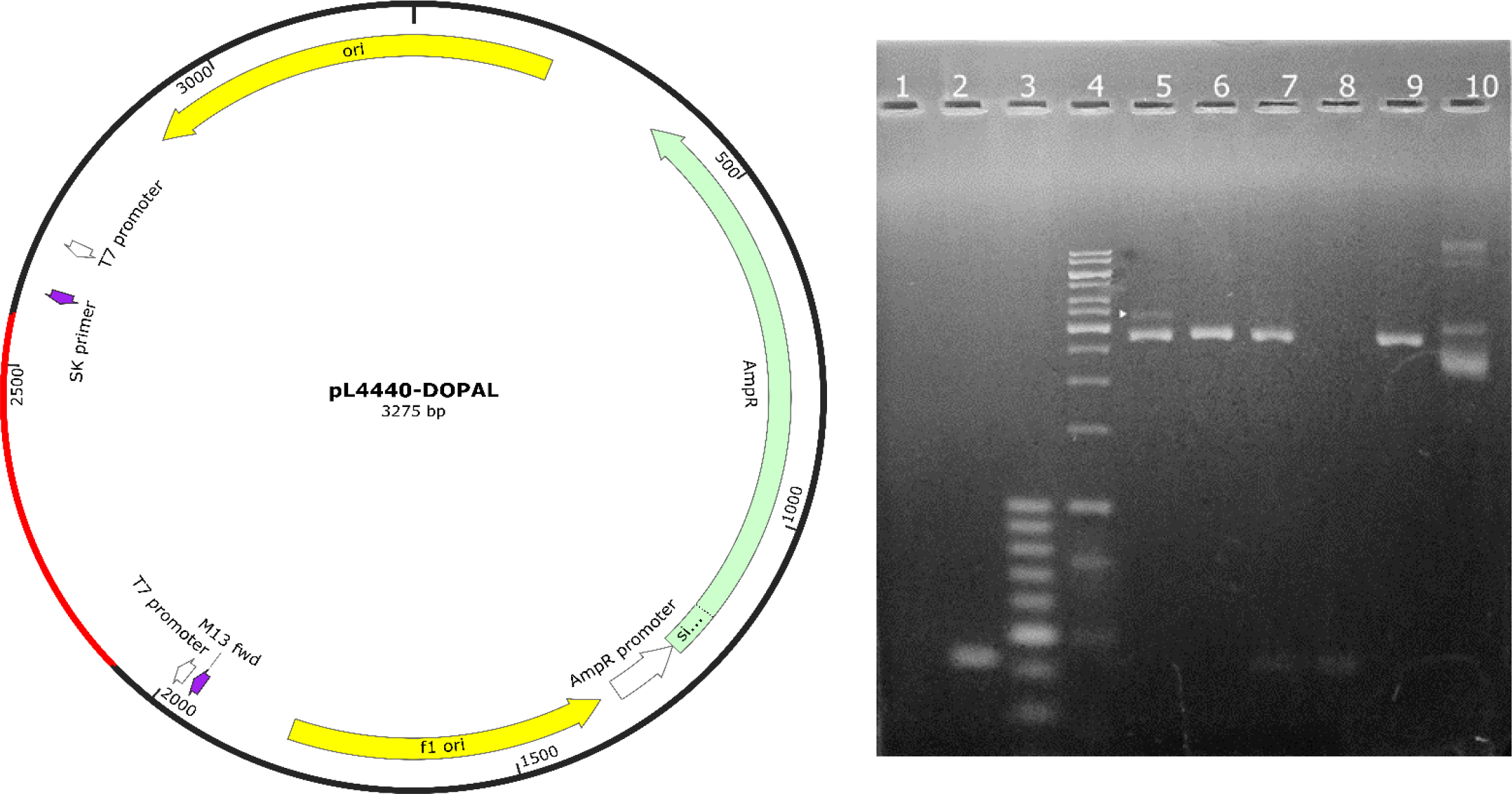
Regarding DOPAL synthase, the pattern bands revealed that the linearization of the pL4440-DOPAL plasmid with the restriction enzyme BamHI produced a band that was 429 bp longer than the linearized pL4440 (Line 5, Figure 3). Similarly, enzymatic digestion with the restriction enzymes PstI and BamHI generated two bands of 2760 bp and 429 bp, corresponding to the pL4440 cloning vector and the DOPAL synthase gene insert, respectively (Line 7, Figure 3). Finally, enzymatic digestion of the insert, amplified with primers containing restriction sites, resulted in a 429 bp band (Line 8, Figure 3).
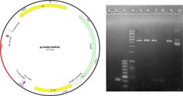
Cloning of DOPAL synthase into pL4440 plasmid and transformation into E. coli HT115. Left. Schematic diagram of the recombinant plasmid pL4440-DOPAL synthase for dsRNA expression in E. coli HT115. Right. Amplification and enzymatic digestion of pL4440-DOPAL plasmid. Lane 1: Non-template control (NTC). Lane 2: DOPAL synthase amplicon (521 bp) from pL4440-DOPAL. Lane 3: 100 bp DNA Ladder. Lane 4: 1 kb DNA Ladder. Lane 5: Simultaneous migration of the pL4440-DOPAL and pL4440 plasmids linearized by BamHI, resulting in a 3171 bp linearized fragment (indicated by the arrow) and 2760 bp, respectively. Lane 6: pL4440 plasmid digested with PstI yielded a 2790 bp fragment. Lane 7: Digestion of pL4440-DOPAL with PstI and BamHI resulted in bands of 2746 bp (pL4440) and 429 bp (DOPAL synthase). Lane 8: Digestion with PstI and BamHI of the DOPAL synthase amplicon containing restriction sites, produced a 429 bp fragment. 9: Digestion of pL4440 with PstI and BamHI produced a 2746 bp fragment. Lane 10: Undigested pL4440 plasmid.
The enzymatic digestion patterns obtained for pL4440-DOPAL synthase also aligned with the expected fragment sizes, confirming the successful insertion of the gene fragment into the plasmid. The successful release of the 429 bp insert, upon digestion with PstI and BamHI, validates proper ligation and, further, supports the structural integrity of the recombinant construct. Given the crucial role of the accurate integration of the insert for ensuring effective dsRNA expression, these results confirm the suitability of the pL4440-DOPAL synthase plasmid for subsequent functional studies.
Detection of dsRNA-GUS and dsRNA-DOPAL synthase by RT-qPCR
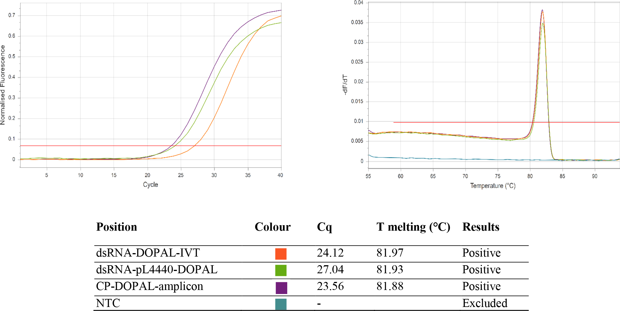
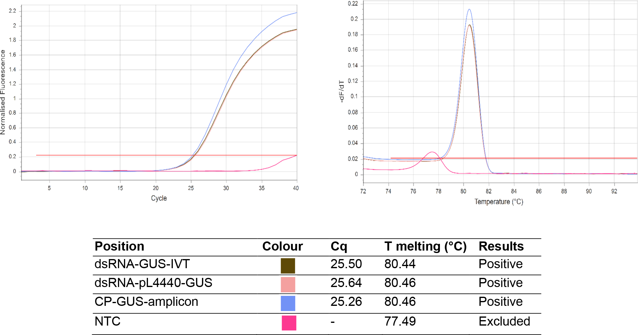
The specificity and successful synthesis of dsRNAs targeting the DOPAL synthase and GUS genes were confirmed in this study using RT-qPCR and dissociation curve analysis (Figures 4 and 5, respectively).
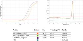
Evaluation of DOPAL synthase gene dsRNA using RT-qPCR. Top left: Amplification curve of the dsRNA produced by IVT and E. coli HT115 pL4440-DOPAL. Top right: Melting curve of the amplified dsRNA DOPAL Synthase products. Bottom: Table with the color codes, Cq values, T melting.
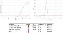
Evaluation of GUS gene dsRNA using RT-qPCR. Top left: Amplification curve of the dsRNA produced by IVT and E. coli HT115 pL4440-GUS. Top right: Melting curve of the amplified dsRNA GUS products. Bottom: Table with the color codes, Cq values, T melting.
For the dsRNA targeting the DOPAL synthase gene, RT-qPCR was performed using purified RNA samples at concentrations of 98.04 ng/µL for dsRNA expressed in E. coli HT115 (dsRNA-pL4440-Dopal) and 60.25 ng/µL for in vitro-transcribed dsRNA (dsRNA-DOPAL-IVT). The Cq values obtained were 24.12 for dsRNA-DOPAL-IVT and 27.04 for dsRNA-pL4440-DOPAL. Although dsRNA-DOPAL-IVT showed lower Cq values compared to dsRNA-pL4440-Dopal, suggesting a possible difference in accessibility for RT-qPCR amplification, statistical analysis using the Student’s t-test (p = 0.4189) indicated no significant differences between the two methods (see Table 2 and 3). This suggests that the observed variation in Cq values is not biologically relevant and that both RNA production approaches yield comparable dsRNA concentrations.
Statistical comparison between dsRNA-Dopal-IVT vs dsRNA-pL4440-Dopal and dsRNA-GUS-IVT vs dsRNApL4440-GUS
For the dsRNA targeting the GUS gene, RT-qPCR was performed using purified RNA samples at concentrations of 40.00 ng/µL for dsRNA expressed in E. coli HT115 (dsRNA-pL4440-GUS) and 50.32 ng/µL for in vitro-transcribed dsRNA (dsRNA-GUS-IVT). The Cq values obtained were 25.50 for dsRNA-GUS-IVT and 25.64 for dsRNA-pL4440-GUS. Statistical analysis using a Student’s t-test (p = 0.7543) confirmed that the differences between the two methods were not statistically significant.
The fluorescence data plotted against the derivative of temperature (dF/dT) of the target genes DOPAL synthase and GUS are similar to their respective positive controls, confirming the specificity of the amplification. In the case of DOPAL synthase, three peaks appear around ~81.9°C, while for GUS, two peaks are observed: a lower peak on the left at 77.49°C, likely corresponding to primer dimer formation, and a main peak at ~80.4°C, associated with the dissociation of the expected amplification products.
Minimal differences in melting temperatures (Tm) were observed between the amplified products of the two genes, with a maximum variation of 0.09°C for DOPAL synthase and 0.02°C for GUS. These small variations could reflect differences in GC content and sequence composition, nevertheless, these remain within the expected range. Considering the CG/AT ratio and the amplicon sequence, the dissociation curve analysis confirms that the observed melting temperatures (Tm variation < 2°C) align with the expected sequences of the dsRNAs from both genes [21, 22]. (Figures 4 and 5).
Chitosan nanoparticles
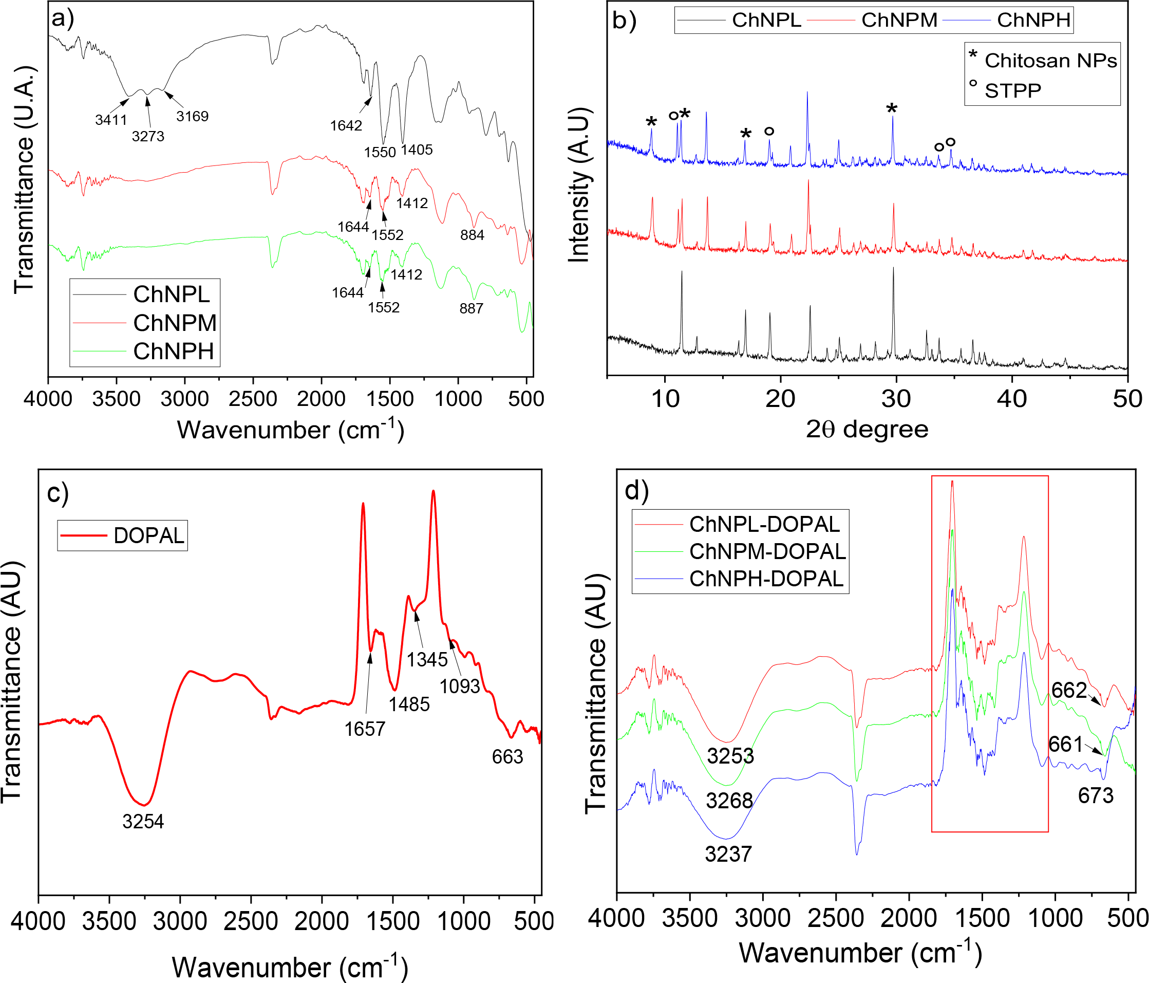
Figure 6a shows the FTIR spectra of the chitosan NPs samples obtained from low, medium, and high molecular weight chitosan. Peaks of the spectra for the three samples coincide or are close to those found by multiple authors who have produced nanoparticles with the same method [23-25]. The peaks at 3 411, 3 273 and 3 169 cm-1, present in the sample ChNPL, would be assigned to hydrogen bonded O-H or N-H stretching modes [26]. However, these peaks are not present in the spectra of the samples ChNPM and ChNPH.
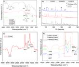
FTIR-ATR (a) and XRD (b) of NPs from chitosan from low, medium and high molecular weight chitosan (ChNPL, ChNPM and ChNPH, respectively). FTIR-ATR of DOPAL synthase-dsARN (c) and the functionalized molecules ChNPL-DOPAL synthase, ChNPM-DOPAL synthase and ChNPH-DOPAL synthase (d)
The peaks at 1 642-1 644 and 1 550-1 552 cm-1 are referred to the NH2 of amide II and CONH2 groups [24, 27] or to the stretching of the N-H group in chitosan/TPP and the N-O stretching of aliphatic nitrogen compounds, respectively [28]. These two peaks confirm the linkage between phosphoric and ammonium ions [29] from Chitosan and TPP respectively. The intensity of both peaks is different, depending on the molecular weight of chitosan used for NPs preparation. ChNPL presents the highest intensities, supposing a stronger interaction between chitosan and TPP compared to the other samples.
The peak at 1 405 - 1 412 cm-1 corresponds to the vibration of the CN group and the peak at 1 117-1 128 cm-1 would correspond to the stretching vibration of P=O in the phosphates [30]. Meanwhile, the peak at 884-887 cm-1, presents in the samples ChNPM and ChNPH, corresponds to P-O along with P-O-P functional groups [30]
Figure 6b shows the x-ray diffractogram of the chitosan nanoparticles obtained with chitosan of various molecular weights. The bands that correspond to Chitosan NPs (2θ= 8.83, 11.38, 16.91,29.67) and STPP (2θ= 11.07, 19.01, 33.63, 34.69) are indicated in the samples diffractograms as * and ° respectively [31]. Chitosan NPs do not show broad peaks as in the case of amorphous chitosan which shows a broad peak at 21.8° [23]. Chitosan NPs has a crystalline structure [31], composed of a dense network structure of interpenetrating polymer chains cross-linked to each other by the oppositely charged ions of STPP.
Chitosan NPs Functionalized with DOPAL synthase dsRNA
Figures 6c and 6d shows the FTIR-ATR of dsRNA for the DOPAL synthase and the functionalized molecules ChNPL-DOPAL, ChNPM-DOPAL and ChNPH-DOPAL, respectively. In the case of naked dsRNA sample, peaks at 3 254 cm-1 are related to O-H and N-H groups, at 1 657 cm-1 to the guanine and uracil structure. Peaks at 1 485 cm-1 correspond to guanine and at 1 093 cm-1 to the PO2- functional group [32].
According to the literature, the way chitosan is functionalized with dsRNA is through electrostatic forces between the positive amino groups of chitosan and the negative charges on the phosphate groups of dsRNA [10]. After functionalization, the peak between 3 254 cm-1 shifted to lower wavenumbers, indicating an interaction between dsRNA and chitosan NPs. The region between 1 000 - 1 600 cm-1 increase the number of peaks because of the interaction. The peak that corresponds to PO2- in the case of dsRNA becomes slightly wider and more intense in the functionalized materials. Apparently, this increase in intensity is referred to the presence of P=O groups in the chitosan NPs (centered at 1 127 - 1 128 cm-1), which overlap with the peak of 1093 cm-1 of the dsRNA corresponding to PO2-.
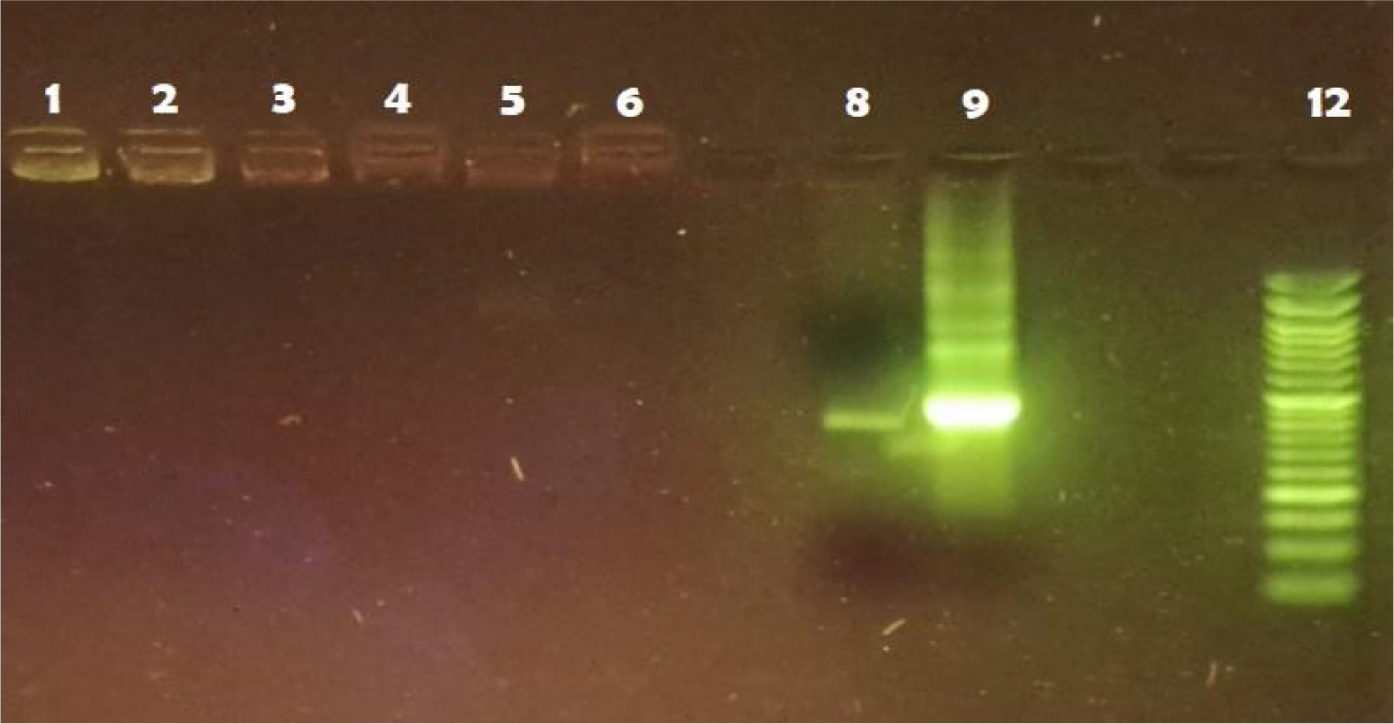
Figure 7 shows the results of the electrophoretic mobility shift assay of the chitosan NPs of different molecular weight functionalized with the DOPAL synthase gene-dsRNA (lanes 1 to 6), as well as the two naked dsRNAs, dsARN-IVT (lane 8) and dsRNA bacterial-non treated with RNAsa T1 (lane 9), and a molecular weight marker of 100 - 3 000 bp (lane 12). The electrophoretic migration of bacterial dsRNA shows RNA molecules of other molecular weights from E. coli that do not correspond to the DOPAL synthase gene, which should be removed after treatment with RNase T1. This indicates that dsRNA-IVT exhibits higher purity.
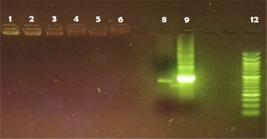
Electrophoretic mobility shift assay (Agarose 2.5%): 1 and 2-ChNPL-DOPAL synthase, 3 and 5-ChNPM-DOPAL synthase, 4 and 6-ChNPH-DOPAL synthase, 8 dsARN-IVT, 9- dsRNA bacterial-non treated with RNAsa T1, 12-Molecular weight marker (100 - 3 000 bp)
This experiment was performed for the purpose of a qualitative comparison of functionalized and non-functionalized RNAi. It is evident that the functionalized materials stayed at the top of the lane, while the DOPAL synthase gene targeted dsRNAs without functionalization migrated towards the middle part of the lane. This shows the functionalization of dsRNAi with NPs from the three molecular weight chitosan. Similar results were found by other authors who functionalized chitosan NPs with dsRNA [1, 7].
Functionalizing dsRNA with chitosan NPs was demostrated and it is the first step to develop a control method for A. aegypti. There are different literature which Chitosan NPs functionalized with dsRNA targeting key genes controlled larvae and adults of A. aegypti, but most of them in laboratory conditions [3, 5, 7, 9]. However, further studies are needed to demostrated its effect over A. aegypti in natural habitats such as water recipies or water bodies.
CONCLUSION
Amplicons of the DOPAL synthase gene from A. aegypti from endemic areas in northwest Peru were isolated, revealed two single nucleotide polymorphisms (SNPs), both A/G replacement, which would not affect the amino acid sequence.
DOPAL synthase-dsRNA was designed and constructed with the isolated amplicons by two methods, IVT and cloning in Escherichia coli HT115 (D3). The RT-qPCR and EMSA analyses confirmed that both methods are effective for obtaining dsRNA from the DOPAL synthase gene. However, the IVT method is superior in terms of speed and ease of execution, and it also facilitates the acquisition of a larger quantity of the target with higher purity.
The analysis of dissociation curves confirms the correspondence of the amplified products with the expected sequences, validating the employed methodology and supporting the continuation of studies in this line of research.
FTIR-ATR spectra and XRD showed the successful production of NPs from three molecular weight-chitosan types (50-190, 190-310 and 310-375 kDa). All NPs exhibited a crystalline structure, similar to the results obtained by other authors.
The interaction of DOPAL synthase -dsRNA and chitosan NPs was proved by FTIR-ATR and EMSA analyses confirm the higher molecular weight of the NP resulting from the interaction of both NPs compared to naked dsRNA.
Findings in this study represent an initial step to identify a promising method to control mosquito A aegypti, however, further studies should be conducted in vivo over mosquitos’ larvae in water recipients or water bodies.
-
Funding:
This research was funded by CANON funding - National University of Tumbes (Resolution N° 0234-2022/UNTUMBES-R)
Acknowledgments:
Experimental works concerning the chitosan NPs preparation and lyophilization process were accomplished by using Large Research Infrastructure ENREGAT at the Institute of Environmental Technology, CEET, VSB-TUO supported by the Ministry of Education, Youth and Sports of the Czech Republic under projects No. LM2018098 and LM2023056 and the equipment of Nanotechnology Centre, CEET, VSB-TUO, respectively.
Data Availability Statement:
Research data are only available upon request for corresponding author.
REFERENCES
- 1 Kolge H, Kadam K, Ghormade V. Chitosan nanocarriers mediated dsRNA delivery in gene silencing for Helicoverpa armigera biocontrol. Pestic Biochem Physiol. 2023;189:105292.
- 2 Airs PM, Bartholomay LC. RNA Interference for mosquito and mosquito-borne disease control. Insects. 2017;8(1).
- 3 Mysore K, Sun L, Tomchaney M, Sullivan G, Adams H, Piscoya AS, et al. siRNA-mediated silencing of doublesex during female development of the dengue vector mosquito Aedes aegypti. PLoS Negl Trop Dis. 2015;9(11):e0004213.
- 4 Mogilicherla K, Roy A. RNAi-chitosan biopesticides for managing forest insect pests: an outlook. Front For Glob Ch. 2023;6.
- 5 Chen J, Lu HR, Zhang L, Liao CH, Han Q. RNA interference-mediated knockdown of 3, 4-dihydroxyphenylacetaldehyde synthase affects larval development and adult survival in the mosquito Aedes aegypti. Parasit Vectors. 2019;12(1):311.
- 6 Das S, Debnath N, Cui Y, Unrine J, Palli SR. Chitosan, carbon quantum dot, and silica nanoparticle mediated dsRNA delivery for gene silencing in Aedes aegypti: A Comparative Analysis. ACS Appl Mater Interfaces. 2015;7(35):19530-5.
- 7 Dhandapani RK, Gurusamy D, Howell JL, Palli SR. Development of CS-TPP-dsRNA nanoparticles to enhance RNAi efficiency in the yellow fever mosquito, Aedes aegypti. Sci Rep. 2019;9(1):8775.
- 8 Mysore K, Andrews E, Li P, Duman-Scheel M. Chitosan/siRNA nanoparticle targeting demonstrates a requirement for single-minded during larval and pupal olfactory system development of the vector mosquito Aedes aegypti. BMC Dev Biol. 2014;14:9.
- 9 Ramesh Kumar D, Saravana Kumar P, Gandhi MR, Al-Dhabi NA, Paulraj MG, Ignacimuthu S. Delivery of chitosan/dsRNA nanoparticles for silencing of wing development vestigial ( vg ) gene in Aedes aegypti mosquitoes. International J Biol Macromol. 2016;86:89-95.
- 10 Zhang X, Mysore K, Flannery E, Michel K, Severson DW, Zhu KY, et al. Chitosan/interfering RNA nanoparticle mediated gene silencing in disease vector mosquito larvae. J Vis Exp. 2015(97).
- 11 Kong M, Chen XG, Xing K, Park HJ. Antimicrobial properties of chitosan and mode of action: a state of the art review. Int J Food Microbiol. 2010;144(1):51-63.
- 12 Acharya R, Saha S, Ray S, Hazra S, Mitra MK, Chakraborty J. siRNA-nanoparticle conjugate in gene silencing: A future cure to deadly diseases? Mater Sci Eng C Mater Biol Appl. 2017;76:1378-400.
- 13 Kolge H, Kadam K, Ghormade V. Chitosan nanocarriers mediated dsRNA delivery in gene silencing for Helicoverpa armigera biocontrol. Pestic Biochem Physiol. 2023;189:105292.
- 14 Timmons L, Court DL, Fire A. Ingestion of bacterially expressed dsRNAs can produce specific and potent genetic interference in Caenorhabditis elegans. Gene. 2001;263(1):103-12.
- 15 Court DL, Gan J, Liang Y-H, Shaw GX, Tropea JE, Costantino N, et al. RNase III: genetics and function; structure and mechanism*. Annu Rev Genet. 2013;47(Volume 47, 2013):405-31.
- 16 Aguado LC, tenOever BR. RNase III nucleases and the evolution of antiviral systems. BioEssays. 2018;40(2):1700173.
- 17 Borkotoky S, Murali A. The highly efficient T7 RNA polymerase: A wonder macromolecule in biological realm. Int J Biol Macromol. 2018;118:49-56.
- 18 Tabor S. Expression using the T7 RNA polymerase/promoter system. Curr Protoc Mol Biol. 1990;11(1):16.2.1-.2.1.
- 19 Whyard S, Erdelyan CNG, Partridge AL, Singh AD, Beebe NW, Capina R. Silencing the buzz: a new approach to population suppression of mosquitoes by feeding larvae double-stranded RNAs. Parasit. Vectors. 2015;8(1):96.
- 20 Timmons L, Fire A. Specific interference by ingested dsRNA. Nat. 1998;395(6705):854.
- 21 Prada-Arismendy J, Castellanos JE. Real time PCR. Application in dengue studies. Colombia Médica. 2011;42(2):243-58.
- 22 Navarro E, Serrano-Heras G, Castaño MJ, Solera J. Real-time PCR detection chemistry. Clinica Chimica Acta. 2015;439:231-50.
- 23 Ali SW, Rajendran S, Joshi M. Synthesis and characterization of chitosan and silver loaded chitosan nanoparticles for bioactive polyester. Carbohydr Polym. 2011;83(2):438-46.
- 24 Lustriane C, Dwivany FM, Suendo V, Reza M. Effect of chitosan and chitosan-nanoparticles on post harvest quality of banana fruits. J Plant Biotechnol. 2018;45(1):36-44.
- 25 Morsy M, Mostafa k, amyn h, El-Ebissy A, Salah A, Youssef M. Synthesis and characterization of freeze dryer chitosan nanoparticles as multi functional eco-friendly finish for fabricating easy care and antibacterial cotton textiles. Egypt J Chem. 2019;0(0):0-.
- 26 Khoerunnisa F, Nurhayati M, Dara F, Rizki R, Nasir M, Aziz HA, et al. Physicochemical properties of TPP-crosslinked chitosan nanoparticles as potential antibacterial agents. Fibers Polym. 2021;22(11):2954-64.
- 27 Hadidi M, Pouramin S, Adinepour F, Haghani S, Jafari SM. Chitosan nanoparticles loaded with clove essential oil: characterization, antioxidant and antibacterial activities. Carbohydr Polym. 2020;236:116075.
- 28 Anand M, Sathyapriya P, Maruthupandy M, Hameedha Beevi A. Synthesis of chitosan nanoparticles by TPP and their potential mosquito larvicidal application. Front Lab Med. 2018;2(2):72-8.
- 29 Kahdestani SA, Shahriari MH, Abdouss M. Synthesis and characterization of chitosan nanoparticles containing teicoplanin using sol-gel. Polym. Bull. 2020;78(2):1133-48.
- 30 Pan C, Qian J, Zhao C, Yang H, Zhao X, Guo H. Study on the relationship between crosslinking degree and properties of TPP crosslinked chitosan nanoparticles. Carbohydr Polym. 2020;241:116349.
- 31 Alehosseini E, Shahiri Tabarestani H, Kharazmi MS, Jafari SM. Physicochemical, thermal, and morphological properties of chitosan nanoparticles produced by ionic gelation. Foods. 2022;11(23).
- 32 Kapusuz D. Exploring the structure of sol-gel-derived hybrids for immobilization of RNA: Influence of RNA content. Colloid Polym Sci. 2020;299(1):63-72.
-
Editor-in-Chief:
Paulo Vitor Farago
-
Associate Editor:
Marcelo Ricardo Vicari