Subacute toxicity of Chrozophora Tinctoria extract: biochemical and histopathological changes in rats
Abstract
The increasing use of medicinal plants in traditional and modern therapies necessitates thorough toxicological evaluations to ensure their safety. Chrozophora genus includes 7-8 species that belong to the Euphorbiaceae family, distributed in Pakistan, India, West Africa, and the Mediterranean with four species found in the Kingdom of Saudi Arabia (KSA). This study evaluates the subacute toxicity of the ethyl acetate extract (EtOAcE) of Chrozophora tinctoria in male and female rats. Over 14 days, rats were daily administered the extract at doses of 50, 100, and 200 mg. Mortality was observed at a rate of one rat per group. Female rats showed a significant reduction in body weight, while organ weights remained unaffected. Kidney function tests revealed significant increases in bilirubin and creatinine levels in male rats at 200 mg, and uric acid levels in female rats at the same dosage. Liver enzyme analysis indicated significant elevations in ALP, ALT, and GGT levels in both sexes at various dosages. Glucose levels increased significantly in female rats at 200 mg/kg, with no significant changes in cortisol levels in either sex. Histopathological examinations demonstrated notable abnormalities in liver, kidney, spleen, and small intestine tissues. These data demonstrate the essentiality of careful evaluation and monitoring when considering C. tinctoria for medicinal use, highlighting the need for further studies on its long-term safety and therapeutic index.
Keywords:
Chrozophora tinctoria; toxicology; oxidative stress; ethyl acetate extract; GC-MS analysis
Resumo
O uso crescente de plantas medicinais em terapias tradicionais e modernas exige avaliações toxicológicas completas para garantir sua segurança. O gênero Chrozophora inclui 7-8 espécies que pertencem à família Euphorbiaceae, distribuídas no Paquistão, na Índia, na África Ocidental e no Mediterrâneo, com quatro espécies encontradas no Reino da Arábia Saudita (KSA). Este estudo avalia a toxicidade subaguda do extrato de acetato de etila (EtOAcE) de Chrozophora tinctoria em ratos machos e fêmeas. Ao longo de 14 dias, os ratos receberam diariamente o extrato em doses de 50, 100 e 200 mg. A mortalidade foi observada a uma taxa de um rato por grupo. As ratas fêmeas mostraram uma redução significativa no peso corporal, enquanto os pesos dos órgãos permaneceram inalterados. Testes de função renal revelaram aumentos significativos nos níveis de bilirrubina e creatinina em ratos machos no grupo com dose de 200 mg, e nos níveis de ácido úrico em ratas fêmeas na mesma dosagem. A análise de enzimas hepáticas indicou elevações significativas nos níveis de ALP, ALT e GGT em ambos os sexos, em várias dosagens. Os níveis de glicose aumentaram significativamente em ratas fêmeas com 200 mg/kg, sem alterações significativas nos níveis de cortisol, em ambos os sexos. Exames histopatológicos demonstraram anormalidades notáveis nos tecidos dos órgãos fígado, rim e baço, e do intestino delgado. Esses dados demonstram a essencialidade de avaliação e monitoramento cuidadosos ao considerar C. tinctoria para uso medicinal, destacando a necessidade de estudos adicionais sobre sua segurança em longo prazo e índice terapêutico.
Palavras-chave:
Chrozophora tinctoria; toxicologia; estresse oxidativo; extrato de acetato de etila; análise GC-MS
1. Introduction
Contemporary alternative medicine increasingly emphasizes the therapeutic use of plants (Phutrakool and Pongpirul, 2022). These medicinal plants are known to trigger the activities of antioxidant enzymes, which play a role in mitigating hepatotoxicity, renal toxicity, and reproductive toxicity (Quan et al., 2020; Xu et al., 2020; Khan et al., 2021). A prevalent misconception is that folk remedies, by being natural, are devoid of toxicity or side effects, especially in comparison with synthetic pharmaceuticals (Karimi et al., 2015). However, concerns over the safety of these plants have prompted toxicological investigations of their safety profiles (Alelign et al., 2020; Nalimu et al., 2022). Despite the plenty of ethnopharmacological reports of the plants widely used in local folk medicine (Aati et al., 2019; Ullah et al., 2020), the potential toxicity of these plants remains under scientific scrutiny. Therefore, any herbal remedy claimed to have therapeutic benefits should be evaluated to detect undesirable effects before market trades.
The Chrozophora genus includes 7-8 species that belong to the Euphorbiaceae family, distributed in Pakistan, India, West Africa, and the Mediterranean, with four species found in the Kingdom of Saudia Arabia (KSA) (Rahman et al., 2004; Abdallah et al., 2015). Chrozophora species are used in folk medicine to relieve mouth ulcers, fever, bone joint pain, wounds, and to eliminate worms in the digestive tract (Sher et al., 2022). Chrozophora tinctoria is an annual summer species that tolerates extreme light, extreme heat, and humidity conditions (Usman et al., 2007). The compositional properties of C. tinctoria vary based on their geographical locations, as indicated in previous studies (Oke-Altuntas et al., 2017; Golkar et al., 2019). These geographical differences can affect the concentration of key chemical compounds in the plant, including the pigments responsible for its dyeing properties, as well as secondary metabolites with potential medicinal benefits. For example, plants growing in regions with different levels of sunlight, temperature, and soil nutrients may produce varying amounts of the blue dye (turnsole) and other bioactive compounds. This variation has important implications for the use of C. tinctoria in traditional medicine, dye production, and other applications. Understanding how environmental factors influence the plant's chemical composition can help optimize its cultivation and improve the quality of its derived products. The antioxidant potential of C. tinctoria enables its use as a therapeutic agent for various conditions, including diarrhea, as an emetic, anthelminthic for treating warts, and as antimicrobial. It has also been proven effective in wound healing in diabetics (Ugurlu and Secmen, 2008; Maurya et al., 2016; Iqbal et al., 2022; Sher et al., 2022). Nevertheless, the species under the Euphorbiaceae family have produced some phytotoxins, such as phorbol esters, alkaloids, glycosides, and ricin (Betancur-Galvis et al., 2002). Moreover, the lack of accurate scientific studies to establish appropriate dosages often leads to misuse of these bioproducts, potentially affecting the vital organs (Ribnicky et al., 2008). In addition, phytochemicals probably interfere with pharmaceutical drugs, reducing their effectiveness or inducing multiple toxic effects, especially in subacute or chronic doses (Williamson, 2003). Despite the various uses of C. tinctoria in phytotherapy, no orderly approach has been made to investigate the toxic effects of C. tinctoria in vivo. Thus, this study aimed to examine the potential toxicity of C. tinctoria extract in rat models.
2. Materials and Methods
2.1. Chemicals and kits
The Ethyl acetate (no. A9346) (the solvent for C. tinctoria extraction) was purchased from ROMIL company (Cambridge, UK). The liver and kidney biomarkers and glucose kits were obtained from Biosystem company (Navghar, India). The cortisol kit was bought from AFG Biosystem (Northbrook, USA).
2.2. Plant material and extract process
Fresh healthy leaves of C. tinctoria were collected in July during the summer of 2023 from Al-Mahed, Al-Madinah, KSA (23°27'48.9”N latitude and 41°17'20.0”E longitude). The C. tinctoria was identified by a taxonomist at the Biology Department, College of Science, Al-Baha University, Al-Baha, KSA. The plant leaves were air-dried at 25 ºC and ground with Commercial Blender (LC Multifunction Grinder, China). The extraction process of C. tinctoria was performed in the Bioproducts Chair laboratory at Zoology Department, College of Science at King Saud University. Approximately 1.5 kg of the pulverized material was macerated in ethyl acetate (EtOAc) for 24 hours at 25°C with periodic stirring to facilitate the extraction of plant compounds. Thereafter the EtOAc extract (EtOAcE) was filtered using muslin cloth and then through a Whatman No. 1 filter paper. The filtrate obtained from the extraction was subjected to evaporation using a rotary evaporator (Heidolph, Germany), maintaining a temperature of 45°C. The weight of the resulting extract was accurately recorded (18 ± 3.5 g). The dried extract of C. tinctoria was stored in clean and dried bottles at 4 °C until used.
2.3. Extract solutions and doses preparation
The extract of C. tinctoria was resuspended in Dimethyl sulfoxide (DMSO) to prepare required doses. To determine the doses used in this experiment, a pilot study was conducted on two rats (one male and one female). They were injected with 2000 mg/kg/bw in 0.5 mL of DMSO to determine the lethal dose of rats. The animals were monitored for a week to observe any sign of toxicity. No signs of toxicity were observed in animals, so we concluded that the LD50 of EtOAcE is higher than 2000 mg/kg/bw. The dose of 50 mg/kg/bw was adopted as a minimum dose based on previous study (Maurya et al., 2016). Based on this, the three doses of EtOAcE of C. tinctoria (50, 100, and 200 mg/kg/bw) were calculated based on 1/10, 1/20, 1/40 of 2000 mg/kg. For sub-acute daily dose, the extract powder (16 g) of EtOAcE was dissolved in fifty milliliters of DMSO at room temperature to make a stock solution of the extract (320 mg/mL). Three sub-stock solutions of the above solution were prepared to obtain 50, 100, and 200 mg/kg/bw in a volume of 0.5 mL of dose/rat.
2.4. Animals and ethical approval
Experiments were conducted in the animal house at King Saud University (KSU). The male and female rats (200±10 g) were housed for acclimatization (two weeks before the experiment) at a temperature of 25 ± 1◦C and a relative humidity of 40-60%, a 12-h light/dark cycle. The animals were given standard feed (ARASCO, KSA) and water libitum throughout the trials. The experiment procedures and handling of animals and biological samples were carried out according to the guidelines approved by the Institutional Review Board (IRB) at KSU (Ethics No: KSU-SE-24-3) and IRB at Imam Abdulrahman bin Faisal University (Ethics No: IRB-2023-10-565).
2.5. Experimental design
According to the Organization for Economic Co-operation and Development OECD (2008) Test No. 407 with slight modifications, fifty rats (25 males and 25 females) were housed in standard-sized polypropylene cages (Length = 410 mm, Breadth = 282 mm, and Height =153 mm) with fine sawdust bedding and polypropylene water bottle (500ml). Each gender of rats was divided into five groups (five animals/group) based on the experimental treatments: Control, DMSO and EtOAcE at 50, 100 and 200 mg/kg/day/body weight (bw). After six hours of fasting, three groups of each gender were orally (via gavage) administered with 50, 100, 200 mg/kg/day/bw of EtOAcE in a volume of 0.5 ml of DMSO for 14 days. The control and DMSO groups were given 0.5 ml/rat of water and DMSO, respectively (Figure 1). The physical, and behavioral status, clinical symptoms, and signs of toxicity in control and treated animals were clearly monitored daily until Day 14. Death and morbidity were also reported.

2.6. Body and organs weights
On day 15, the body weights were recorded for all male and female rats. Thereafter, animals were euthanized under anesthesia by IP injection of 200 mg/kg sodium lidocaine solution. During the necropsy, the animals were comprehensively examined, including their external body surface, oral, abdominal, and thoracic cavities, then the organs were collected and weighed. The relative weight (RW) of the liver, kidneys and spleen is calculated by dividing the wet organ weight (OW) by the body weight (BW) of the animal at sacrifice, then multiplying it by one hundred (RW= OW/BW at day 15 × 100%).
2.7. Biochemical analysis
Blood was aspirated from male and female rats directly from the animal's heart in uncoated tubes and kept overnight at 4 ◦C. Then, the serum was separated by using a refrigerated centrifuge at 1000 rpm for 10 min. The aspartate aminotransferase (AST), alanine transaminase (ALT), alkaline phosphatase (ALP), gamma-glutamyl transferase (GGT), serum creatinine, blood urea nitrogen, uric acid, bilirubin, glucose, and cortisol levels were analyzed according to the manufacturer's instructions.
2.8. Histopathology
After autopsy, a piece of liver, kidney, spleen, and small intestine tissues of male and female rats were prepared for histopathological evaluation. The formalin-fixed organ tissues were embedded in paraffin wax, sectioned (5 μm), extended on slides, and stained with hematoxylin-eosin.
2.9. GC-MS analysis
The GC-MS analysis of EtOAcE of C. tinctoria were examined using a single quadrupole gas chromatograph-mass spectrometer (Shimadzu GCMS-QP2010 SE) according to (Alsohaimi et al. 2020).
2.10. Statistical analysis
The statistical analysis of all parameters was performed by GraphPad Prism (version 10.2.2, Graph Pad Software Inc., San Diego, CA, USA). The data were analyzed using one-way ANOVA followed by Tukey's multiple comparisons test. Statistical significance was determined at p < 0.05, and the results were expressed as mean ± standard deviation (SD).
3. Results
3.1. Subacute toxic effect of C. tinctoria
The mortality recorded during the fourteen days of the subacute exposure to EtOAcE of C. tinctoria is one rat per treated group (50, 100, and 200 mg) of male and female animals.
3.2. The body and organs weights
The body weight of female rats significantly decreased (p < 0.0001) after 14 days of the treatment by EtOAcE, while no changes were observed in the body weight of the counter gender (Figure 2). In addition, the EtOAcE of C. tinctoria doses did not significantly influence the relative weight of the liver, kidneys, and spleen throughout the experiment (Figure 3-5).

Body weight significantly decreased (p < 0.0001) after 14 days of the EtOAcE treatment in the female rats but not in males.
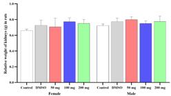
C. tinctoria does not significantly influence the relative weight of the kidneys at day 15.


3.3. Kidney function
The effect of EtOAcE of C. tinctoria on kidney function markers of the male and female rats is presented in Tables 1 and 2, respectively. The bilirubin and creatinine levels in male rats significantly elevated in the 200 mg/ kg group compared to the control group (Table 1). However, there was a marked increase in the uric acid levels only in the 200 mg female group in comparison with DMSO and 50 mg groups (Table 2).
3.4. Liver enzymes
In male rats, ALP levels significantly increased at the lower doses of 50 mg and 100 mg of the EtOAcE. Furthermore, higher doses (200 mg and 100 mg) of the EtOAcE caused GGT levels to be notably elevated compared to the control group (Table 3). In contrast, the ALT levels were exclusively elevated in the 100 mg treated male group compared to the control group.
Conversely, the female rats showed a remarkable decline in the ALP levels at the dose of 100 mg of the EtOAcE compared to the control group (Table 4). However, a significant increase in ALT levels was observed with a higher dose of the EtOAcE (200 mg) than the male rats. Interestingly, the GGT levels were noticeably increased in all EtOAcE-treated groups compared to the control and DMSO groups. These variations in hepatic response between male and female rats to the EtOAcE suggest the presence of gender-specific differences in toxicity tolerance, which could raise questions about the underlying regulatory mechanisms.
3.5. Glucose and cortisol levels
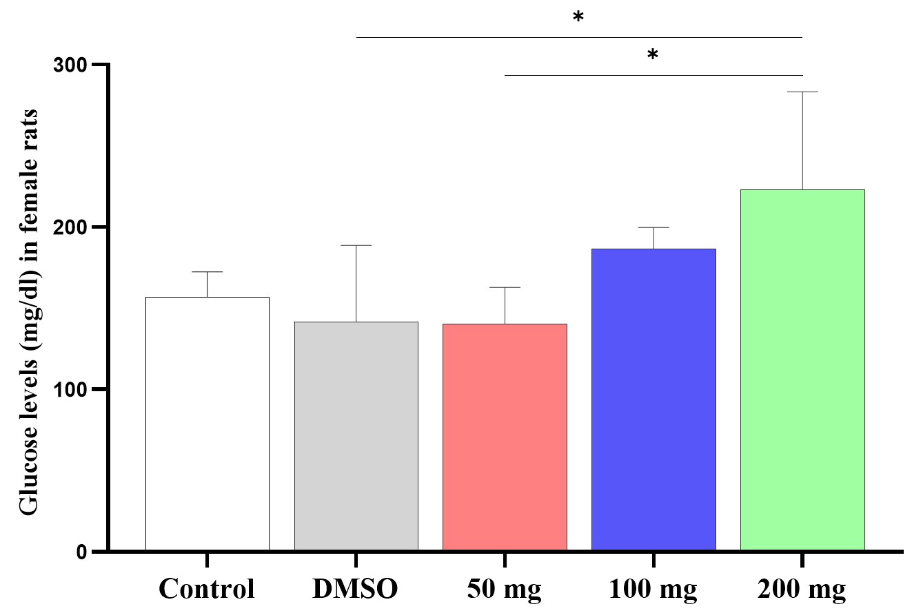
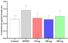
The glucose levels at day 15 were increased in female rats treated with 200 mg of the EtOAcE for 14 days compared to DMSO (p < 0.047) and 50 mg (p < 0.042) groups (Figure 6). By contrast, the male EtOAcE-treated groups after 14 days of treatment did not show any significant difference (p > 0.05) in glucose concentrations among groups (Figure 7). Similarly, the serum cortisol levels remained unchanged (p > 0.05) in both male and female groups (Figures 8 and 9).

The glucose levels after 14 days of the EtOAcE treatment were increased in female rats treated with 200 mg compared to DMSO (p < 0.047) and 50 mg (p < 0.042) groups.

The male rats treated with the EtOAcE for 14 days did not show any significant difference (p > 0.05) in glucose concentrations among groups.

3.6. Histopathological studies
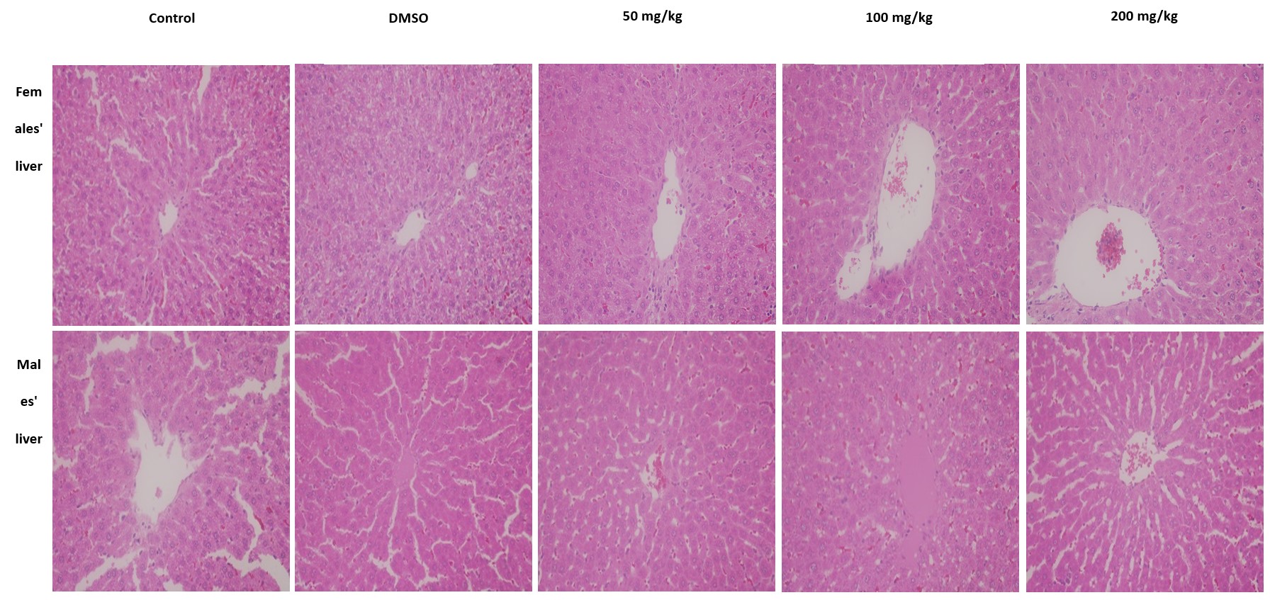
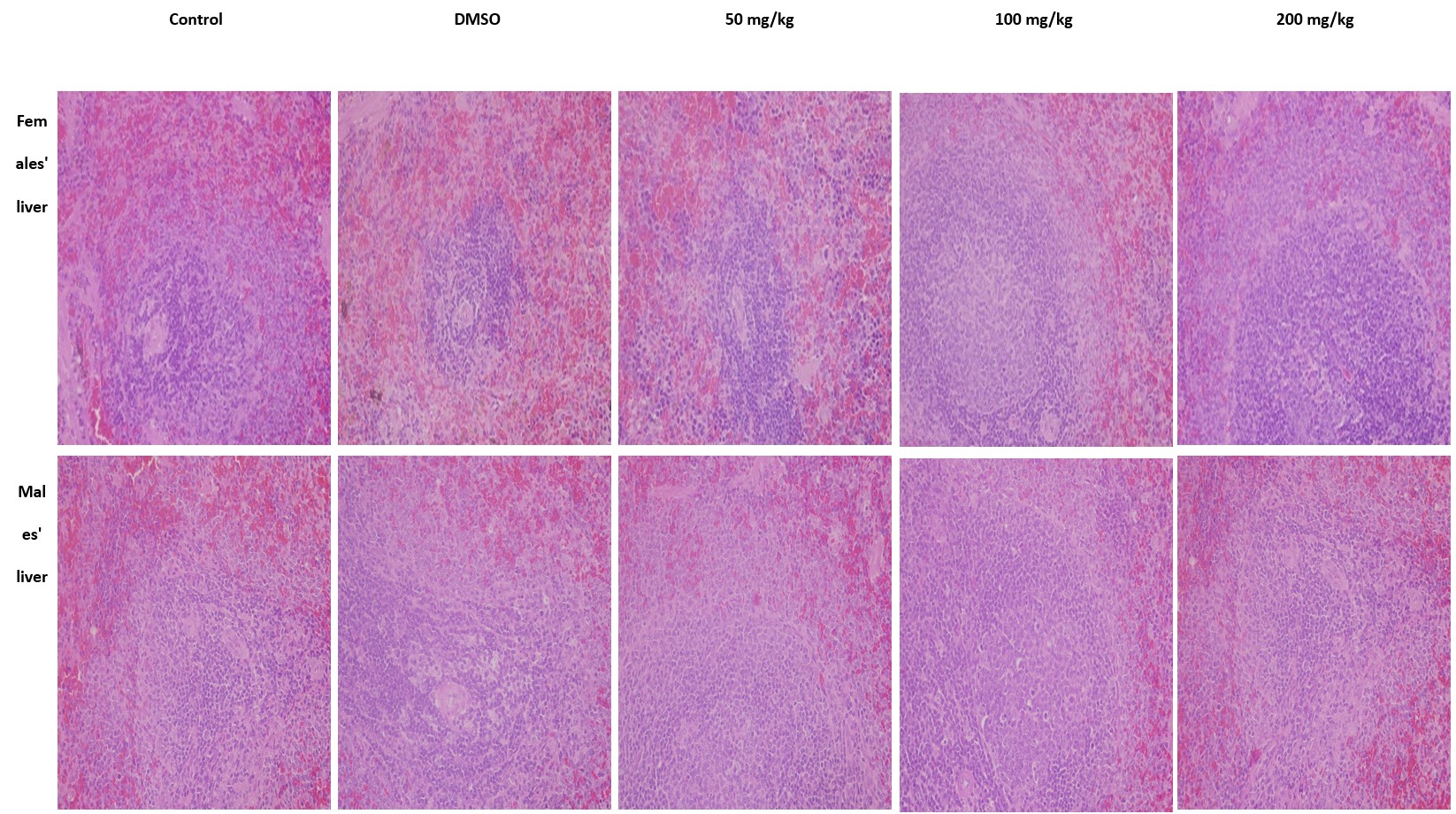
The histological sections of the liver obtained from male and female EtOAcE-treated rats exhibited several abnormalities, including congestion in the central vein with red blood and inflammatory cells, infiltration of inflammatory cells into the parenchyma and sinusoids, sinusoids dilation, and cytoplasmic vacuolization in hepatocytes (Figure 10). Renal histological sections showed narrowing of Bowman's space, vacuolization in the renal tubules, and congestion and shrinkage in the glomerulus in male and female rats treated with EtOAcE compared to the control and DMSO groups (Figure 11). In addition, the histological sections of the spleen of animals treated with EtOAcE showed lymphocyte hyperplasia in the white pulp compared to the control and DMSO groups (Figure 12). The histological sections of the small intestine in the male and female rats treated with EtOAcE of C. tinctoria displayed congestion in blood vessels and some inflammatory cells in the connective tissue of the lamina propria (Figure 13).
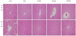
Hepatic histological sections of male and female rats treated with C. tinctoria extract for 14 days. (H&E × 40).

Renal histological sections of male and female rats treated with C. tinctoria extract for 14 days. (H&E × 40).

Spleen histological sections of male and female rats treated with C. tinctoria extract for 14 days. (H&E × 40).

Histological sections of small intestine in male and female rats treated with C. tinctoria extract for 14 days. (H&E × 40).
3.7. GC–MS analysis
The GC–MS spectra of the EtOAcE of C. tinctoria displayed peaks corresponding to the presence of 13 different compounds. Through spectral analysis using data libraries and molecular weights, the compound names are presented in (Table 5). The characterization of these compounds using mass spectrometry revealed four major bioactive compounds: 9,12-Octadecadienoic acid (Z,Z)- (27.42%), α-Tocopheryl acetate (19.01%), n-Hexadecanoic acid (14.98%), and 8,11,14-Eicosatrienoic acid (Z,Z,Z)- (9.36%).
4. Discussion
Chrozophora tinctoria has traditionally been used as a remedy for a variety of ailments, including diarrhea, vomiting, its anthelmintic properties, and the treatment of warts (Maurya et al., 2016; Iqbal et al., 2022; Sher et al., 2022). The use of herbal remedies often requires consistent daily administration over an extended period, especially for chronic diseases. Therefore, it is crucial to assess the potential adverse effects of these herbal substances on different physiological systems within the body (Alelign et al., 2020; Nalimu et al., 2022). This study aimed to evaluate the potential subacute toxicity of the EtOAcE of C. tinctoria in both male and female rats. The administration of the EtOAcE resulted in notable alterations in body weight, uric acid, bilirubin, creatinine, ALP, ALT, GGT, and glucose levels. These biochemical changes in serum biomarkers were accompanied by histological changes in the liver, kidneys, spleen, and small intestine, indicating a correlation between biochemical and histological toxicity markers.
The significant decrease in body weight in female rats, but not in males, suggests sex-specific susceptibility to the extract. This difference may be attributed to variations in metabolism, hormonal regulation, and detoxification processes between sexes. Previous studies have highlighted that females often exhibit different metabolic responses to toxins compared to males (Zuo et al., 2022; Tassinari et al., 2023). The unchanged relative weights (RW) of the liver, kidneys, and spleen suggest that toxicity may not manifest through changes in organ mass but rather through functional impairments and histopathological alterations, as seen in our findings.
Elevations in liver enzymes (ALT, GGT) in both male and female rats indicate hepatic stress or damage due to C. tinctoria extract. ALP, ALT, AST and GGT are enzymes released into the bloodstream during liver damage, and their elevated levels are indicative of hepatocellular damage and cholestasis (Iluz-Freundlich et al., 2020). The significant increase in ALT and GGT, particularly at higher dosages, suggests that the extract may cause significant liver injury. The decrease in ALP levels in females at the 100 mg/kg dosage, though contrary to the increases observed in other markers, might reflect a complex response mechanism in female liver metabolism, potentially influenced by hormonal variations. The reduction in ALP levels in female rats at this specific dosage might be explained by a few potential mechanisms. One possibility is that the EtOAcE of C. tinctoria exerts a selective inhibitory effect on ALP production or release in the liver. This could occur through a downregulation of ALP gene expression or through direct inhibition of the enzyme's activity. Certain bioactive compounds in natural products are known to interact with enzymatic pathways, leading to altered enzyme levels (Li et al., 2024). Additionally, sex-specific hormonal influences could play a role in this observed effect (Waxman and Holloway, 2009). Furthermore, disparities in the absorption of bioactive constituents may be a principal factor underlying the sex-specific differences in herbal product toxicity (Zuo et al., 2022).
The significant increase in bilirubin and creatinine levels in male rats at the highest dosage (200 mg/kg) suggests impaired kidney function, indicating renal stress or damage. Bilirubin and creatinine are reliable biomarkers for assessing renal function, and their elevation is indicative of compromised kidney filtration and function (Fevery, 2008; Jegnie et al., 2024). In female rats, the increased uric acid levels at the same dosage point to similar renal dysfunction. These findings are consistent with previous research showing nephrotoxicity of some various herbal products (Yang et al., 2018; Xu et al., 2020; Pearson et al., 2022). Many scientific studies underscore the necessity of considering sex as a biological variable in toxicological assessments (Gochfeld, 2017; Allegra et al., 2023).
The increase in glucose levels in female rats treated with the highest dosage of C. tinctoria extract suggests an impact on glucose homeostasis, either by increase insulin resistance or by directly influencing pancreatic function. The fatty acids present in C. tinctoria extract may interfere with normal insulin signaling pathways, thereby promoting inflammation and oxidative stress. This interference can disrupt the regulatory mechanisms of glucose uptake and utilization, potentially resulting in elevated blood glucose levels especially in female rats. The metabolic disturbances caused by fatty acids could contribute to the development of insulin resistance and related metabolic disorders (Sears and Perry, 2015). The absence of significant changes in cortisol levels across all groups implies that the hypothalamic-pituitary-adrenal (HPA) axis might not be directly affected by the extract in a manner that influences circulating cortisol. However, this does not rule out the possibility of stress responses at the cellular level that do not manifest as changes in serum cortisol.
The congestion, inflammation, and cytoplasmic vacuolization in the liver, along with tubular vacuolization and glomerular changes in the kidney, support the hepato-nephrotoxicity of the extract. Additionally, lymphocyte hyperplasia in the spleen suggests immunomodulation due to inflammation. Intestinal histopathology showed vascular congestion and inflammation, contributing to diarrhea, which is common with bioactive herbal extracts.
Plant collection may vary across seasons, plant growth stages, and environmental conditions, potentially affecting the presence of active compounds. Additionally, processes like freezing and thawing of extracts, oxidation, degradation, hydrolysis, thermal instability, and photodegradation can lead to the loss of compound activity or changes in extract solubility. To address these challenges, generating a fingerprint of extracts or fractions is crucial for monitoring production stability over time and ensuring consistency upon recollection. Quality control is paramount in herbal medicine development, and fingerprinting techniques, such as HPLC, IR, HPTLC, and GC-MS, are widely used for this purpose. For instance, (Alagrafi et al., 2017) utilized GC-MS to establish a fingerprint of Rhazya stricta, identifying 15 compounds as markers for the extract. In our study, we present fingerprints of the EtOAcE of C. tinctoria, crucial for monitoring stability, comparing compositions, and facilitating further studies for active compound isolation and characterization, as well as conducting future in vivo and in vitro research.
5. Conclusion
The EtOAcE of C. tinctoria demonstrated notable biochemical and histological toxicity in both male and female rats, with sex-specific differences in susceptibility and metabolic responses. These findings emphasize the importance of thorough toxicological assessments of herbal remedies to ensure their safe use, particularly in the context of chronic administration for long-term health conditions. Future studies should explore the molecular mechanisms underlying these toxic effects and assess the long-term consequences of chronic exposure. Reprotoxic and teratological analyses are necessary to obtain comprehensive evaluations of the EtOAcE of C. tinctoria. Additionally, the therapeutic index of C. tinctoria needs to be carefully evaluated to ensure its safe use in medicinal applications. These findings underscore the need for researchers to delve into the toxic byproducts present in this plant and explore the potency of other ingredients as pharmaceutical drugs.
Acknowledgements
The authors are grateful to Imam Abdulrahman bin Faisal University in Dammam, Saudi Arabia, for providing financial support for this project under grant number 2017-102-Sci.
References
-
AATI, H., EL-GAMAL, A., SHAHEEN, H. and KAYSER, O., 2019. Traditional use of ethnomedicinal native plants in the Kingdom of Saudi Arabia. Journal of Ethnobiology and Ethnomedicine, vol. 15, no. 1, pp. 2. http://doi.org/10.1186/s13002-018-0263-2 PMid:30626417.
» http://doi.org/10.1186/s13002-018-0263-2 -
ABDALLAH, H.M., ALMOWALLAD, F.M., ESMAT, A., SHEHATA, I.A. and ABDEL-SATTAR, E.A., 2015. Anti-inflammatory activity of flavonoids from Chrozophora tinctoria. Phytochemistry Letters, vol. 13, pp. 74-80. http://doi.org/10.1016/j.phytol.2015.05.008
» http://doi.org/10.1016/j.phytol.2015.05.008 -
ALAGRAFI, F.S., ALAWAD, A.O., ABUTAHA, N.M., NASR, F.A., ALHAZZAA, O.A., ALHARBI, S.N., ALKHRAYEF, M.N., HAMMAD, M., ALHAMDAN, Z.A., ALENAZI, A.D. and WADAAN, M.A., 2017. In vitro induction of human embryonal carcinoma differentiation by a crude extract of Rhazya stricta. BMC Complementary and Alternative Medicine, vol. 17, no. 1, pp. 342. http://doi.org/10.1186/s12906-017-1852-7 PMid:28662725.
» http://doi.org/10.1186/s12906-017-1852-7 -
ALELIGN, T., CHALCHISA, D., FEKADU, N., SOLOMON, D., SISAY, T., DEBELLA, A. and PETROS, B., 2020. Evaluation of acute and sub-acute toxicity of selected traditional antiurolithiatic medicinal plant extracts in Wistar albino rats. Toxicology Reports, vol. 7, pp. 1356-1365. http://doi.org/10.1016/j.toxrep.2020.10.001 PMid:33102139.
» http://doi.org/10.1016/j.toxrep.2020.10.001 -
ALLEGRA, S., CHIARA, F., DI GRAZIA, D., GASPARI, M. and DE FRANCIA, S., 2023. Evaluation of sex differences in preclinical pharmacology research: how far is left to go? Pharmaceuticals (Basel, Switzerland), vol. 16, no. 6, pp. 786. http://doi.org/10.3390/ph16060786 PMid:37375734.
» http://doi.org/10.3390/ph16060786 -
ALSOHAIMI, I.H., KHAN, M.R., ALI, H.M., AZAM, M. and ALAMMARI, A.M., 2020. Solvent extraction and gas chromatography-mass spectrometric determination of probable carcinogen 1,4-dioxane in cosmetic products. Scientific Reports, vol. 10, no. 1, pp. 5214. http://doi.org/10.1038/s41598-020-62149-x PMid:32251326.
» http://doi.org/10.1038/s41598-020-62149-x -
BETANCUR-GALVIS, L.A., MORALES, G.E., FORERO, J.E. and ROLDAN, J., 2002. Cytotoxic and antiviral activities of Colombian medicinal plant extracts of the Euphorbia genus. Memorias do Instituto Oswaldo Cruz, vol. 97, no. 4, pp. 541-546. http://doi.org/10.1590/S0074-02762002000400017 PMid:12118288.
» http://doi.org/10.1590/S0074-02762002000400017 -
FEVERY, J., 2008. Bilirubin in clinical practice: A review. Liver International, vol. 28, no. 5, pp. 592-605. http://doi.org/10.1111/j.1478-3231.2008.01716.x PMid:18433389.
» http://doi.org/10.1111/j.1478-3231.2008.01716.x -
GOCHFELD, M., 2017. Sex differences in human and animal toxicology. Toxicologic Pathology, vol. 45, no. 1, pp. 172-189. http://doi.org/10.1177/0192623316677327 PMid:27895264.
» http://doi.org/10.1177/0192623316677327 -
GOLKAR, P., TAGHIZADEH, M. and JALALI, S.A.H., 2019. Determination of phenolic compounds, antioxidant and anticancer activity of Chrozophora tinctoria accessions collected from different regions of Iran. Journal of Food Biochemistry, vol. 43, no. 11, pp. e13036. http://doi.org/10.1111/jfbc.13036 PMid:31495949.
» http://doi.org/10.1111/jfbc.13036 -
ILUZ-FREUNDLICH, D., ZHANG, M., UHANOVA, J. and MINUK, G.Y., 2020. The relative expression of hepatocellular and cholestatic liver enzymes in adult patients with liver disease. Annals of Hepatology, vol. 19, no. 2, pp. 204-208. http://doi.org/10.1016/j.aohep.2019.08.004 PMid:31628070.
» http://doi.org/10.1016/j.aohep.2019.08.004 -
IQBAL, A., SHER, A.A., MUHAMMAD, N., BADSHAH, S.L., EMWAS, A.H. and JAREMKO, M., 2022. Extraction and fractionation of prokinetic phytochemicals from Chrozophora tinctoria and their bioactivities. Molecules (Basel, Switzerland), vol. 27, no. 13, pp. 4321. http://doi.org/10.3390/molecules27134321 PMid:35807565.
» http://doi.org/10.3390/molecules27134321 -
JEGNIE, M., ABULA, T., SISAY, B., ABEBE, A., DEGU, S. and AFEWORK, M., 2024. Toxicological evaluation of chronic oral administration of Justicia schimperiana (Hochst. ex Nees) T. Anderson leaf 80% methanolic extract in Wistar albino rats. Toxicology Reports, vol. 12, pp. 158-167. http://doi.org/10.1016/j.toxrep.2024.01.010 PMid:38304696.
» http://doi.org/10.1016/j.toxrep.2024.01.010 - KARIMI, A., MAJLESI, M. and RAFIEIAN-KOPAEI, M., 2015. Herbal versus synthetic drugs: beliefs and facts. Journal of Nephropharmacology, vol. 4, no. 1, pp. 27-30. PMid:28197471.
-
KHAN, M.F., ALQAHTANI, A.S., ALMARFADI, O.M., ULLAH, R., NASR, F.A., NOMAN, O.M., SIDDIQUI, N.A., SHAHAT, A.A. and AHAMAD, S.R., 2021. The reproductive toxicity associated with Dodonaea viscosa, a folk medicinal plant in Saudi Arabia. Evidence-Based Complementary and Alternative Medicine, vol. 2021, no. 1, pp. 6689110. http://doi.org/10.1155/2021/6689110 PMid:33510808.
» http://doi.org/10.1155/2021/6689110 -
LI, Z., WU, J., ZHAO, Y., SONG, J. and WEN, Y., 2024. Natural products and dietary interventions on liver enzymes: an umbrella review and evidence map. Frontiers in Nutrition, vol. 11, pp. 1300860. http://doi.org/10.3389/fnut.2024.1300860 PMid:38371505.
» http://doi.org/10.3389/fnut.2024.1300860 -
MAURYA, H., SEMWAL, M. and DUBEY, S.K., 2016. Pharmacological evaluation of Chrozophora tinctoria as wound healing potential in diabetic rat’s model. BioMed Research International, vol. 2016, no. 1, pp. 7475124. http://doi.org/10.1155/2016/7475124 PMid:28097147.
» http://doi.org/10.1155/2016/7475124 -
NALIMU, F., OLORO, J., PETER, E.L. and OGWANG, P.E., 2022. Acute and sub-acute oral toxicity of aqueous whole leaf and green rind extracts of Aloe vera in Wistar rats. BMC Complementary Medicine and Therapies., vol. 22, no. 1, pp. 16. http://doi.org/10.1186/s12906-021-03470-4 PMid:35031035.
» http://doi.org/10.1186/s12906-021-03470-4 -
OKE-ALTUNTAS, F., IPEKCIOGLU, S., YAGLIOGLU, A.S., BEHCET, L. and DEMIRTAS, I., 2017. Phytochemical analysis, antiproliferative and antioxidant activities of Chrozophora tinctoria: a natural dye plant. Pharmaceutical Biology, vol. 55, no. 1, pp. 966-973. http://doi.org/10.1080/13880209.2016.1277767 PMid:28164719.
» http://doi.org/10.1080/13880209.2016.1277767 - ORGANIZATION FOR ECONOMIC COOPERATION AND DEVELOPMENT – OECD, 2008. Test No. 407: Repeated Dose 28-day Oral Toxicity Study in Rodents Paris: OECD Publishing.
-
PEARSON, A., GAFNER, S., RIDER, C.V., EMBRY, M., FERGUSON, S.S. and MITCHELL, C.A., 2022. Plant vs. kidney: evaluating nephrotoxicity of botanicals with the latest toxicological tools. Current Opinion in Toxicology, vol. 32, pp. 100371. http://doi.org/10.1016/j.cotox.2022.100371 PMid:36311298.
» http://doi.org/10.1016/j.cotox.2022.100371 -
PHUTRAKOOL, P. and PONGPIRUL, K., 2022. Acceptance and use of complementary and alternative medicine among medical specialists: A 15-year systematic review and data synthesis. Systematic Reviews, vol. 11, no. 1, pp. 10. http://doi.org/10.1186/s13643-021-01882-4 PMid:35027078.
» http://doi.org/10.1186/s13643-021-01882-4 -
QUAN, N.V., DANG XUAN, T. and TESCHKE, R., 2020. Potential hepatotoxins found in herbal medicinal products: a systematic review. International Journal of Molecular Sciences, vol. 21, no. 14, pp. 5011. http://doi.org/10.3390/ijms21145011 PMid:32708570.
» http://doi.org/10.3390/ijms21145011 -
RAHMAN, M.A., MOSSA, J.S., AL-SAID, M.S. and AL-YAHYA, M.A., 2004. Medicinal plant diversity in the flora of Saudi Arabia: A report on seven plant families. Fitoterapia, vol. 75, no. 2, pp. 149-161. http://doi.org/10.1016/j.fitote.2003.12.012 PMid:15030919.
» http://doi.org/10.1016/j.fitote.2003.12.012 -
RIBNICKY, D.M., POULEV, A., SCHMIDT, B., CEFALU, W.T. and RASKIN, I., 2008. Evaluation of botanicals for improving human health. The American Journal of Clinical Nutrition, vol. 87, no. 2, pp. 472S-475S. http://doi.org/10.1093/ajcn/87.2.472S PMid:18258641.
» http://doi.org/10.1093/ajcn/87.2.472S -
SEARS, B. and PERRY, M., 2015. The role of fatty acids in insulin resistance. Lipids in Health and Disease, vol. 14, no. 121, pp. 121. http://doi.org/10.1186/s12944-015-0123-1 PMid:26415887.
» http://doi.org/10.1186/s12944-015-0123-1 -
SHER, A.A., IQBAL, A., MUHAMMAD, N., BADSHAH, S.L., EMWAS, A.H. and JAREMKO, M., 2022. Prokinetic and laxative effects of Chrozophora tinctoria whole plant extract. Molecules (Basel, Switzerland), vol. 27, no. 7, pp. 2143. http://doi.org/10.3390/molecules27072143 PMid:35408541.
» http://doi.org/10.3390/molecules27072143 -
TASSINARI, R., TAMMARO, A., MARTINELLI, A., VALERI, M. and MARANGHI, F., 2023. Sex-specific effects of short-term oral administration of food-grade titanium dioxide nanoparticles in the liver and kidneys of adult rats. Toxics, vol. 11, no. 9, pp. 776. http://doi.org/10.3390/toxics11090776 PMid:37755786.
» http://doi.org/10.3390/toxics11090776 -
UGURLU, E. and SECMEN, O., 2008. Medicinal plants popularly used in the villages of Yunt Mountain (Manisa-Turkey). Fitoterapia, vol. 79, no. 2, pp. 126-131. http://doi.org/10.1016/j.fitote.2007.07.016 PMid:17878061.
» http://doi.org/10.1016/j.fitote.2007.07.016 -
ULLAH, R., ALQAHTANI, A.S., NOMAN, O.M.A., ALQAHTANI, A.M., IBENMOUSSA, S. and BOURHIA, M., 2020. A review on ethno-medicinal plants used in traditional medicine in the Kingdom of Saudi Arabia. Saudi Journal of Biological Sciences, vol. 27, no. 10, pp. 2706-2718. http://doi.org/10.1016/j.sjbs.2020.06.020 PMid:32994730.
» http://doi.org/10.1016/j.sjbs.2020.06.020 -
USMAN, H., MUSA, Y.M., AHMADU, A.A. and TIJJANI, M.A., 2007. Phytochemical and antimicrobial effects of Chrozophora senegalensis. African Journal of Traditional, Complementary, and Alternative Medicines, vol. 4, no. 4, pp. 488-494. http://doi.org/10.4314/ajtcam.v4i4.31242 PMid:20161917.
» http://doi.org/10.4314/ajtcam.v4i4.31242 -
WAXMAN, D.J. and HOLLOWAY, M.G., 2009. Sex differences in the expression of hepatic drug metabolizing enzymes. Molecular Pharmacology, vol. 76, no. 2, pp. 215-228. http://doi.org/10.1124/mol.109.056705 PMid:19483103.
» http://doi.org/10.1124/mol.109.056705 -
WILLIAMSON, E.M., 2003. Drug interactions between herbal and prescription medicines. Drug Safety, vol. 26, no. 15, pp. 1075-1092. http://doi.org/10.2165/00002018-200326150-00002 PMid:14640772.
» http://doi.org/10.2165/00002018-200326150-00002 -
XU, X., ZHU, R., YING, J., ZHAO, M., WU, X., CAO, G. and WANG, K., 2020. Nephrotoxicity of herbal medicine and its prevention. Frontiers in Pharmacology, vol. 11, pp. 569551. http://doi.org/10.3389/fphar.2020.569551 PMid:33178019.
» http://doi.org/10.3389/fphar.2020.569551 -
YANG, B., XIE, Y., GUO, M., ROSNER, M.H., YANG, H. and RONCO, C., 2018. Nephrotoxicity and Chinese herbal medicine. Clinical Journal of the American Society of Nephrology; CJASN, vol. 13, no. 10, pp. 1605-1611. http://doi.org/10.2215/CJN.11571017 PMid:29615394.
» http://doi.org/10.2215/CJN.11571017 -
ZUO, M.T., GONG, M.D., MA, X., XU, W.B., WANG, Z.Y., TANG, M.H., WU, Y. and LIU, Z.Y., 2022. Sex differences in the in vivo exposure process of multiple components of Gelsemium elegans in rats. Metabolites, vol. 13, no. 1, pp. 33. http://doi.org/10.3390/metabo13010033 PMid:36676958.
» http://doi.org/10.3390/metabo13010033