Tumor classification of adenoid cystic carcinoma: systematic review and meta-analysis
Abstract
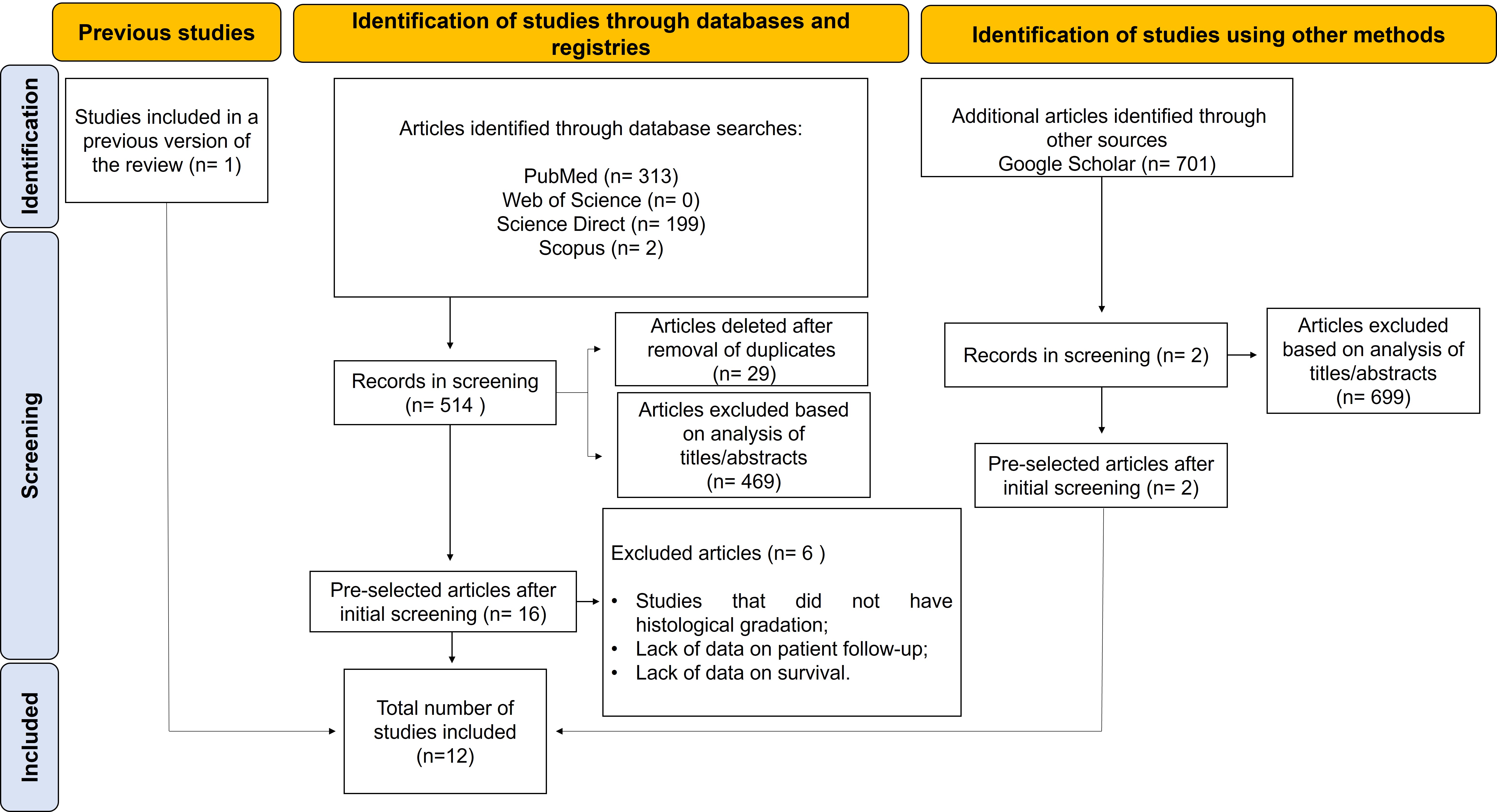
Adenoid Cystic Carcinoma - ACC is a common neoplasm in major and minor salivary glands with a high risk of metastasis. Thus, the objective of the present study was to perform an analysis to better understand the histological grading systems of the ACC and its influence on tumor prognosis in terms of overall survival, disease-free and metastasis-free. This is a systematic review, with meta-analysis, based on the PRISMA parameters. The search was carried out by two independent examiners, and searches were performed in the PubMed, Web of Science, Scopus, Science Direct, and Google Scholar databases. Of the 12 articles included in the systematic review, only 7 met the eligibility criteria for entry into the meta-analysis, with a statistically significant association between tumors with a solid pattern and death from the disease (DOD) (p <0.001; 95% CI 2.84 [1.94-4.15]; I 2 61%). Regarding the evaluation of the quality of studies by the Quality in Prognostic Studies (QUIPS), six studies were of high quality, five articles received moderate quality, and one article received low quality. It was concluded that the presence of any solid component contributes to a worse prognosis and lower specific survival of the disease over the 10 years.
Keywords:
adenoid cystic carcinoma; survival; neoplasm grading; salivary glands
Resumo
O Carcinoma Adenóide Cístico - ACC é uma neoplasia comum em glândulas salivares maiores e menores com alto risco de metástase. Assim, o objetivo do presente estudo foi realizar uma análise para melhor compreender os sistemas de graduação histológica do ACC e sua influência no prognóstico tumoral em termos de sobrevida global, livre de doença e livre de metástase. Trata-se de uma revisão sistemática, com meta-análise, baseada nos parâmetros PRISMA. A busca foi realizada por dois examinadores independentes, e as buscas foram realizadas nas bases de dados PubMed, Web of Science, Scopus, Science Direct e Google Scholar. Dos 12 artigos incluídos na revisão sistemática, apenas 7 preencheram os critérios de elegibilidade para entrada na meta-análise, com associação estatisticamente significativa entre tumores com padrão sólido e morte pela doença (DOD) (p < 0,001; IC 95% 2,84 [1,94-4,15]; I 2 61%). Em relação à avaliação da qualidade dos estudos pelo Quality in Prognostic Studies (QUIPS), seis estudos foram de alta qualidade, cinco artigos receberam qualidade moderada e um artigo recebeu qualidade baixa. Concluiu-se que a presença de qualquer componente sólido contribui para um pior prognóstico e menor sobrevida específica da doença ao longo dos 10 anos.
Palavras-chave:
carcinoma adenoide cístico; sobrevida; gradação de tumores; glândulas salivares
1. Introduction
Adenoid cystic carcinoma (ACC) is a slow-growing malignancy with a high risk of late distant metastasis. It is one of the most common salivary gland malignancies, accounting for 10-15% of all salivary gland tumors (van der Wal et al., 2002). ACC can arise in the major salivary glands such as parotid, submandibular, sublingual, lacrimal glands or in the minor salivary glands distributed throughout the oral cavity (Xu et al., 2017). The treatment of choice is surgical removal, followed by radiotherapy and chemotherapy, depending on the stage of the tumor and the presence of perineural invasion and positive surgical margins (van Weert et al., 2015). The ACC shows a slow and indolent growth pattern with an optimistic 5 years overall survival rate of 68-90%. However, long-term results revealed a decline in 10 and 15 years overall survival of 52% and 28%, respectively, primarily as a result of perineural invasion, local control failure, and distant metastases (Fang et al., 2022).
The original histopathological term for salivary gland ACC was “cylindroma” based on the histological appearance of epithelial/secretory cell casts and a hyaline stroma. Tumor cells have hyperchromatic angled nuclei and minimal cytoplasm, usually clear or eosinophilic (Dillon et al., 2016).
It is composed of epithelial and myoepithelial cells arranged in morphological growth patterns of tubular, cribriform and/or solid architecture, being the solid component with the highest chances of systemic dissemination and with a worse prognosis (Xuan et al., 2022).
Because the solid component has higher rates of late metastases and a poor prognosis, several gradation systems are currently applied, but it is still difficult to measure the proportion of solid components and objectively distinguish the solid and mixed components of the tumor (Zhu et al., 2021).
As for chemotherapy there are no standard recommendations since the growth of ACC is slow, thus reducing the cytotoxic effect of QT, but it is still used due to the lack of effective treatment alternatives for the disseminated disease. While, in patients treated surgically and with radiotherapy, they had a higher survival rate of local control when compared to patients treated only with surgical removal (Zupancic et al., 2023).
Currently, two histological grading systems are described by Perzin et al. (1978), Szanto et al. (1984) and Spiro et al. (1974). The Perzin/Szanto system considers high-grade ACC with more than 30% of the tumor composed of a solid component. In the Spiro system, the presence of more than 50% of solid parts in the tumor is considered high-grade. However, the amount of tumor to be investigated is not well defined in these studies (van Weert et al., 2015). The Perzin/Szanto and Spiro systems were tools for analysis only for overall survival (Morita et al., 2021).
The solid component has been studied over the years and new criteria for the solid tumor component, minAmax (maximal minor axis) in conjunction with van Weert, are independent prognostic tools for overall survival, disease-free and free of distant metastases (Morita et al., 2021).
Therefore, the aim of the present study was to perform an analysis to better understand the histological grading systems of ACC and its influence on tumor prognosis in terms of overall survival, disease-free and free of distant metastases, considering that the solid component is associated with a higher risk of systemic spread and poor prognosis, with consequently poor patient survival. The growing exploration of the topic by the scientific community is important, as studies will allow better conclusions about this pathology, its implications for health, types of treatment and consequent patient survival.
2. Materials and Methods
2.1. Research question
The research question of this systematic review was formulated according to the PICO (population, intervention, control and outcome) according to Page et al (2021) strategy in which it helps to formulate accurate and relevant research questions for the study: Is the histopathological grading of Adenoid Cystic Carcinoma (ACC) a prognostic predictor for survival?
2.2. Search strategy
The present systematic review was conducted according to the Artificial Intelligence Tool for systematic reviews and meta-analysis (PRISMA). The review was registered in the International Prospective Systems Thematic Reviews (PROSPERO) under number CRD42023406011. Searches were performed in the following electronic databases without restrictions on language or date of publication: PubMed, Web of Science, Scopus, Science Direct, Google Scholar. All stages of the study were evaluated by a reviewer with experience in the preparation of systematic reviews (G.M.F.). A manual search in the reference list of included articles was performed to optimize and expand the search (Table 1).
2.3. Eligibility criteria
The following eligibility criteria were applied to select the articles: (I) ACC in the oral cavity, (II) cross-sectional study, (III) case-control study, and (IV) cohort study. The following studies were excluded: (I) systematic review and literature articles, (II) case reports, (III) conference proceedings, (IV) letters to the reader, (V) books and book chapters, and (VI) studies on ACC not involving the oral cavity, studies that did not use histopathological classification, and studies without data on patient monitoring and survival.
2.4. Selection of studies
Articles that did not meet the eligibility criteria and duplicate articles were removed. In the first stage, the articles were selected through the screening of titles and abstracts. Then, all studies whose titles and abstracts were considered relevant were selected for reading the full text. Finally, the articles selected by the evaluators were included in the database. We used the Rayyan tool for systematic reviews (Qatar Foundation) to manage the selected articles and delete duplicates. All steps were conducted by two independent reviewers, and a third reviewer was consulted in case of disagreement.
2.5. Data extraction
Eligible studies were reviewed and the following data were extracted: authors, year of publication, country, number of cases, type of study, target audience, histological grading, TNM staging system (T: tumor, N: regional lymph nodes, and M: distant metastasis), surgical margins, radiotherapy, and chemotherapy. The articles were independently selected by two reviewers (D.C.S.O. and L.B.V.N.). Any disagreement was resolved by consensus.
2.6. Assessment of risk of bias of included studies
The risks of bias of full-text articles were independently assessed by two researchers (D.C.S.O. and L.B.V.N.) using the Quality in Prognostic Studies (QUIPS) tool (Hayden et al., 2013). Based on the score of each article in five domains: (1) study participation, (2) study attrition, (3) measurement of prognostic factors, (4) measurement of outcomes, (5) analysis and presentation of statistics. Since in prognostic studies, the prediction of disease-specific survival is based on a combination of several possible prognostic factors.
The five domains of QUIPS received maximum scores of 15 points each, equally divided by domains. The scores established for all items were 5 (low risk of bias), 2.5 (moderate risk of bias), and 0 (high risk of bias), except for domain 1 (patient selection bias), which was scored as follows: Up to 3 (low risk of bias), 1.5 (moderate risk of bias), and 0 (high risk of bias). A maximum total score of 75 was established based on the sum of all scores per item. A priori, a score ≥ 60 points was classified as high quality of the study, 45-60 as moderate quality, and < 45 as low quality (Samuels et al., 2017). Finally, scores were determined to synthesize these data: High quality (+) when the risk of bias was low; moderate (+/-) quality when risk of bias was moderate and low quality (-) when risk of bias was high.
2.7. Data analysis
The data collected from the results based on the studies selected for meta-analysis were entered and analyzed with the RAYYAN for systematic review tool (Qatar Foundation). Several results were evaluated, such as: (1) independent histological grading; (2) tumor staging (T1/T2 or T3/T4); (3) lymph node staging (N0 – negative or N+ – positive); (4) surgical margins (positive/negative or indeterminate); (5) postoperative radiotherapy (dose in Gy) and (6) chemotherapy for distant metastasis when positive. The meta-analysis was performed using the Review Software Manager® (RevMan, 2014) to estimate the correlation between histopathological grade (grade I/II and Grade III) and the frequency of deaths from the disease. Heterogeneity between studies was estimated using the Cochran Q test (Dersimonian and Laird, 1986; Higgins et al., 2011).
3. Results
3.1. Selection of studies
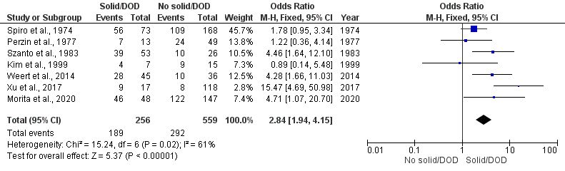
Using the search strategy developed in this systematic review, a total of 1,216 articles were retrieved from the databases. After removing duplications, 1,187 articles remained for title and abstract screening. At this stage, only 16 articles were considered potentially eligible and their text was read independently by two researchers (D.C.S.O. and L.B.V.N.). After the analysis, 12 articles presented inclusion criteria and were selected for this review (Figure 1). An inter-rater Cohen's Kappa index was created, which resulted in a value of K=0.962.
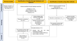
PRISMA flowchart of the selection process of articles for inclusion in the systematic review. Source: Personal archive.
3.2. Histological classification
The search resulted in 12 observational articles, 11 cross-sectional studies and 01 cohort study. These studies come from Europe (n=2), Asia (n=5), and North America (n=5). The sample had 1454 cases of ACC, with a predominance of females (n=820) to the detriment of males (n=634) in a ratio of 1.29:1. The most frequent age group is the 5th decade of life. All articles were published in English. Univariate and multivariate tables were used as a method of analysis for the study evaluating the relationship between the type of histological gradation and survival.
Table 1 shows the relationship between the histological gradation of the ACC where 07 histopathological grading systems were found, namely: Kim et al. (1999), Perzin et al. (1978), Szanto et al. (1984), Spiro et al. (1974), van Weert et al. (2015), Morita et al. (2021) and Xu et al. (2017). In the study by Patey and Thackray (1958), there was no data information in univariate and multivariate tables that were used as a method of analysis of the research.
There was a predominance of low and high-grade tumors, which prevailed in terms of the presence of solid areas. Histologically, ACC is a biphasic tumor composed of ductal and myoepithelial cells, forming tubular, cribriform, and solid growth patterns (Figure 2A, 2B and 2C). The presence of a solid component, but not its percentage, appears to be prognosically significant (Xu et al., 2017).

Histological aspects of ACC stained with HE (hematoxylin-eosin). There was a predominance of low and high grade tumors, with the prevalence of the classification according to the presence of solid areas. (A) Cribriform (100X); (B) Tubular (100X); (C) Solid (100X). Source: Personal archive (CESMAC Pathology Laboratory).
Regarding tumor staging, most articles presented T3/T4 staging (n=582) and T1/T2 staging (n=631) and lymph node metastasis (n=107). Surgical margins were positive (n=281). Chemotherapy was used in 63 cases, mainly in patients with distant metastases, such as cisplatin and 5-FU. As for radiotherapy, it was used in 484 cases, of which 219 were used as an adjuvant in the postoperative period (Zhang et al., 2013; Szanto et al., 1984). The highest dose of radiotherapy with 70 Gy for tumors with positive margins (Zhu et al., 2021) in another study the dose ranged from 57 Gy to 66 Gy (Zhang et al., 2013); at a daily dose of 1.8 to 2 Gy between 7 and 8 weeks (Table 2).
Prevalence of the number of cases who underwent surgery, radiotherapy, and/or chemotherapy of ACC.
Regarding overall survival and disease-free survival, it can be observed that tumors with high transformation are predominantly the solid histological type, with this a worse survival is notorious (Table 3).
3.3. Meta-analysis
The meta-analysis was performed among the histopathological gradations of adenoid cystic carcinoma in the literature, the cribiform/tubular histological patterns were grouped as non-solid patterns, while the mixed and solid patterns were grouped as solid. A higher frequency of deaths from the disease (DOD) was observed in the solid pattern (p<0.001) and the heterogeneity presented between the studies was (I^2 = 61%), as shown in Figure 3.

Forest plot graph of the relationship between the presence of a solid tumor and deaths from the disease. Source: Personal archive.
Regarding the quantity of studies included in the systematic review, six studies were of high quality, five were of moderate quality, and one study was of low quality. Only studies that presented their own histological gradation were included in the meta-analysis (Table 4).
4. Discussion
ACC can affect all ages, but it is more common in women between the fifth and sixth decade of life, it is represented as a slow-growing neoplasm, but with a high chance of distant metastasis. Its diagnosis is controversial due to the difficulty in histopathological differentiation between polymorphous adenocarcinoma in the cribriform aspect and basal cell adenocarcinoma when solid (Zupancic et al., 2023).
Approximately 40% of patients with ACC develop metastatic disease. Risk factors for metastasis include local recurrence, presence of solid tumor, tumor size greater than 3 cm, and regional lymph node involvement. The most common sites of metastases are the lungs, followed by bones, liver, skin, and breast. Metastases to the brain result from invasion along the cranial nerves (Dillon et al., 2016).
Seven histopathological grading systems were found including Kim et al. (1999), Perzin et al. (1978), Szanto et al. (1984), Spiro et al. (1974), van Weert et al. (2015); Xu et al. (2017) and Morita et al. (2021). All of them involve the major and minor salivary glands. However, these systems are controversial and there is no established histopathological protocol.
In most studies, tumors were divided into high and low grade and generally classified according to the presence of a solid area in the three existing patterns: Cribriform, tubular, and solid. Solid components that coexist with tubular or cribriform patterns indicate a worse prognosis. This correlation between histopathological classification and survival is still controversial in the literature (Zhu et al., 2021).
Morphologically, ACC is diagnosed by histopathological patterns. The cribriform pattern is characterized by islands of scattered pseudocysts of small, hyperchromatic myoepithelial neoplastic cells. This pattern usually has a good prognosis. The tubular pattern consists of pseudoducts and tubules, which are lined by one or several layers of myoepithelial cells, often surrounded by a hyaline stroma (van Weert et al., 2015). When compared to the cribriform and tubular type, the solid type is formed by islands of small, hyperchromatic neoplastic cells. It is considered a high-grade tumor with poor prognosis and survival (Fordice et al., 1999).
The classification system developed by Morita, minAmax, considers a size of solid nests greater than 0.20 mm as an indicator of worse prognosis. However, this gradation system cannot be applied to small specimens and is therefore limited to cases of larger tumors (Morita et al., 2021).
The presence of the solid component coexisting with the tubular and cribriform patterns indicates a poor prognosis. Exploration of the systems is necessary as they have found that the existence of any solid component and high-grade transformation is considered an aggressive disease pattern, which is associated with a poor prognosis. Although the three patterns exhibit well-defined characteristics, most tumors consist of a mixture of these three (Zhu et al., 2021).
The definitions of the Perzin/Szanto classification system subdivide tumors into grade I (predominantly tubular and non-solid), grade II (predominantly cribriform, < 30% solid) and grade III (solid component > 30%). In the Spiro system, tumors are classified as grade I (occasionally solid tubular or cribriform patterns), grade II (substantial solid > 50%), and grade III (solid only). The high-grade ACC consists of the grade III classification of Perzin/Szanto and Spiro et al. (1974) (van Weert et al., 2015).
The studies by Sung et al. (2003), Patey and Thackray (1958), Perzin et al. (1978) and Szanto et al. (1984) indicate that distant metastases develop mainly in solid subtypes.
In the present meta-analysis, the solid pattern or grade III was associated with overall and specific survival of poor disease across all classification systems (Szanto et al., 1984; Spiro et al., 1974; Morita et al., 2021; van Weert et al., 2015; Perzin et al., 1978). Poor disease-free survival is due to frequent local recurrences or distant metastases (van Weert et al., 2015).
Perineural invasion was frequently reported in the studies by Zhu et al. (2021), Xuan et al. (2022), van Weert et al. (2015), Zhang et al. (2013), Zupancic et al. (2023), Morita et al. (2021), Spiro et al. (1974) and Szanto et al. (1984). Considered a characteristic of the disease; however, studies do not allow us to conclude to what extent it is related to the prognosis of the disease.
The importance of describing its existence lies in the fact that perineural invasion can extend through the nerves at a considerable distance from the primary tumor; however, perineural invasion as a prognostic factor is still considered ambiguous (Barrett and Speight, 2009). On the other hand, Zupancic et al. (2023) reports that perineural invasion is associated in most cases with positive surgical margins and poor local tumor control, resulting in a poor prognosis. Many authors state that perineural invasion occurs particularly along larger nerves and is strongly associated with lower overall and disease-free survival rates (Zupancic et al., 2023).
Treatment of ACC varies depending on your location. A surgical approach is more appropriate in the early stages, when the tumor is smaller. Radiation therapy is an alternative treatment choice when the tumor is inaccessible or, combined with surgery, when surgical margins are compromised (Wang and Goodman, 1991).
The treatment modalities of choice are surgery followed by radiation therapy. Although widely employed, radiotherapy was generally used as an adjunctive treatment during the postoperative period in cases where surgical margins were positive and lymph node involvement and perineural invasion were present, with combined treatment (radiotherapy + surgery) significantly improving overall survival (Dillon et al., 2016). Modified radical neck dissection is reserved for patients with lymph node involvement in whom 5 to 10 years recurrence rates range from 30% to 75% despite adherence to good surgical technique (Zupancic et al., 2023).
The highest doses of radiation therapy observed in the study were 70 Gy for tumors with positive margins or a range of 57 to 66 Gy. The daily dose is 1.8 to 2 Gy for 7 to 8 weeks. Thus, surgery combined with adjuvant radiotherapy using doses above 60 Gy should be considered the standard of care (Chen et al., 2006). Chemotherapy is limited only to palliative cases of due to the diversity of histological subtypes and the rarity of the disease (Papaspyrou et al., 2011). In addition, ACC is considered chemoresistant. However, some studies indicate a good response to treatment with paclitaxel, tamoxifen, and cetuximab (Till and Martins, 2008).
In the present review, chemotherapy was rarely used, especially in cases of distant metastases. The main drugs of choice were cisplatin and 5-fluorouracil, corroborating studies in the literature with promising results (Ross et al., 2009; Ghosal et al., 2011). This type of treatment is often chosen for palliative care or more advanced cases with promising results; however, radiation therapy and surgery remain the gold standard (Andry et al., 2012).
The studies by Morita et al. (2021) and van Weert et al. (2015) were the only ones that provided statistically significant results showing that solid tumors have poor metastasis-free survival. Within this context, the minAmax system was considered a useful tool for the prognosis of overall survival, disease-free survival, and metastasis-free survival (Morita et al., 2021).
In view of the poor long-term prognosis of ACC and its slow growth, a survival rate of at least 15 to 20 years should be considered. In addition, the development of metastases more than 5 years after initial treatment is observed in patients with ACC (Chen et al., 2008). Late and distant metastases are responsible for a low long-term survival rate, with patients having a poor prognosis and 80-90% dying within 10-15 years (Hamper et al., 1990).
Our study has some limitations. ACC is a disease with a poor prognosis, depending on its histopathological degree and clinical manifestation. Therefore, heterogeneity was observed in the presentation of overall survival and disease-free survival data in the 12 studies included in this review. Some studies have not reported data on tumor stage, lymph node stage (Spiro et al., 1974; Perzin et al., 1978; Szanto et al., 1984; Du et al., 2016), perineural invasion or surgical margins (Spiro et al., 1974; Perzin et al., 1978; Brackrock et al., 2005). These studies were also unclear as to the doses of radiotherapy and which chemotherapy drugs were used for each type of treatment.
In addition, the system proposed by van Weert et al. (2015) was more powerful in predicting disease-specific survival and compared low and high grade tumors according to the presence of the solid component; this generates bias on the part of the researchers, claiming that their classification system is more sensitive in predicting patient survival, although all classification systems show statistically significant associations with the variables analyzed. The articles included in the meta-analysis had high, moderate quality and low risk of bias. The present review suggests that future studies of classification systems should better explain the stratification of results to reduce heterogeneity and allow the association of histopathological classification with lymph node and perineural invasion, thus providing greater scientific evidence (van Weert et al., 2015).
5. Conclusion
In this review, tumor classification systems suggest that the presence of the solid component is associated with a poor prognosis. ACC is a frequent tumor with a poor prognosis at 10 years of follow-up. Thus, further prognostic studies of this tumor are needed. The lowest rates of disease-specific survival and distant metastasis-free survival were associated with high tumor grade, perineural invasion, lymph node involvement, and positive surgical margins.
References
-
ANDRY, G., HAMOIR, M., LOCATI, L.D., LICITRA, L. and LANGENDIJK, J.A., 2012. Management of salivary gland tumors. Expert Review of Anticancer Therapy, vol. 12, no. 9, pp. 1161-1168. http://doi.org/10.1586/era.12.92 PMid:23098116.
» http://doi.org/10.1586/era.12.92 -
BARRETT, A.W. and SPEIGHT, P.M., 2009. Perineural invasion in adenoid cystic carcinoma of the salivary glands: a valid prognostic indicator? Oral Oncology, vol. 45, no. 11, pp. 936-940. http://doi.org/10.1016/j.oraloncology.2009.07.001 PMid:19692291.
» http://doi.org/10.1016/j.oraloncology.2009.07.001 - BRACKROCK, S., KRÜLL, A., RÖSER, K., SCHWARZ, R., RIETHDORF, L. and ALBERTI, W., 2005. Neutron therapy, prognostic factors and dedifferentiation of adenoid cystic carcinomas (ACC) of salivary glands. Anticancer Research, vol. 25, no. 2B, pp. 1321-1326. PMid:15865085.
-
CHEN, A.M., BUCCI, M.K., WEINBERG, V., GARCIA, J., QUIVEY, J.M., SCHECHTER, N.R., PHILLIPS, T.L., FU, K.K. and EISELE, D.W., 2006. Adenoid cystic carcinoma of the head and neck treated by surgery with or without postoperative radiation therapy: prognostic features of recurrence. International Journal of Radiation Oncology, Biology, Physics, vol. 66, no. 1, pp. 152-159. http://doi.org/10.1016/j.ijrobp.2006.04.014 PMid:16904520.
» http://doi.org/10.1016/j.ijrobp.2006.04.014 -
CHEN, A.M., GARCIA, J., GRANCHI, P.J., JOHNSON, J. and EISELE, D.W., 2008. Late recurrence from salivary gland cancer: when does "cure" mean cure? Cancer, vol. 112, no. 2, pp. 340-344. http://doi.org/10.1002/cncr.23165 PMid:18008358.
» http://doi.org/10.1002/cncr.23165 -
DERSIMONIAN, R. and LAIRD, N., 1986. Meta-analysis in clinical trials. Controlled Clinical Trials, vol. 7, no. 3, pp. 177-188. http://doi.org/10.1016/0197-2456(86)90046-2 PMid:3802833.
» http://doi.org/10.1016/0197-2456(86)90046-2 -
DILLON, P.M., CHAKRABORTY, S., MOSKALUK, C.A., JOSHI, P.J. and THOMAS, C.Y., 2016. Adenoid cystic carcinoma: a review of recent advances, molecular targets, and clinical trials. Head & Neck, vol. 38, no. 4, pp. 620-627. http://doi.org/10.1002/hed.23925 PMid:25487882.
» http://doi.org/10.1002/hed.23925 -
DU, F., ZHOU, C.X. and GAO, Y., 2016. Myoepithelial differentiation in cribriform, tubular and solid pattern of adenoid cystic carcinoma: a potential involvement in histological grading and prognosis. Annals of Diagnostic Pathology, vol. 22, pp. 12-17. http://doi.org/10.1016/j.anndiagpath.2016.03.001 PMid:27180054.
» http://doi.org/10.1016/j.anndiagpath.2016.03.001 -
FANG, Y., PENG, Z., WANG, Y., GAO, K., LIU, Y., FAN, R., ZHANG, H., XIE, Z. and JIANG, W., 2022. Current opinions on diagnosis and treatment of adenoid cystic carcinoma. Oral Oncology, vol. 130, pp. 105945. http://doi.org/10.1016/j.oraloncology.2022.105945 PMid:35662026.
» http://doi.org/10.1016/j.oraloncology.2022.105945 -
FORDICE, J., KERSHAW, C., EL-NAGGAR, A. and GOEPFERT, H., 1999. Adenoid cystic carcinoma of the head and neck: predictors of morbidity and mortality. Archives of Otolaryngology--Head & Neck Surgery, vol. 125, no. 2, pp. 149-152. http://doi.org/10.1001/archotol.125.2.149 PMid:10037280.
» http://doi.org/10.1001/archotol.125.2.149 -
GHOSAL, N., MAIS, K., SHENJERE, P., JULYAN, P., HASTINGS, D., WARD, T., RYDER, W.D., BRUCE, I., HOMER, J. and SLEVIN, N.J., 2011. Phase II study of cisplatin and imatinib in advanced salivary adenoid cystic carcinoma. British Journal of Oral & Maxillofacial Surgery, vol. 49, no. 7, pp. 510-515. http://doi.org/10.1016/j.bjoms.2010.09.013 PMid:21071117.
» http://doi.org/10.1016/j.bjoms.2010.09.013 -
HAMPER, K., LAZAR, F., DIETEL, M., CASELITZ, J., BERGER, J., ARPS, H., FALKMER, U., AUER, G. and SEIFERT, G., 1990. Prognostic factors for adenoid cystic carcinoma of the head and neck: a retrospective evaluation of 96 cases. Journal of Oral Pathology & Medicine, vol. 19, no. 3, pp. 101-107. http://doi.org/10.1111/j.1600-0714.1990.tb00806.x PMid:2160530.
» http://doi.org/10.1111/j.1600-0714.1990.tb00806.x -
HAYDEN, J.A., VAN DER WINDT, D.A., CARTWRIGHT, J.L., CÔTÉ, P. and BOMBARDIER, C., 2013. Assessing bias in studies of prognostic factors. Annals of Internal Medicine, vol. 158, no. 4, pp. 280-286. http://doi.org/10.7326/0003-4819-158-4-201302190-00009 PMid:23420236.
» http://doi.org/10.7326/0003-4819-158-4-201302190-00009 -
HIGGINS, J.P.T., ALTMAN, D.G., GØTZSCHE, P.C., JÜNI, P., MOHER, D., OXMAN, A.D., SAVOVIC, J., SCHULZ, K.F., WEEKS, L. and STERNE, J.A., 2011. The Cochrane Collaboration’s tool for assessing risk of bias in randomised trials. BMJ, vol. 343, no. 2, pp. d5928. http://doi.org/10.1136/bmj.d5928 PMid:22008217.
» http://doi.org/10.1136/bmj.d5928 -
KIM, G.E., PARK, H.C., KEUM, K.C., LEE, C.G., SUH, C.O., HUR, W.J., KIM, K.M. and HONG, W.P., 1999. Adenoid cystic carcinoma of the maxillary antrum. American Journal of Otolaryngology, vol. 20, no. 2, pp. 77-84. http://doi.org/10.1016/S0196-0709(99)90015-7 PMid:10203156.
» http://doi.org/10.1016/S0196-0709(99)90015-7 -
MORITA, N., MURASE, T., UEDA, K., NAGAO, T., KUSAFUKA, K., NAKAGURO, M., URANO, M., TAGUCHI, K.I., YAMAMOTO, H., KANO, S., TADA, Y., TSUKAHARA, K., OKAMI, K., ONITSUKA, T., FUJIMOTO, Y., KAWAKITA, D., SAKURAI, K., NAGAO, T., HANAI, N., KAWATA, R., HATO, N., OTSUKI, N., NIBU, K.I. and INAGAKI, H., 2021. Pathological evaluation of tumor grade for salivary adenoid cystic carcinoma: a proposal of an objective grading system. Cancer Science, vol. 112, no. 3, pp. 1184-1195. http://doi.org/10.1111/cas.14790 PMid:33377247.
» http://doi.org/10.1111/cas.14790 -
PAGE, M.J., MCKENZIE, J.E., BOSSUYT, P.M., BOUTRON, I., HOFFMANN, T.C., MULROW, C.D., SHAMSEER, L., TETZLAFF, J.M., AKL, E.A., BRENNAN, S.E., CHOU, R., GLANVILLE, J., GRIMSHAW, J.M., HRÓBJARTSSON, A., LALU, M.M., LI, T., LODER, E.W., MAYO-WILSON, E., MCDONALD, S., MCGUINNESS, L.A., STEWART, L.A., THOMAS, J., TRICCO, A.C., WELCH, V.A., WHITING, P. and MOHER, D., 2021. The PRISMA 2020 statement: an updated guideline for reporting systematic reviews. Systematic Reviews, vol. 10, no. 1, pp. 89. http://doi.org/10.1186/s13643-021-01626-4 PMid:33781348.
» http://doi.org/10.1186/s13643-021-01626-4 -
PAPASPYROU, G., HOCH, S., RINALDO, A., RODRIGO, J.P., TAKES, R.P., VAN HERPEN, C., WERNER, J.A. and FERLITO, A., 2011. Chemotherapy and targeted therapy in adenoid cystic carcinoma of the head and neck: a review. Head & Neck, vol. 33, no. 6, pp. 905-911. http://doi.org/10.1002/hed.21458 PMid:20652885.
» http://doi.org/10.1002/hed.21458 -
PATEY, D.H. and THACKRAY, A.C., 1958. The treatment of parotid tumours in the light of a pathological study of parotidectomy material. British Journal of Surgery, vol. 45, no. 193, pp. 477-487. http://doi.org/10.1002/bjs.18004519314 PMid:13536351.
» http://doi.org/10.1002/bjs.18004519314 -
PERZIN, K.H., GULLANE, P. and CLAIRMONT, A.C., 1978. Adenoid cystic carcinomas arising in salivary glands: a correlation of histologic features and clinical course. Cancer, vol. 42, no. 1, pp. 265-282. http://doi.org/10.1002/1097-0142(197807)42:1<265::AID-CNCR2820420141>3.0.CO;2-Z PMid:208752.
» http://doi.org/10.1002/1097-0142(197807)42:1<265::AID-CNCR2820420141>3.0.CO;2-Z - REVMAN, 2014. ReviewManager (RevMan) [software]. The Cochrane Collaboration.
-
ROSS, P.J., TEOH, E.M., A’HERN, R.P., RHYS-EVANS, P.H., HARRINGTON, K.J., NUTTING, C.M. and GORE, M.E., 2009. Epirubicin, cisplatin and protracted venous infusion 5-fluorouracil chemotherapy for advanced salivary adenoid cystic carcinoma. Clinical Oncology, vol. 21, no. 4, pp. 311-314. http://doi.org/10.1016/j.clon.2008.12.009 PMid:19201585.
» http://doi.org/10.1016/j.clon.2008.12.009 -
SAMUELS, N., VAN DE GRAAF, R.A., DE JONGE, R.C.J., REISS, I.K.M. and VERMEULEN, M.J., 2017. Risk factors for necrotizing enterocolitis in neonates: a systematic review of prognostic studies. BMC Pediatrics, vol. 17, no. 1, pp. 105. http://doi.org/10.1186/s12887-017-0847-3 PMid:28410573.
» http://doi.org/10.1186/s12887-017-0847-3 -
SEETHALA, R.R., HUNT, J.L., BALOCH, Z.W., LIVOLSI, V.A. and LEON BARNES, E., 2007. Adenoid cystic carcinoma with high-grade transformation: a report of 11 cases and a review of the literature. The American Journal of Surgical Pathology, vol. 31, no. 11, pp. 1683-1694. http://doi.org/10.1097/PAS.0b013e3180dc928c PMid:18059225.
» http://doi.org/10.1097/PAS.0b013e3180dc928c -
SPIRO, R.H., HUVOS, A.G. and STRONG, E.W., 1974. Adenoid cystic carcinoma of salivary origin: A clinicopathologic study of 242 cases. American Journal of Surgery, vol. 128, no. 4, pp. 512-520. http://doi.org/10.1016/0002-9610(74)90265-7 PMid:4371368.
» http://doi.org/10.1016/0002-9610(74)90265-7 -
SUNG, M.W., KIM, K.H., KIM, J.W., MIN, Y.G., SEONG, W.J., ROH, J.L., LEE, S.J., KWON, T.K. and PARK, S.W., 2003. Clinicopathologic predictors and impact of distant metastasis from adenoid cystic carcinoma of the head and neck. Archives of Otolaryngology--Head & Neck Surgery, vol. 129, no. 11, pp. 1193-1197. http://doi.org/10.1001/archotol.129.11.1193 PMid:14623749.
» http://doi.org/10.1001/archotol.129.11.1193 -
SZANTO, P.A., LUNA, M.A., TORTOLEDO, M.E. and WHITE, R.A., 1984. Histologic grading of adenoid cystic carcinoma of the salivary glands. Cancer, vol. 54, no. 6, pp. 1062-1069. http://doi.org/10.1002/1097-0142(19840915)54:6<1062::AID-CNCR2820540622>3.0.CO;2-E PMid:6088017.
» http://doi.org/10.1002/1097-0142(19840915)54:6<1062::AID-CNCR2820540622>3.0.CO;2-E -
TILL, B.G. and MARTINS, R.G., 2008. Response to paclitaxel in adenoid cystic carcinoma of the salivary glands. Head & Neck, vol. 30, no. 6, pp. 810-814. http://doi.org/10.1002/hed.20731 PMid:17979110.
» http://doi.org/10.1002/hed.20731 -
VAN DER WAL, J.E., BECKING, A.G., SNOW, G.B. and VAN DER WAAL, I., 2002. Distant metastases of adenoid cystic carcinoma of the salivary glands and the value of diagnostic examinations during follow-up. Head & Neck, vol. 24, no. 8, pp. 779-783. http://doi.org/10.1002/hed.10126 PMid:12203804.
» http://doi.org/10.1002/hed.10126 -
VAN WEERT, S., VAN DER WAAL, I., WITTE, B.I., LEEMANS, C.R. and BLOEMENA, E., 2015. Histopathological grading of adenoid cystic carcinoma of the head and neck: analysis of currently used grading systems and proposal for a simplified grading scheme. Oral Oncology, vol. 51, no. 1, pp. 71-76. http://doi.org/10.1016/j.oraloncology.2014.10.007 PMid:25456010.
» http://doi.org/10.1016/j.oraloncology.2014.10.007 -
WANG, C.C. and GOODMAN, M.L., 1991. Photon irradiation of unresectable carcinomas of salivary glands. International Journal of Radiation Oncology, Biology, Physics, vol. 21, no. 3, pp. 569-576. http://doi.org/10.1016/0360-3016(91)90672-Q PMid:1651301.
» http://doi.org/10.1016/0360-3016(91)90672-Q -
XU, B., DRILL, E., HO, A., HO, A., DUNN, L., PRIETO-GRANADA, C.N., CHAN, T., GANLY, I., GHOSSEIN, R. and KATABI, N., 2017. Predictors of outcome in adenoid cystic carcinoma of salivary glands: a clinicopathologic study with correlation between myb fusion and protein expression. The American Journal of Surgical Pathology, vol. 41, no. 10, pp. 1422-1432. http://doi.org/10.1097/PAS.0000000000000918 PMid:28719465.
» http://doi.org/10.1097/PAS.0000000000000918 -
XUAN, L., YUAN, J., ZHANG, H., ZHANG, Y. and LIU, H., 2022. Dominant cell type analysis predicts head and neck adenoid cystic carcinoma outcomes. Annals of Diagnostic Pathology, vol. 56, pp. 151867. http://doi.org/10.1016/j.anndiagpath.2021.151867 PMid:34826781.
» http://doi.org/10.1016/j.anndiagpath.2021.151867 -
ZHANG, C.Y., XIA, R.H., HAN, J., WANG, B.S., TIAN, W.D., ZHONG, L.P., TIAN, Z., WANG, L.Z., HU, Y.H. and LI, J., 2013. Adenoid cystic carcinoma of the head and neck: clinicopathologic analysis of 218 cases in a Chinese population. Oral Surgery, Oral Medicine, Oral Pathology and Oral Radiology, vol. 115, no. 3, pp. 368-375. http://doi.org/10.1016/j.oooo.2012.11.018 PMid:23453028.
» http://doi.org/10.1016/j.oooo.2012.11.018 -
ZHU, Y., ZHU, X., XUE, X., ZHANG, Y., HU, C., LIU, W. and LU, H., 2021. Exploration of high-grade transformation and postoperative radiotherapy on prognostic analysis for primary adenoid cystic carcinoma of the head and neck. Frontiers in Oncology, vol. 11, pp. 647172. http://doi.org/10.3389/fonc.2021.647172 PMid:33898317.
» http://doi.org/10.3389/fonc.2021.647172 -
ZUPANCIC, M., NÄSMAN, A., BERGLUND, A., DALIANIS, T. and FRIESLAND, S., 2023. Adenoid Cystic Carcinoma (AdCC): a clinical survey of a large patient cohort. Cancers, vol. 15, no. 5, pp. 1499. http://doi.org/10.3390/cancers15051499 PMid:36900288.
» http://doi.org/10.3390/cancers15051499