Embryos extract from germinated yellow soybean (Glycine max [L.] Merrill) inhibits promastigotes form of Leishmania amazonensis
Abstract
We investigated the effect of extracts of the germinated seeds of yellow soybean cultivar BRS 258 (Glycine max [L.] Merrill) against Leishmania amazonensis promastigotes. The yellow soybean embryos extract (YSEE) was fractionated on a Sephacryl S-100 gel filtration column coupled to AKTA. Then, the fractions were applied on a 1D SDS-PAGE gels and analyzed using a Micromass ESI-Q-ToF mass spectrometer coupled to a NanoUPLC. The anti-Leishmania properties of the soybean and the Jack bean (Canavalia ensiformis) urease were evaluated by measuring promastigotes mitochondrial activity using the MTT method. The YSEE reduced significantly L. amazonensis promastigotes cell viability (94.9%) and this inhibition can be related to an embryo-specific urease. We suggest that this enzyme was able to reduce the cell viability of L. amazonensis, since an anti-Leishmania activity was confirmed with an isolated urease of the legume Jack bean. The results suggest a possible relationship of soybean embryos with urease and leishmanicidal activities. Up to now, no data were found in the literature that demonstrates an anti-Leishmaniaactivity from an embryo-specific urease of soybean.
Keywords:
Leishmania amazonensis; soybean; urease
Resumo
Investigamos o efeito de extratos de sementes germinadas de soja amarela cultivar BRS 258 (Glycine max [L.] Merrill) contra promastigotas de Leishmania amazonensis. O extrato de embriões de soja amarela (YSEE) foi fracionado em coluna de filtração em gel Sephacryl S-100 acoplada a AKTA. Em seguida, as frações foram aplicadas em géis 1D SDS-PAGE e analisadas utilizando um espectrômetro de massa Micromass ESI-Q-ToF acoplado a um NanoUPLC. As propriedades anti-Leishmania da urease da soja e do feijão-de-porco (Canavalia ensiformis) foram avaliadas medindo a atividade mitocondrial de promastigotas pelo método MTT. O YSEE reduziu significativamente a viabilidade celular dos promastigotas de L. amazonensis (94,9%) e esta inibição pode estar relacionada a uma urease específica do embrião. Sugerimos que esta enzima foi capaz de reduzir a viabilidade celular de L. amazonensis, uma vez que foi confirmada atividade anti-Leishmania com uma urease isolada da leguminosa feijão-de-porco. Os resultados sugerem uma possível relação dos embriões de soja com as atividades ureásica e leishmanicida. Até o momento, não foram encontrados dados na literatura que demonstrem atividade anti-Leishmaniaà partir de uma urease específica de embrião de soja.
Palavras-chave:
Leishmania amazonensis; soja; urease
1. Introduction
Leishmaniasis are neglected infectious diseases, caused by the intracellular protozoa of approximately 20 species of the genus Leishmania (Kato, 2025). Endemic in almost 100 countries mainly in subtropical and tropical areas and the estimated total risk population is approximately one billion individuals (Burza et al., 2018; De Vries and Schallig, 2022; Sheikh et al., 2024; Kato, 2025). Each year, an estimated 700,000–1 million new cases occur, and the overall prevalence is 12 million cases (Sheikh et al., 2024; De Vries and Schallig, 2022). Currently, drugs used for the treatment of leishmaniasis are pentavalent antimonials, amphotericin B and miltefosine. However, none of the available drugs for leishmaniasis treatment can be considered ideal, due to their high toxicity, long duration of treatment, severe adverse reactions, and emergence of drug resistance (Menezes et al., 2015; Hendrickx et al., 2019; Sheikh et al., 2024). An interesting alternative for the discovery of new therapeutic agents is prospecting natural products from different sources, such as functional foods, which possess a wide range of secondary metabolites, mainly phenolic compounds such as phytoalexins and phytoanticipins (Caleja et al., 2017; Parthasarathy et al., 2021).
The soybean (Glycine max [L.] Merrill) is an herbaceous plant originating in northwestern China and their leguminous seeds are economically important. Soybean consists of different phytochemicals, such as polyphenolic compounds, phytates, protease inhibitors, saponins, lectins, oligosaccharides and a defense peptide with insecticidal and antifungal activities, the soybean embryo-specific urease (Carlini and Ligabue-Braun, 2016; Çakir et al., 2019; Chatterjee et al., 2018).
The germination process of legumes may promote modifications in chemical compounds, and these changes may vary depending on the type of plant, seed variety, and germination conditions (Sangronis and Machado, 2007). Furthermore, germination can improve the quality of legumes by enhancing protein digestibility and mineral bioavailability, increasing the content of soluble protein, dietary fiber and reducing the levels of non-nutritional factors (Kocyigit et al., 2023). Besides, this process has been reported to lead to the production of secondary plant metabolites such as anthocyanins and flavonoids (Aguilera et al., 2013). Moreover, the germination generates changes in isoflavones isomers, well as enhancement of antioxidant activity and storage proteins. In seeds, the proteins have multifunctional effects, acting as an energy source and providing amino acids during germination. In addition, these proteins possess antimicrobial activity derived from protein degradation molecules, such as peptides and amino acids, involved in the defense mechanism of plants (Bau et al., 2000; Trugo et al., 2000; Zhu et al., 2005). Therefore, the objective of this study was to investigate the effect of extracts from the germinated seeds of yellow soybean cultivar (Glycine max [L.] Merrill) against Leishmania amazonensis promastigotes.
2. Material and Methods
2.1. Plant material
Glycine max seeds (cultivar BRS 258) were kindly donated by the Empresa Brasileira de Pesquisa Agropecuária (EMBRAPA - Soja, Londrina, PR, Brazil).
2.2. Sample preparation
The soybean (Glycine max [L.] Merrill) seeds were weighed and then milled in a mechanical disk mill in 7.5 mm mesh until obtaining fine powder. Afterwards, they were frozen in a freezer (-22 ± 2 ºC) until analysis. This sample was denominated ungerminated seed. For germination process, the soybeans were weighed and then sanitized with 2% sodium hypochlorite solution for 1 minute, washed with sterile distilled water for 10 minutes and soaked for 6 h in sterile distilled water. After this, the water was discarded, and the seeds placed to germinate for 48 h in cotton and filter paper soaked in sterile distilled water. From imbibition to the end of 48 h of germination, the seeds were stored in a B.O.D (Biochemical Oxygen Demand) incubator at 28 ± 2 °C in the absence of light (Brasil, 2009; Fortes and Bortolini, 2005). The germinated seeds were divided into three groups: completely germinated seeds, embryos and cotyledons. All samples were frozen (-22 ± 2 °C) until analysis. The non-germinated seeds, the germinated seeds, the embryos and the cotyledons were macerated with the aid of degree and pistil in 30 mMTris-HCl pH 8.0 buffer (Castro-Rubio et al., 2005), then shaken vigorously in a tube shaker, sonicated for 10 minutes in an ultrasound bath and again vigorously shaken to facilitate homogenization. Finally, the samples were centrifuged at 4000 g for 30 minutes at 4 °C (Paucar-Menacho et al., 2010). The precipitate formed was discarded and the supernatant, named crude extract, was filtered using a 0.22 μM pore syringe filter to ensure sample sterilization and frozen at -22 ± 2 °C until analysis.
2.3. Protein concentration assay
The determination of protein concentration was measured following the protocol described by Bradford (Bradford, 1976) using 0.1 g% bovine serum albumin as standard. The reaction was read spectrophotometrically at 595 nm and the results expressed in mg/mL protein.
2.4. Fractionation of crude extract
The crude extract of BRS 258 seeds was precipitated with ammonium sulfate in the concentration of 35%. The precipitate obtained after centrifugation (4000 g for 30 minutes at 4 °C) was resuspended in distilled water. The 35% ammonium sulfate precipitated embryo sample was fractionated using a Sephacryl S-100 gel filtration column coupled to AKTA Prime Plus chromatography system (Amersham Pharmacia Biotech), at room temperature. Elution was monitored by absorbance at 280 nm. The column was pre-equilibrated in buffer (30 mMTris-HCl, 150 mMNaCl, pH 8.0) under continuous flow of 1.0 mL/min. Fractions of 5 mL per tube were collected. Samples eluted from the Sephacryl S-100 column were then dialyzed against 50 mM sodium phosphate buffer (PBS) pH 7.4.
2.5. Polyacrylamide gel electrophoresis
Samples (30 μL) eluted from the Sephacryl S-100 column were added with sample buffer (30 μL) and boiled for 10 min. Then, were subjected to gel electrophoresis (15% SDS-PAGE, 75 mm wide) (Laemmli, 1970) in a BioRad vertical gel system at a constant current of 20 mA for 45 minutes. The proteins were stained with colloidal Coomassie blue G-250 (CBB-G250) (Neuhoff et al., 1988) and gel images were scanned with Canon Scan using Photoshop CS3 software. Molecular weight standard: Page Ruler Prestained Protein Ladder (Fermentas-Life Science).
2.6. Mass spectrometry protein analysis
Sample derived from 1D SDS-PAGE gel, lane 2, major peak of protein eluted from the column with higher anti-Leishmania amazonensis activity) was analyzed using a Micromass ESI-Q-ToF mass spectrometer (Waters Corporation – USA) coupled to a NanoUPLC (NanoAcquity – Waters) located in the Proteomics and Mass Spectrometry Unit, Health Sciences Center (Federal University of Rio de Janeiro, Rio de Janeiro, Brazil). The settings for the mass spectrometry platform and the configuration of chromatographic runs were established according to guidelines recommended by MIAPE (Minimum Information About Proteomics Experiments; http://psidev.info/miape). The raw data obtained in the spectrometric identification were processed using Data Explorer 2.4 software to facilitate the search and identification of proteins using MASCOT software. The functional classification of the identified proteins was performed by searching the following databases: <http://www.ncbi.nlm.nih.gov>, <http://www.uniprot.org> and www.genome.jp/ kegg.
2.7. Parasite culture
Leishmania amazonensis (MHOM/BR/75/LTB0016) promastigotes were cultivated at 26 °C in Schneider’s Drosophila medium (pH 6.9) supplemented with 10% bovine fetal serum (v/v), 100 µg/mL streptomycin and 100 U / mL penicillin. Parasite maintenance was promoted by passages every 3 days of culture (Albuquerque et al., 2020).
2.8. Anti-promastigote activity
The leishmanial properties of the soybean were evaluated by measuring promastigotes mitochondrial activity using the MTT method with 3-(4,5-dimethylthiazol-2-yl)-2,5-diphenyltetrazolium bromide (Albuquerque et al., 2020; Mosmann, 1983). Briefly, stationary-phase promastigotes were treated with different concentrations of tested samples for 72 h at 26 °C and were then incubated with MTT (0.5 mg/mL) for 4 h. The eluted samples from the Sephacryl S-100 column were dialyzed against 50 mM sodium phosphate buffer (PBS) pH 7.4 and 30 µL of each fraction were dried and resuspended in Milli-Q water to be tested later. The absorbance at 570 nm was measured with ELISA reader (BIORAD 680XR) and percentage inhibition of cell viability was calculated using untreated parasite (control) as 100% of viable cells. The IC50 value was determined by logarithmic regression analysis using GraphPad Prism 6.
2.9. Anti-promastigote activity of Jack bean urease
The leishmanicidal properties of a purified urease from the legume Canavalia ensiformis (Jack bean) (Sigma-Aldrich) were evaluated against promastigote forms at different concentrations (1.25, 2.5, and 5.0 µg/mL) using MTT assay, as previously described in section 2.8.
2.10. Statistical analysis
Data were analyzed using Student's t-test when comparing two groups or one-way ANOVA for more than two groups using the software GraphPad Prism 6 (GraphPad Software, La Jolla, CA, USA). The results were considered significant when p≤ 0.05. The data are expressed as the mean ± standard error. All experiments were performed in three independent trials.
3. Results
3.1. Anti-promastigote activity of soybean crude extracts
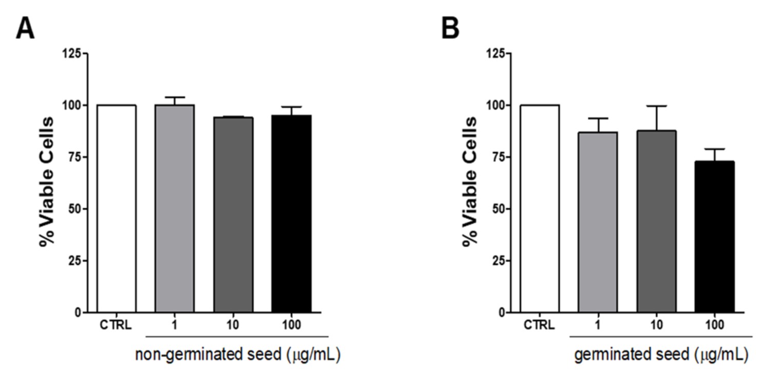
The activity of non-germinated and germinated crude extracts of soybeans BRS 258 cultivar on L. amazonensis promastigotes survival was assayed (Figures 1a and 1b). The anti-Leishmania activity of these extracts was tested by the cell viability assay performed with three different concentrations (1, 10 and 100 μg/mL). However, our results demonstrated that there was not significant difference between the samples extracts and the control. However, Figure 1b suggests that there is an inhibition of cell viability when the concentration of 100 μg/mL was used. The presence of soybean embryos present in this sample could justify this possible inhibition.
Effect of the BRS 258 soybean extract on the L. amazonensis promastigotes. Promastigotes (106 cells / mL) were grown in the present or absence of (a) non-germinated seed and (b) germinated seed, at the indicated concentrations, during 72 h. Parasite viability was measured using the MTT assay. The results were expressed as the percentage of viable promastigotes in relation to the untreated promastigote (CTRL) and are shown as the mean ± (SEM) of three independent experiments performed in triplicate.
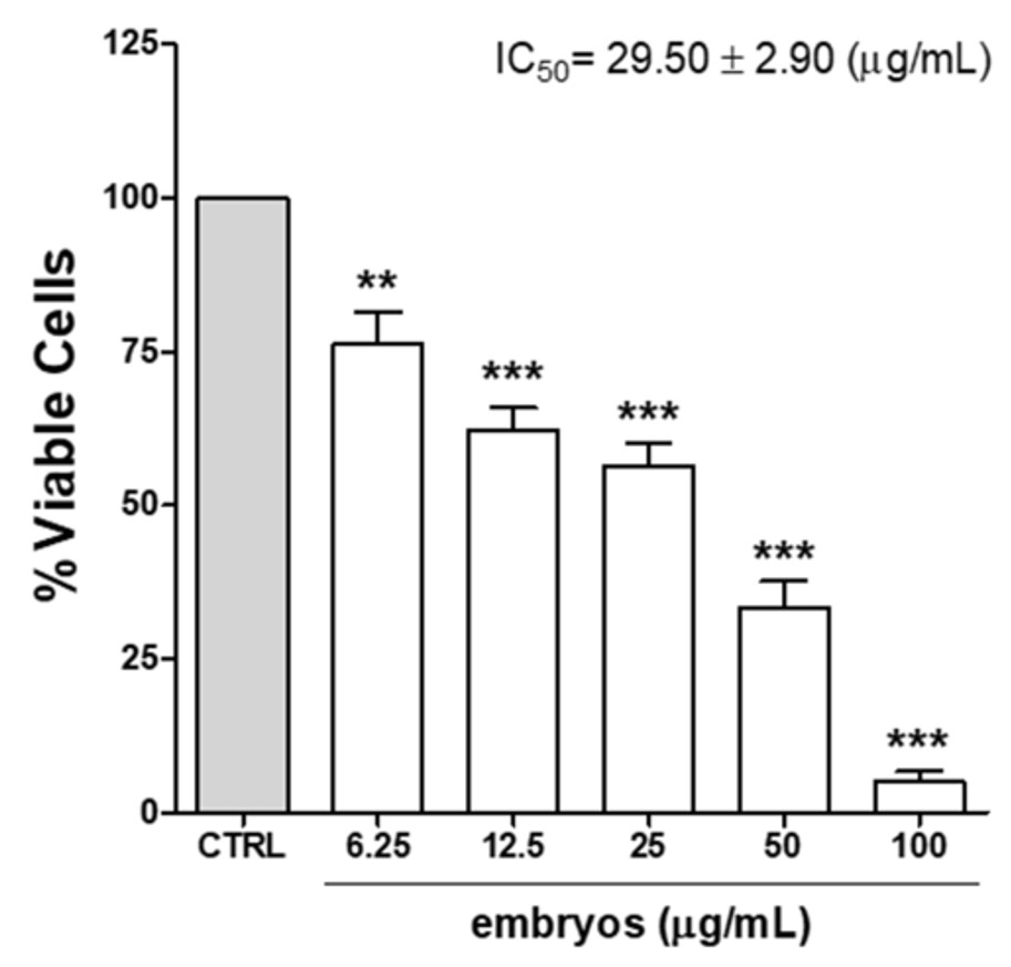
However, this could be verified when the extracts of soybeans BRS 258 embryos were tested (Figure 2). Our results demonstrated an anti-Leishmania activity of soybean embryos extract with IC50 value of 29.50 ± 2.90 μg/mL. Promastigotes were inhibited in a dose-dependent manner achieving 94.9% of inhibition at the highest concentration of 100 μg/mL.
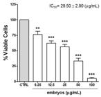
Effect of the BRS 258 soybean embryo extract in the L. amazonensis promastigotes. Promastigotes (106 cells/mL) were grown in the presence or absence of embryos extract during 72 h. Parasite viability was measured using the MTT assay. The results were expressed as the percentage of viable promastigotes in relation to the untreated promastigote (CTRL) and are shown as the mean ± (SEM) of three independent experiments performed in triplicate. ** p<0.001 and *** p<0.0001, compared to the control. IC50= inhibitory concentration for 50% of the cell population.
3.2. Description and anti-promastigote activity of protein fractions isolated of soybeans
After identification of the potential anti-L.amazonensis effect of the crude extract of the embryos of the BRS 258 soybean cultivar, we started the process of isolating the active substances. The protein of crude embryo extract was precipitated with 35% ammonium sulfate, then, subjected to Sephacryl S-100 gel filtration chromatography. The protein eluted from the column were collected and then, the cellular viability was tested using L. amazonensis promastigotes.
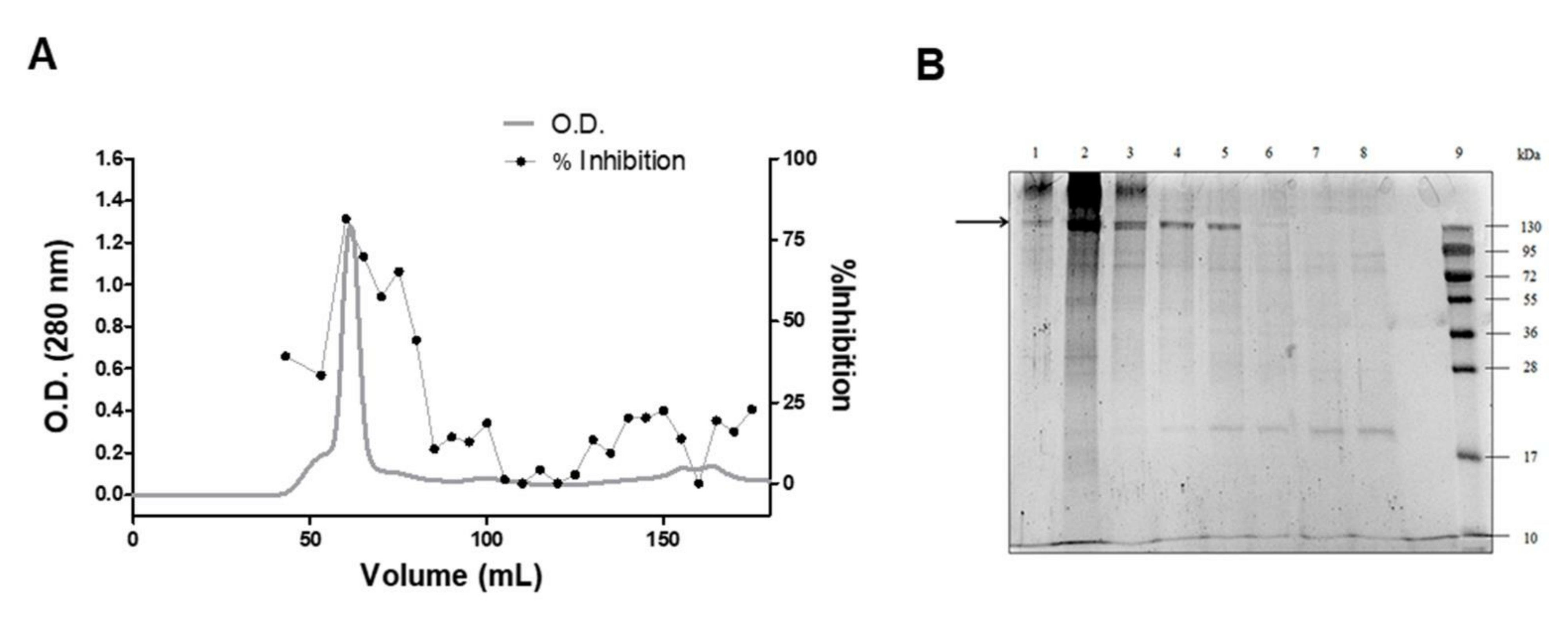
Our results demonstrated that the highest percentage of inhibition of cell viability (81.5%) corresponded to the major peak eluted from the column (Figure 3a). Fractions that inhibited 81.5% of viable cells were subjected to denaturing electrophoresis (SDS-PAGE) using a 15% acrylamide gel (Figure 3b). This is evidenced by the presence of two main protein bands around 130 kDa, with higher intensity in lane 2, gradually decreasing toward lane 5. The fraction showing the highest percentage of inhibition corresponded to the peak containing the greatest amount of protein bands.
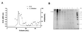
Fractionation, inhibitory activity and protein profile of BRS 258 soybean embryo extract. (a) Sephacryl S-100 gel filtration cromatography of the BRS 258 soybean embryo extract, after precipitation of this extract with 35% of ammonium sulfate. L. amazonensis promastigotes (106 cells / mL) were grown in the presence of obtained fractions, during 72 h and parasite viability was measured using the MTT assay. The major fraction of the peak was responsible for 80% of reduction of promastigotes cell viability. (b) Representative SDS-PAGE 15% of the samples after Sephacryl S-100 fractionation. Lane 1: sample with L. amazonensis activity, prior to major protein peak (extracted in the volume of 53 mL); lane 2: major peak protein eluted from the column with higher anti-L. amazonensis activity (extracted in the volume of 60 mL); lanes 3, 4 and 5: active samples against L. amazonensis after the major peak (extracted in the volumes of 65, 70 and 75 mL); lanes 6, 7 and 8: non-active samples against L. amazonensis after the major peak (extracted in the volumes of 80, 85 and 90 mL); lane 9: Molecular Weight Standards (from 10 to 130kDa). Gel was stained with Coomassie Blue.
Protein bands corresponding to the 130 kDa molecular weight of the SDS-PAGE gel (Figure 3b), were analyzed by mass spectrometry (ESI-Q-TOF). Eighteen proteins with molecular weights of approximately 90 kDa were identified (Table 1). From total proteins identified, 13 (72%) are involved in lipid metabolism (lipoxygenases and phospholipase), 02 as sucrose synthase (11%) are involved in carbohydrate metabolism, 02 (11%) as unclassified proteins (eukaryotic translation initiation factor 3 subunit B-like and 26S proteasome non-ATPase regulatory subunit 2 1A-like). In addition, to corroborate our results, an embryo-specific urease has been identified.
Proteins identified by mass spectrometry (ESI-Q-TOF) of the bands with approximately 130 kDa (15% SDS-PAGE).
3.3. Anti-promastigote activity of Jack bean urease
Among all proteins found by mass spectrometry (ESI-Q-TOF), one unclassified protein was described as an embryo-specific urease enzyme. After finding that the ureases are multifunctional enzymes and may be involved with the plant defense system, we tested the leishmanicidal activity of a purified legume Jack bean urease, by the cell viability assay. Our results demonstrated that 1.25, 2.5 and 5.0 μg/mL of Jack bean urease were able to reduce 67.95, 71.80 and 64.57% the cell viability of the promastigote forms of the parasite, respectively (Figure 4).
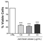
Effect of the Jack bean urease in the L. amazonensis promastigotes. Promastigotes (106 cells/mL) were grown in the presence or absence of jack bean urease during 72 h. Parasite viability was measured using the MTT assay. The results were expressed as the percentage of viable promastigotes in relation to the untreated promastigote (CTRL) and are shown as the mean ± (SEM) of three independent experiments performed in triplicate. *** p<0.0001, compared to the control.
4. Discussion
Soybean [Glycine max (L.) Merr.] belongs to the family Fabaceae and subfamily Papilionaceae and is a rich source of high-quality proteins containing all the essential amino acids found in animal proteins, cholesterol free and with low saturated fat (Tidke et al., 2015; Kumar et al., 2023). Sahu et al. (2024) described that 12 isoflavone-derivatives and 16 phenolic compounds were found in soybean seeds, including chlorogenic acid, p-coumaric acid, caffeic acid and ferulic acid, which have advantageous antioxidant activities to human health. The major compounds found were vanillic acid, hesperidin, syringic acid, gallic acid, caffeic acid, hydroxybenzoic acid, myricetin and rutin. It has also been described pharmacological properties that include anticancer, anti-hypercholesterolaemic, anti-diabetic, oestrogenic, anti-hyperlipidaemic, anti-inflammatory, anti-obesity, anti-hypertensive, anti-mutagenic, immunomodulatory, anti-osteoporotic, antiviral, hepatoprotective, antimicrobial, goitrogenic anti-skin ageing, wound healing, neuroprotective and anti-photoageing activities (Sahu et al., 2024).
However, there are no studies demonstrating the anti-Leishmania amazonensis effect of soybean. Therefore, our study showed, for the first time, that soybean embryo extract inhibits L. amazonensis promastigotes survival.
In order to elucidate the effect on parasite growth, we first evaluated the effect of non-germinated and germinated soybeans, cotyledons and embryos crude extracts in the promastigote forms of L. amazonensis. Soybeans embryos extract (100 µg/mL) inhibited 80% of the promastigote’s growth. Soybeans embryos extract was more effective against promastigotes presenting a dose-dependent effect and an IC50of 29.50 µg/mL in 72 h of treatment. In the plant kingdom, including the legume family, phytoalexins are produced in response to infections caused by pathogens. In this group of substances are the isoflavonoids, which are described as antifungal and anti-Leishmania activities (Braga et al. 2007; Sartorelli et al., 2009). However, there are few studies showing an anti-Leishmania activity isolated from legumes. It has been shown that soybean germinated embryo extract has isoflavone aglycones, such as daidzein, genistein and glycitein (Jeong et al., 2019), however, in the BRS 258 soybean cultivar, there is low levels of isoflavones (Paucar-Menacho et al., 2010) and besides that, we do not detect the presence of flavonoids in these samples after using the thin layer chromatography (data not shown). Kim et al. (2013) demonstrated that during germination of soy seed, there are a significant increase in the contents of free amino acids, tocopherol, soyasaponins, isoflavones, and proteins (Paucar-Menacho et al., 2010).
Natural products are a major source for the discovery of new treatments of leishmaniasis (Gervazoni et al., 2020). Naddaf and Haddad (2020) demonstrated the leishmanicidal effect of apigenin, a flavonoid present in plants such as parsley, thyme and oregano (Naddaf and Haddad, 2020). The same was described by Kigondu et al. (2009), which evaluated the anti-Leishmania activity of aqueous and methanolic extracts from three plants of the Fabaceae family (Kigondu et al., 2009). At the concentration of 100 μg/mL, the aqueous extracts were able to reduce the cellular viability of the promastigote form of Leishmania major to 15.8%. Differently from this result, soybean BRS 258 embryo aqueous extract reduced the cellular viability of L. amazonensis, reaching 95% using the same protein concentration (100 μg/mL). This data is of extreme relevance, once since to date no anti-Leishmania activity has been described in the literature from embryos soybean extracts.
After analyzing by mass spectrometry (ESI-Q-TOF) the proteins bands corresponding to the 130 kDa molecular weight of the 15% SDS-PAGE gel, we identified proteins like the lipoxygenases. These ones are present in developing seeds of soybean and are ubiquitous enzymes that catalyze the hydroperoxidation of polyunsaturated lipids (Felton et al., 1994; Sabljic et al., 2020). It is possible that seed lipoxygenases regulate pathogen infection responses and are also involved in diverse aspects of plant physiology like growth and development, senescence and wounding (Bau et al., 2000; Sabljic et al., 2020). Regarding pest resistance, Sabljic et al. 2020 suggest that soybean plants respond against stink bug attack by increasing LOX expression after 24 h following treatment. On the other hand, short period of germination (72 h) reduced lipoxygenase activities (Sabljic et al., 2020; Bau et al., 2000).
Phospholipids play roles in structures for cell membranes, and cellular regulation, such as signal transduction, cytoskeletal, vesicle trafficking, and secretion. The phospholipase D is an enzyme responsible for synthesizing phosphatidic acid from membrane phospholipids. In plants, this enzyme has effects in biochemical response and defense signaling, and in parasites, such as in Leishmania, the phospholipase D plays a central role in transitioning between environments of the host and parasites cells (Li and Wang, 2019; Plonski et al., 2018). In this study, phospholipase D alpha 1 was identified in the soybean embryos crude extracts. Blum et al. (2001), demonstrated in L. donovani promastigotes that phospholipase D plays a role in the adjustment of these parasites to osmotic stress, which is important and necessary for their survival and propagation. However, biochemical events related to alterations in lipid composition accompany apoptosis cells, as alterations in mitochondrial outer membrane potential and exposure of phosphatidylserine in the outer leaflet of the plasma membrane. Lipids are potential biomarkers associated with drug resistance, and the potential of enzymes involved in lipid synthesis and metabolism serve as targets for anti-trypanosomal drugs (Gannavaram and Debrabant, 2012; Gutierrez Guarnizo et al., 2021; Leroux et al., 2023). Thus, we suggest that the increase in lipid enzymes, such as phospholipase D and lipoxygenases may lead to changes in the lipid homeostasis of L. amazonensis promastigotes.
Another protein identified is an embryo-specific urease. This enzyme has been described with different biological properties, like high insecticidal, antibacterial and antifungal action, both independent to the ureolytic activity, suggesting that ureases may be linked to the immune system of plants (Follmer et al., 2004; Martinelli et al., 2017; Grahl et al., 2020). According to Stanisçuaski and Carlini (2012), ureases are widely distributed in legume seeds and the accumulation of these proteins during seed maturation is suggestive of an important physiological role possibly related to the plant defense (Stanisçuaski and Carlini 2012).
Soybean has more than one type of urease, one ubiquitous and one embryo specific. Besides the function related to the hydrolysis of urea, these enzymes have several properties that are independent of their enzymatic activity, presents entomotoxic and antimicrobial actions (Follmer et al., 2004; Grahl et al., 2020). After finding that the ureases are multifunctional enzymes, and may be involved with the plant defense system, it was verified whether the sample with anti-Leishmania activity could have an ureolytic action. Therefore, the anti-protozoal effect can be attributed to a possible urease enzyme present in the sample, which was observed after measuring the ureolytic activity of the active sample. This data is of great importance, since no toxicity has been reported to Leishmania from ureases. Thus, it was necessary to test the effect of a standard urease on the cell viability of L. amazonensis. Jack bean urease was also able to reduce the cell viability of the promastigote forms, in which the sensitivity of the parasite to the enzyme was higher compared to the sample from soybean germinated embryo extract, possibly because it was a purified enzyme. This data indicates that urease may be responsible for the anti-Leishmania amazonensis action described in this work.
5. Conclusion
In conclusion, the extract of the BRS 258 soybean embryos significantly reduced the cell viability of promastigotes of L. amazonensis and an embryo-specific urease was identified amongst the fractions isolated by chromatography. Simultaneously, an anti-Leishmania activity from an isolated urease of the legume Jack bean was confirmed, suggesting a possible relationship of soybean embryos with urease and leishmanicidal activities.
Data Availability Statement
Research data is only available upon request.
Acknowledgements
This study was supported by Coordenação de Aperfeiçoamento de Pessoal de Nível Superior / Brazil (CAPES – Financial Code 001) e Conselho Nacional de Desenvolvimento Científico e Tecnológico / Brazil (CNPq, Edital Universal – processo 402481/2016-0).
-
#These authors contributed equally to this work
References
-
AGUILERA, Y.,DÍAZ, M.F., JIMÉNEZ, T., BENÍTEZ, V., HERRERA, T., CUADRADO, C., MARTÍN-PEDROSA, M. and MARTÍN-CABREJAS, M.A., 2013. Changes in nonnutritional factors and antioxidant activity during germination of nonconventional legumes. Journal of Agricultural and Food Chemistry, vol. 61, no. 34, pp. 8120-8125. http://doi.org/10.1021/jf4022652PMid:23909570.
» http://doi.org/10.1021/jf4022652 -
ALBUQUERQUE, R.D.D.G., OLIVEIRA, A.P., FERREIRA, C., ALVES PASSOS, C.L., FIALHO, E., SOARES, D.C., AMARAL, V.F., BEZERRA, G.B., ESTEVES, R.S., SANTOS, M.G., ALBERT, A.L.M. and ROCHA, L., 2020. Anti-Leishmania amazonensis activity of the terpenoid fraction from Eugenia pruniformis leaves. Anais da Academia Brasileira de Ciências, vol. 92, no. 4, pp. e20201181. http://doi.org/10.1590/0001-3765202020201181 PMid:33295583.
» http://doi.org/10.1590/0001-3765202020201181 -
BAU, H.-M., VILLAUME, C.H. and MEJEAN, L., 2000. Effects of soybean (Glycine max) germination on biologically active components, nutritional values of seeds, and biological characteristics in rats. Die Nahrung, vol. 44, no. 1, pp. 2-6. http://doi.org/10.1002/(SICI)1521-3803(20000101)44:1<2::AID-FOOD2>3.0.CO;2-9 PMid:10702991.
» http://doi.org/10.1002/(SICI)1521-3803(20000101)44:1<2::AID-FOOD2>3.0.CO;2-9 -
BLUM, J.J., LEHMAN, J.A., HORN, J.M. and GOMEZ-CAMBRONERO, J., 2001.Phospholipase D (PLD) is present in Leishmaniadonovani and its activity increases in response to acute osmotic stress. The Journal of Eukaryotic Microbiology, vol. 48, no. 1, pp. 102-110. http://doi.org/10.1111/j.1550-7408.2001.tb00421.x PMid:11249184.
» http://doi.org/10.1111/j.1550-7408.2001.tb00421.x -
BRADFORD, M.M., 1976. A rapid and sensitive method for the quantification of microgram quantities of protein utilizing the principle of protein-dye binding. Analytical Biochemistry, vol. 72, no. 1-2, pp. 248-254. http://doi.org/10.1016/0003-2697(76)90527-3 PMid:942051.
» http://doi.org/10.1016/0003-2697(76)90527-3 -
BRAGA, F.G., BOUZADA, M.L.M., FABRI, L.R., MATOS, M.O., MOREIRA, F.O., SCIO, E. and COIMBRA, E.S., 2007. Antileishmanial and antifungal activity of plants used in traditional medicine in Brazil. Journal of Ethnopharmacology, vol. 111, no. 2, pp. 396-402. http://doi.org/10.1016/j.jep.2006.12.006PMid:17234373.
» http://doi.org/10.1016/j.jep.2006.12.006 - BRASIL. Ministério da Agricultura, Pecuária e Abastecimento – MAPA, 2009. Regras para análise de sementes. Brasília: Mapa/ACS. pp.15-395.
-
BURZA, S., CROFT, L.S. and BOELAERT, M., 2018. Leishmaniasis. Lancet, vol. 392, no. 10151, pp. 951-970. http://doi.org/10.1016/S0140-6736(18)31204-2 PMid:30126638.
» http://doi.org/10.1016/S0140-6736(18)31204-2 -
ÇAKIR, Ö.,UÇARLI, C., TARHAN, C., PEKMEZ, M. and TURGUT-KARA, N., 2019. Nutritional and health benefits of legumes and their distinctive genomic properties. Food Science and Technology, vol. 39, no. 1, pp. 1-12. http://doi.org/10.1590/fst.42117
» http://doi.org/10.1590/fst.42117 -
CALEJA, C., RIBEIRO, A., BARREIRO, M.F. and FERREIRA, I.C.F.R., 2017. Phenolic compounds as nutraceuticals or functional food ingredients. Current Pharmaceutical Design, vol. 23, no. 19, pp. 2787-2806. http://doi.org/10.2174/1381612822666161227153906 PMid:28025943.
» http://doi.org/10.2174/1381612822666161227153906 -
CARLINI, C.R. and LIGABUE-BRAUN, R., 2016.Ureases as multifunctional toxic proteins: A review. Toxicon, vol. 110, pp. 90-109. http://doi.org/10.1016/j.toxicon.2015.11.020 PMid:26690979.
» http://doi.org/10.1016/j.toxicon.2015.11.020 -
CASTRO-RUBIO, F., GARCÍA, M.C., RODRÍGUEZ, R. and MARINA, M.L., 2005. Simple and inexpensive method for the reliable determination of additions of soybean proteins in heat-processed meat products: an alternative to the AOAC official method. Journal of Agricultural and Food Chemistry, vol. 53, no. 2, pp. 220-226. http://doi.org/10.1021/jf049557e PMid:15656653.
» http://doi.org/10.1021/jf049557e -
CHATTERJEE, C., GLEDDIE, S. and XIAO, C.W., 2018.Soybean bioactive peptides and their functional properties. Nutrients, vol. 10, no. 9, pp. 1211. http://doi.org/10.3390/nu10091211 PMid:30200502.
» http://doi.org/10.3390/nu10091211 -
DE VRIES, H.J.C. and SCHALLIG, H.D., 2022. Cutaneous Leishmaniasis: A 2022 updated narrative review into diagnosis and management developments. American Journal of Clinical Dermatology, vol. 23, no. 6, pp. 823-840. http://doi.org/10.1007/s40257-022-00726-8 PMid:36103050.
» http://doi.org/10.1007/s40257-022-00726-8 -
FELTON, G.W., SUMMERS, C.B. and MUELLER, A.J., 1994. Oxidative responses in soybean foliage to herbivory by bean leaf beetle and three-cornered alfalfa hopper. Journal of Chemical Ecology, vol. 20, no. 3, pp. 639-650. http://doi.org/10.1007/BF02059604 PMid:24242118.
» http://doi.org/10.1007/BF02059604 -
FOLLMER, C., REAL-GUERRA, R., WASSERMAN, G.E., OLIVERA-SEVERO, D. and CARLINI, C.R., 2004.Jackbean, soybean and Bacillus pasteurii ureases: biological effects unrelated to ureolytic activity. European Journal of Biochemistry, vol. 271, no. 7, pp. 1357-1363. http://doi.org/10.1111/j.1432-1033.2004.04046.xPMid:15030486.
» http://doi.org/10.1111/j.1432-1033.2004.04046.x - FORTES, A.M.T.andBORTOLINI, M.F., 2005. Efeitos alelopáticos sobre a germinação de sementes de soja (Glycine max L. Merrill). Ciências Agrárias, vol. 26, no. 1, pp. 5-10. http://doi.org/10.5433/1679-0359.2005v26n1p5
-
GANNAVARAM, S. and DEBRABANT, A., 2012. Programmed cell death in Leishmania: biochemical evidence and role in parasite infectivity. Frontiers in Cellular and Infection Microbiology, vol. 2, pp. 95.http://doi.org/10.3389/fcimb.2012.00095 PMid:22919685.
» http://doi.org/10.3389/fcimb.2012.00095 -
GERVAZONI, L.F.O., BARCELLOS, G.B., FERREIRA-PAES, T. and ALMEIDA-AMARAL, E.E., 2020. Use of natural products in Leishmaniasis Chemotherapy: an overview. Frontiers in Chemistry, vol. 8, pp. 579891. http://doi.org/10.3389/fchem.2020.579891
» http://doi.org/10.3389/fchem.2020.579891 -
GRAHL, M.V.C., LOPES, F.C., MARTINELLI, A.H.S., CARLINI, C.R. and FRUTTERO, L.L., 2020. Structure-function insights of Jaburetox and Soyuretox: novel intrinsically disordered polypeptides derived from plant ureases. Molecules, vol. 25, no. 22, pp. 5338. http://doi.org/10.3390/molecules25225338 PMid:33207637.
» http://doi.org/10.3390/molecules25225338 -
GUTIERREZ GUARNIZO, S.A., TIKHONOVA, E.B., ZABET-MOGHADDAM, M., ZHANG, K., MUSKUS, C., KARAMYSHEV, A. and KARAMYSHEVA, Z.N., 2021. Drug-Induced lipid remodeling in Leishmania parasites. Microorganisms, vol. 9, no. 4, pp. 790. http://doi.org/10.3390/microorganisms9040790 PMid:33918954.
» http://doi.org/10.3390/microorganisms9040790 -
HENDRICKX, S., CALJON, G. and MAES, L., 2019. Need for sustainable approaches in antileishmanial drug discovery. Parasitology Research, vol. 118, no. 10, pp. 2743-2752. http://doi.org/10.1007/s00436-019-06443-2 PMid:31473855.
» http://doi.org/10.1007/s00436-019-06443-2 -
JEONG, H., LEE, J., KIM, S., PARK, S., YANG, H., AHN, B.H., JANG, C.Y., JEONG, H.C., LEE, S.J., KIM, S.L., SEO, W.D., SOHN, J. and CHANG, M., 2019. Characterization of soybean germinated embryo extract as an estrogen receptor subtype-selective and tissue-specific modulator. Journal of Medicinal Food, vol. 22, no. 2, pp. 186-195. http://doi.org/10.1089/jmf.2018.4250 PMid:30585749.
» http://doi.org/10.1089/jmf.2018.4250 -
KATO, H., 2025. Epidemiology of Leishmaniasis: risk factors for its pathology and infection. Parasitology International, vol. 105, pp. 102999. http://doi.org/10.1016/j.parint.2024.102999
» http://doi.org/10.1016/j.parint.2024.102999 -
KIGONDU, E.V.M., RUKUNGA, G.M., KERIKO, M.J., TONUI, W.K., GATHIRWA, J.W., KIRIRA, P.G., IRUNGU, B.N., INGONGA, J.M. and NDIEGE, I.O., 2009. Anti-parasitic activity and cytotoxicity of selected medicinal plants from Kenya. Journal of Ethnopharmacology, vol. 123, no. 3, pp. 504-509. http://doi.org/10.1016/j.jep.2009.02.008 PMid:19501282.
» http://doi.org/10.1016/j.jep.2009.02.008 -
KIM, S.L., LEE, J.E., KWON, Y.U., KIM, W.H., JUNG, G.H., KIM, D.W., LEE, C.K., LEE, Y.Y., KIM, M.J., KIM, Y.H., HWANG, T.Y. and CHUNG, I.M., 2013. Introduction and nutritional evaluation of germinated soy germ. Food Chemistry, vol. 136, no. 2, pp. 491-500. http://doi.org/10.1016/j.foodchem.2012.08.022 PMid:23122089.
» http://doi.org/10.1016/j.foodchem.2012.08.022 -
KOCYIGIT, E., KOCAADAM-BOZKURT, B., BOZKURT, O., AGAGÜNDÜZ, D. and CAPASSO, R., 2023. Plant toxic proteins: their biological activities, mechanism of action and removal strategies. Toxins, vol. 15, no. 6, pp. 356. http://doi.org/10.3390/toxins15060356 PMid:37368657.
» http://doi.org/10.3390/toxins15060356 -
KUMAR, M., SUHAG, R., HASAN, M., DHUMAL, S., RADHA.,PANDISELVAM, R., SENAPATHY, M., SAMPATHRAJAN, V., PUNIA, S., SAYED, A.A.S., SINGH, S. and KENNEDY, J.F., 2023. Black soybean (Glycine max (L.) Merr): paving the way toward new nutraceutical. Critical Reviews in Food Science and Nutrition, vol. 63, no. 23, pp. 6208-6234. http://doi.org/10.1080/10408398.2022.2029825 PMid:35139704.
» http://doi.org/10.1080/10408398.2022.2029825 -
LAEMMLI, U.K., 1970. Cleavage of structural proteins during the assembly of the head of bacteriophage T4. Nature, vol. 227, no. 5259, pp. 680-685. http://doi.org/10.1038/227680a0 PMid:5432063.
» http://doi.org/10.1038/227680a0 -
LEROUX, M., LUQUAIN-COSTAZ, C., LAWTON, P., AZZOUZ-MAACHE, S. and DELTON, I., 2023. Fatty acid composition and metabolism in Leishmania parasite species: potential biomarkers or drug targets for Leishmaniasis? International Journal of Molecular Sciences, vol. 24, no. 5, pp. 4702. http://doi.org/10.3390/ijms24054702 PMid:36902138.
» http://doi.org/10.3390/ijms24054702 -
LI, J. and WANG, X., 2019. Phospholipase D and phosphatidic acid in plant immunity. Plant Science, vol. 279, pp. 45-50. http://doi.org/10.1016/j.plantsci.2018.05.021 PMid:30709492.
» http://doi.org/10.1016/j.plantsci.2018.05.021 -
MARTINELLI, A.H.S., LOPES, F.C., BROLL, V., DEFFERRARI, M.S., LIGABUE-BRAUN, R., KAPPAUN, K., TICHOTA, D.M., FRUTTERO, L.L., MOYETTA, N.R., DEMARTINI, D.R., POSTAL, M., MEDEIROS-SILVA, M., BECKER-RITT, A.B., PASQUALI, G. and CARLINI, C.R., 2017. Soybean ubiquitous urease with purification facilitator: an addition to the moonlighting studies toolbox. Process Biochemistry, vol. 53, pp. 245-258. http://doi.org/10.1016/j.procbio.2016.12.003
» http://doi.org/10.1016/j.procbio.2016.12.003 -
MENEZES, J.P., GUEDES, C.E., PETERSEN, A.L., FRAGA, D.B. and VERAS, P.S., 2015. Advances in development of new treatment for Leishmaniasis. BioMed Research International, vol. 2015, pp. 815023. http://doi.org/10.1155/2015/815023 PMid:26078965.
» http://doi.org/10.1155/2015/815023 -
MOSMANN, T., 1983.Rapid colorimetric assay for cellular growth and survival: application to proliferation and cytotoxicity assays. Journal of Immunological Methods, vol. 65, no. 1-2, pp. 55-63. http://doi.org/10.1016/0022-1759(83)90303-4 PMid:6606682.
» http://doi.org/10.1016/0022-1759(83)90303-4 -
NADDAF, N. and HADDAD, S., 2020. Apigenin effect against Leishmaniatropica amastigotes in vitro. Journal of Parasitic Diseases: Official Organ of the Indian Society for Parasitology, vol. 44, no. 3, pp. 574-578. http://doi.org/10.1007/s12639-020-01230-8 PMid:32801509.
» http://doi.org/10.1007/s12639-020-01230-8 -
NEUHOFF, V., AROLD, N., TAUBE, D. and EHRHARDT, W., 1988. Improved staining of proteins in polyacrylamide gels including isoelectric focusing gels with clear background at nanogram sensitivity using Coomassie Briliant Blue G-250 and R-250. Electrophoresis, vol. 9, no. 6, pp. 255-262. http://doi.org/10.1002/elps.1150090603 PMid:2466658.
» http://doi.org/10.1002/elps.1150090603 -
PARTHASARATHY, A., BORREGO, E.J., SAVKA, M.A., DOBSON, R.C.J. and HUDSON, A.O., 2021. Amino acid–derived defense metabolites from plants: a potential source to facilitate novel antimicrobial development. The Journal of Biological Chemistry, vol. 296, pp. 100438. http://doi.org/10.1016/j.jbc.2021.100438 PMid:33610552.
» http://doi.org/10.1016/j.jbc.2021.100438 -
PAUCAR-MENACHO, L.M., BERHOW, M.A., MANDARINO, J.M.G., CHANG, Y.K. and GONZALEZ DE MEJIA, E., 2010. Effects of time and temperature on bioactive compounds in germinated Brazilian soybean cultivar BRS 258. Food Research International, vol. 43, no. 7, pp. 1856-1865. http://doi.org/10.1016/j.foodres.2009.09.016
» http://doi.org/10.1016/j.foodres.2009.09.016 -
PLONSKI, N.M., BISSONI, B., ARACHCHILAGE, M.H., ROMSTEDT, K., KOOIJMAN, E.E. and PIONTKIVSKA, H., 2018. Shedding light on lipid metabolism in Kinetoplastida: A phylogenetic analysis of phospholipase D protein homologs. Gene, vol. 656, pp. 95-105. http://doi.org/10.1016/j.gene.2018.02.063 PMid:29501621.
» http://doi.org/10.1016/j.gene.2018.02.063 - SABLJIC, I., BARNETO, J.A., BALESTRASSE, K.B., ZAVALA, J.A. and PAGANO, E.A., 2020. Role of reactive oxygen species and isoflavonoids in soybean resistance to the attack of the southern green stink bug. Peer J, vol. 8, pp. e9956. http://doi.org/10.7717/peerj.9956. PMid:32995095.
-
SAHU, M., KAMAKSHI, SAHOO, J., SWAIN, S.R., CHAUHAN, M., GOYAL, R., GUPTA, S. and KAUR, K., 2024. Phytochemical investigation and characterisation of methanolic extract of Glycine max seeds using LCMS/MS and in silico studies for wound healing activity. Journal of Mass Spectrometry, vol. 59, no. 7, pp. e5045. http://doi.org/10.1002/jms.5045 PMid:38837562.
» http://doi.org/10.1002/jms.5045 - SANGRONIS, E. and MACHADO, C.J., 2007. Influence of germination on the nutritional quality of Phaseolus vulgaris and Cajanuscajan. Food Science and Technology, vol. 40, no. 1, pp. 116-120. http://doi.org/10.1016/j.lwt.2005.08.003.
-
SARTORELLI, P., CARVALHO, C.S., REIMÃO, J.Q., FERREIRA, M.J.andTEMPONE, A.G., 2009. Antiparasitic activity of biochanin A, an isolated isoflavone from fruits of Cassia fistula (Leguminosae). Parasitology Research, vol. 104, no. 2, pp. 311-314. http://doi.org/10.1007/s00436-008-1193-z PMid:18810492.
» http://doi.org/10.1007/s00436-008-1193-z -
SHEIKH, S.Y., HASSAN, F., SHUKLA, D., BALA, S., FARUQUI, T., AKHTER, Y., KHAN, A.R. and NASIBULLAH, M., 2024. A review on potential therapeutic targets for the treatment of leishmaniasis. Parasitology International, vol. 100, pp. 102863. http://doi.org/10.1016/j.parint.2024.102863 PMid:38272301.
» http://doi.org/10.1016/j.parint.2024.102863 -
STANISÇUASKI, F. and CARLINI, C.R., 2012. Plant ureases and related peptides: understanding their entomotoxic properties. Toxins, vol. 4, no. 2, pp. 55-67. http://doi.org/10.3390/toxins4020055 PMid:22474566.
» http://doi.org/10.3390/toxins4020055 -
TIDKE, S.A., RAMAKRISHNA, D., KIRAN, S., KOSTURKOVA, G. and RAVISHANKAR, G.A., 2015. Nutraceutical potential of soybean: review. Asia Pacific Journal of Clinical Nutrition, vol. 7, no. 2, pp. 22-32. http://doi.org/10.3923/ajcn.2015.22.32
» http://doi.org/10.3923/ajcn.2015.22.32 -
TRUGO, L.C., DONANGELO, C.M., TRUGO, N.M. and BACH KNUDSEN, K.E., 2000. Effect of heat treatment on nutritional quality of germinated legume seeds. Journal of Agricultural and Food Chemistry, vol. 48, no. 6, pp. 2082-2086. http://doi.org/10.1021/jf9913920 PMid:10888502.
» http://doi.org/10.1021/jf9913920 -
ZHU, D., HETTIARACHCHY, N.S., HORAX, R. and CHEN, P., 2005. Isoflavone contents in germinated soybean seeds. Plant Foods for Human Nutrition, vol. 60, no. 3, pp. 147-151. http://doi.org/10.1007/s11130-005-6931-0 PMid:16187018.
» http://doi.org/10.1007/s11130-005-6931-0
Edited by
-
Editor:
Takako Matsumura Tundisi