Cytological development, calcium dynamic and metabolite profile of stress-induced tobacco (Nicotiana tabacum L.) microspore under calcium chloride treatment
Abstract
Exogenous calcium is pivotal in activating microspore embryogenesis via calcium signaling. Altering the external calcium levels in the medium has been documented to impact the success rate of microspore embryogenesis initiation. However, the cellular effects on the formation of embryogenic tobacco microspore and its cellular changes during stress treatment remain elusive. We examined microspore cytological development, specifically vacuolation types and first division pattern. We also used Fluo 3-AM staining to observe the calcium dynamics, including accumulation and distribution of intracellular calcium ions (Ca2+) under fluorescent microscope. Metabolomic analysis of induced microspores was carried out using gas chromatography-mass spectrometry (GC-MS). The result showed that at 2.0 mM CaCl2.2H2O treatment, 57.06 ± 6.95% of microspores exhibited star-like structures, and 47.39 ± 8.84% underwent symmetric first division, twice as effective as 1.0 mM. Fluo 3-AM staining revealed increased Ca2+ fluorescence on day one, persisting in 2.0 mM treatment by day three. By day six, fluorescence in 1.0 mM and 2.0 mM treatments significantly differed. Intracellular Ca2+ signals rose with 1.0 mM CaCl2.2H2O and fell with 2.0 mM CaCl2.2H2O. The metabolomic analysis detected 34 metabolites, including amino acids, organic acids, and fatty acids, with predominant oleic acid, palmitic acid, and stearic acids. We conclude that two millimolar of calcium chloride was optimum for inducing embryogenic microspore formation in tobacco. Our findings underscore the significant role of exogenous calcium in microspore embryogenesis, elucidating its impact on cellular dynamics and metabolite profiles. These insights contribute to hold potential implications for enhancing crop breeding strategies.
Keywords:
calcium dynamic; metabolomic; microspore embryogenesis; tobacco
Resumo
O cálcio exógeno é fundamental na ativação da embriogênese de micrósporos por meio da sinalização de cálcio. Foi documentado que a alteração dos níveis externos de cálcio no meio afeta a taxa de sucesso do início da embriogênese do micrósporo. No entanto, os efeitos celulares sobre a formação de micrósporos embriogênicos de tabaco e suas alterações celulares durante o tratamento de estresse permanecem indefinidos. Examinamos o desenvolvimento citológico dos micrósporos, especificamente os tipos de vacuolação e o padrão da primeira divisão. Também usamos a coloração Fluo 3-AM para observar a dinâmica do cálcio, incluindo o acúmulo e a distribuição de íons de cálcio intracelular (Ca2+) sob microscópio fluorescente. A análise metabolômica dos micrósporos induzidos foi realizada por meio de cromatografia gasosa e espectrometria de massa (GC-MS). O resultado mostrou que, no tratamento com CaCl2.2H2O a 2,0 mM, 57,06 ± 6,95% dos micrósporos exibiram estruturas estreladas e 47,39 ± 8,84% sofreram a primeira divisão simétrica, duas vezes mais eficaz do que a 1,0 mM. A coloração com Fluo 3-AM revelou aumento da fluorescência de Ca2+ no primeiro dia, persistindo no tratamento com 2,0 mM no terceiro dia. No sexto dia, a fluorescência nos tratamentos de 1,0 mM e 2,0 mM diferiu significativamente. Os sinais de Ca2+ intracelular aumentaram com 1,0 mM de CaCl2.2H2O e diminuíram com 2,0 mM de CaCl2.2H2O. A análise metabolômica detectou 34 metabólitos, incluindo aminoácidos, ácidos orgânicos e ácidos graxos, com predominância de ácido oleico, ácido palmítico e ácido esteárico. Concluímos que dois milimolares de cloreto de cálcio foram ideais para induzir a formação de micrósporos embriogênicos no tabaco. Nossas descobertas destacam a função significativa do cálcio exógeno na embriogênese de micrósporos, elucidando seu impacto na dinâmica celular e nos perfis de metabólitos. Essas percepções contribuem para manter possíveis implicações no aprimoramento das estratégias de melhoramento de culturas.
Palavras-chave:
dinâmica de cálcio; metabolômica; embriogênese de micrósporos; tabaco
1. Introduction
Calcium is crucial in activating microspore embryogenesis as a signaling molecule in response to stress (Pauls et al., 2006). To alter the developmental pathway, a combination of heat stress and starvation medium is typically used in tobacco microspore culture (Touraev and Heberle-Bors, 2003; Touraev et al., 1996a, b). Heat stress tends to increase the fluidity of the plasma membrane (Dubas et al., 2013), making it more permeable to extracellular calcium ions (Ca2+), subsequently increasing intracellular Ca2+ in microspores (Parra-Vega et al., 2015; Rivas-Sendra et al., 2017). The increase of intracellular Ca2+ is translated and mediated by the calcium-binding protein, which then triggers downstream effects leading to mitosis and changes in gene expression (Tsuwamoto and Takahata, 2008), redirecting the microspore developmental pathway towards embryogenesis (Seguí-Simarro et al., 2005).
The influx of extracellular Ca2+ ions through the plasma membrane is required to stimulate microspore embryogenesis (Pauls et al., 2006; Dubas et al., 2013). Adding calcium ion channel blockers to the culture medium, such as verapamil (Reynolds 2000) and caffeine (Parra-Vega et al., 2015), inhibited microspore embryogenesis. Rivas-Sendra et al. (2017) reported that after one day of Brassica napus L. microspores induced using heat stress, many microspores had drastically increased intracellular calcium signals compared to the fresh microspores.
Modified exogenous calcium concentration in the culture medium has been reported to affect the efficiency of microspore embryogenesis induction (Reynolds, 2000). However, whether the difference in exogenous calcium affects the concentration of intracellular Ca2+, calcium signaling, and embryogenic response in microspores remains unclear. Considering the critical role of calcium in microspore embryogenesis, especially related to its concentration within the cell, it is essential to study the accumulation and distribution of intracellular Ca2+ in microspores with exogenous calcium treatment to address this question. Here, we report the calcium dynamics of intracellular Ca2+, metabolite profile, and the cytological development regarding the embryogenic responses during the early stage of tobacco microspore embryogenesis with modified exogenous calcium concentration. The results provide new insights that high extracellular calcium moderates the undesirable effects of prolonged heat stress by altering the relative abundance of plasma membrane constituents, thereby increasing embryogenic microspores. These findings enhance our understanding of extracellular calcium's role in microspore embryogenesis and offer new strategies to optimize this process in other important plants.
2. Material & Methods
2.1. Plant material preparation
In this study, tobacco (Nicotiana tabacum L.) cv Manilo was used. The seeds were obtained from the National Research and Innovation Agency, Indonesia. Tobacco cultivation and maintenance were conducted in natural condition in Made Village, Kudu Subdistrict, Jombang Regency, East Java, Indonesia. Flower buds, as explant sources, were harvested approximately two to three months after planting.
2.2. Determination of flower bud size and microspore developmental stage
The size of the flower buds was used as a morphological marker for selecting flower buds containing microspores at the appropriate developmental stage. Flower buds sized 9–14 mm was divided into six groups to observe and determine the developmental stage of microspore.
The microspore developmental stage was determined by observing the nuclei of microspores stained with 4',6-diamidino-2-phenylindole (DAPI) under a fluorescent microscope. The microspore isolation method was modified by Touraev and Heberle-Bors (2003). Initially, microspores were isolated using a glass rod method. Ten anthers from two tobacco flower buds were placed in a 1.5 ml tube containing B medium (Kyo and Harada, 1986). The anthers were gently crushed to release the microspores. The suspension was transferred to a new tube and centrifuged at 250 g for 3 minutes. The pellet was fixed with a mixture of 70% ethanol, glacial acetic acid, formaldehyde (90:5:5) and then centrifuged at 250 g for 3 minutes. The pellet was rehydrated with 1 ml of 70% ethanol and centrifuged again. This procedure was repeated three times. Thirty microliter DAPI solution (Partec) was mixed with the microspore isolate in a 1.5 mL tube and stored in the refrigerator overnight. The sample was observed under a fluorescent microscope (Olympus BX-43). Each sample was observed for a minimum of 500 microspores. Flower buds with over 50% uninucleate microspore developmental stages were used as explant sources for microspore culture.
2.3. Sterilization and isolated microspore culture
Flower buds at the appropriate developmental stage were sterilized for 1 minute in 70% ethanol and then rinsed twice with sterile distilled water. Subsequently, they were sterilized for 10 minutes in a 50% (v/v) commercial bleaching solution with the addition of one drop of Tween-80, followed by rinsing three times with sterile distilled water. Anthers extracted from ten flower buds were placed in a sterile 10 mL tube containing 1 mL of medium B. The anthers were gently crushed using a sterile glass rod to release the microspores. The suspension was transferred to an empty sterile 1.5 mL tube and then centrifuged at 250 g for 3 minutes. The supernatant was removed using a micropipette, the pellet was suspended in 1 ml of medium B, and centrifuged again. This procedure was repeated three times until the greenish layer above the whitish pellet layer disappeared. The microspore isolate in the form of a whitish pellet was resuspended with 3 mL of treatment medium B and then placed in a 60×15 mm Petri dish, sealed with parafilm, and incubated at 33ºC in the dark for six days.
2.4. Observation of cytological development
Cytological development of microspores, including microspore vacuolation and development, was observed on the sixth day of culture using an inverted microscope (Olympus IX51 with DP20 camera) with five replications for each treatment. Microspores were photographed, and quantification was performed with samples from five fields of view in each Petri dish. Subsequently, observation of the microspore development stage was conducted using a destructive method. The microspore suspension in the medium was transferred to a 1.5 mL tube and then centrifuged at 250 g for 3 minutes. The microspore isolate was stained with DAPI and then observed using a fluorescent microscope (Olympus BX-43).
2.5. Calcium staining and detection
The calcium staining and detection procedures were carried out based on Qu et al. (2012). Fresh microspores and microspores cultured under calcium chloride treatment for 1, 3, and 6 days were stained with Fluo 3-AM (Sigma-Aldrich). A total of 48 µL of microspore suspension in medium B was mixed with 2 µL of the one mM Fluo 3-AM solution and 20% Pluronic F-127 (1:1). The mixture was then incubated for 15, 30, and 60 minutes at 37ºC in the dark. After incubation, microspores were washed with B medium and centrifuged at 250 g for 1 minute three times. The culture was then incubated for 30 minutes at room temperature. The observation was carried out using a fluorescent microscope. The obtained data consisted of images, which were then analyzed using ImageJ software to determine the mean relative fluorescence intensity.
2.6. Sample extraction and Gas Chromatography-Mass Spectrometry (GC-MS) analysis
The extraction and GC-MS method were performed based on Hosp et al. (2006). About fifty milligrams, both fresh and CaCl2-treated samples in medium B, were homogenized using a mortar, then frozen and stored in a deep freezer (-86 ºC) until the samples were ready for extraction.
The samples were extracted in 1400 μl of methanol (100%) using a thermomixer for 15 minutes at 70°C, then mixed with distilled water (1:1). To separate polar and non-polar metabolites, 750 μl of chloroform was added. After centrifugation at 2200 × g, the upper methanol/water phase was collected and then dried using a vacuum. The obtained residue was stored in a deep freezer (-86 ºC) until the samples were ready for derivatization.
The residue was re-dissolved and derivatized using 40 μl of methoxyamine hydrochloride (20 mg/ml in pyridine) for 90 minutes at 37°C, followed by treatment for 30 minutes with a mixture of 60 μl MSTFA (N-methyl-N-(trimethylsilyl)trifluoroacetamide) and 8 μl n-alkane at 37°C. One microliter sample was then injected into the Agilent 7890B GC system with HP-5MS column at 230 ºC, using a continuous flow of helium gas as the carrier phase (1 mL/minute). The initial temperature was held at 80 ºC and increased by 10 ºC per minute until reaching 150 ºC, held for 4 minutes, then increased by 5 ºC per minute until reaching 260 ºC. The final temperature was maintained for 5 minutes. Compound identification was based on comparing mass spectra with those in the NIST14.L library.
2.7. Data analysis
The statistical analysis performed using one-way ANOVA and Kruskal-Wallis’s test at a confidence level of 95%. IBM SPSS software version 22 was employed for the statistical analysis.
3. Results
3.1. The effect of calcium chloride on microspore cytological development
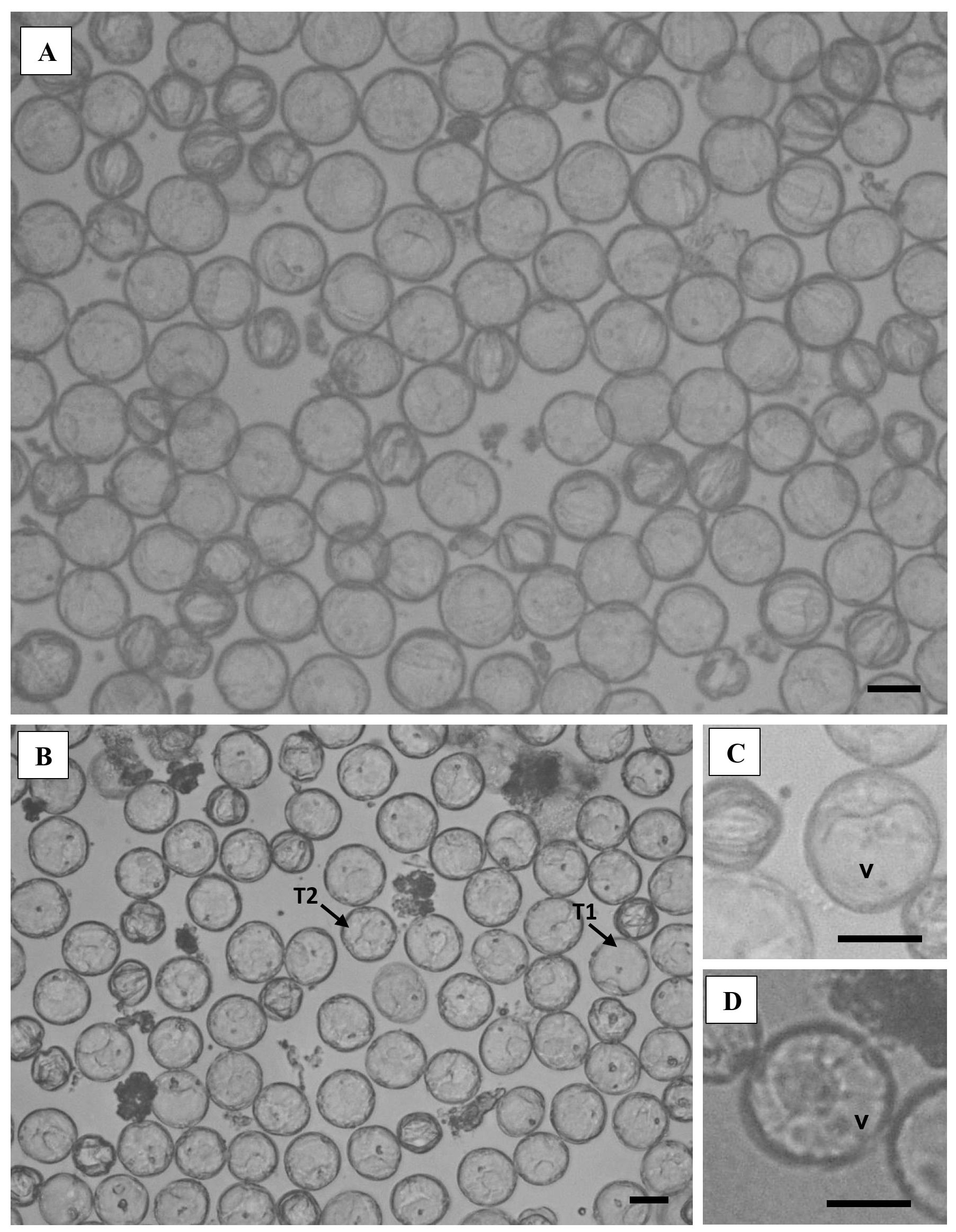
Based on DAPI staining results, tobacco cv Manilo flower buds with length of 12 mm are the most suitable initial material for inducing microspore embryogenesis, with a mean percentage of late uninucleate microspores at 94.51 ± 1.45%. Freshly isolated microspores exhibit cytological characteristics with large vacuoles that nearly fill the cell volume (see Figure 1A). Following a six-day incubation period at 33°C in B medium with varying concentrations of calcium chloride, microspores exhibited diverse cytological development, as evidenced by the observed variations in vacuolation types and first division patterns (see Figure 1B).

Cytological development of microspores in B medium. A. Fresh microspores. B-D. Microspores after incubation at 33ºC for 6 days. C. Type 1 microspore, with a large vacuole (T1). D. Type 2 microspore, with fragmented vacuole/star-like structure (T2). v: vacuole. Scale bar: 20 µm.
Two distinct types of tobacco microspore vacuolation were observed in this study. The first type of microspore, designated Type 1, exhibited a prominent central vacuole (see Figure 1C). The second type, designated Type 2, displayed a fragmented vacuole and scattered cytoplasmic threads, which were referred to as a star-like structure (see Figure 1D). The mean percentage of type 2 microspores exhibited a linear increase in correlation with the elevation in calcium chloride concentration up to 2 mM. This result is twice that observed with the addition of 1 mM and 1.5 mM calcium chloride.
Microspore cytological development was observed using DAPI staining, which revealed the presence of uninucleate, asymmetrically binucleate, and symmetrically binucleate microspores in all treatments (see Figure 2). Microspores that undergo asymmetric division have different fluorescent intensities in generative nuclei and vegetative nuclei (see Figure 2). The smaller generative nucleus has a stronger intensity than the vegetative nucleus. Symmetrically dividing microspores show two equal-sized nuclei whose fluorescent intensity is similar to that of vegetative nuclei. In the presence of 1.5 mM and 2 mM calcium chloride, the mean percentage of symmetrically binucleate microspores was the highest at 31.37 ± 3.84% and 47.39 ± 8.84%, respectively (as shown in Table 1).

Microspore development in B medium after incubation at 33ºC for 6 days as determined using DAPI staining. A. Uninucleate; B. Asymmetric binucleate; C. Symmetric binucleate. vn: vegetative nucleus. gn: generative nucleus. Arrow: nucleus. Scale bar: 25 µm.
Percentage of microspore cytological development based on vacuolation type and DAPI staining on B medium with calcium chloride treatment after incubation at 33ºC for 6 days.
3.2. Effect of calcium chloride on calcium dynamic of stress-induced tobacco microspores
In this study, Fluo 3-AM (acetoxymethyl) ester was employed to identify the Ca2+ signal in microspores. The greater the intensity of the yellowish-green fluorescent signal visible under a fluorescent microscope, the higher the concentration of calcium ions in the cell. The optimal incubation period for the Fluo 3-AM staining procedure in tobacco cv Manilo microspores was determined to be 60 minutes.
The visualization of Fluo 3-AM staining on microspores after one day and three days of incubation revealed the presence of calcium signals in both the vacuole and cytosol of microspores in all treatments, with no significant differences observed (as shown in Table 2) (see Figure 3). On the sixth day of incubation, calcium signals were observed in all treatments, including those of type 2 microspores, which exhibited no apparent difference in distribution. Nevertheless, the mean fluorescent intensity exhibited notable variation among the treatments (see Figure 4). Microspores treated with higher calcium chloride concentrations (1.5 and 2 mM) exhibited a relatively lower fluorescent intensity compared to the other treatments after the sixth day of incubation (as shown in Table 2).
Mean relative fluorescent intensity of Ca2+ signal in calcium chloride-treated tobacco microspores after incubation at 33ºC for 1, 3, and 6 days using Fluo 3-AM staining.

Fluo 3-AM staining of microspores of tobacco cv Manilo with calcium chloride treatment during embryogenesis induction in B medium at 33ºC. A. Fresh microspores; B. 0 mM CaCl2; C. 0.5 mM CaCl2; D. 1 mM CaCl2; E. 1.5 mM CaCl2; F. 2 mM CaCl2. H-0: fresh microspores. H-1: day 1. H-3: day 3. H-6: day 6. v: vacuole. s: cytosol. Scale bar: 20 µm.

Relative fluorescent intensity of Ca2+ signal in fresh and calcium chloride-treated tobacco microspores after incubation at 33ºC for 1, 3, and 6 days using Fluo 3-AM staining. A. 0 mM CaCl2; B. 0.5 mM CaCl2; C. 1 mM CaCl2; D. 1.5 mM CaCl2; E. 2 mM CaCl2. Different letter notations indicate significant difference at p-value: 0.05 based on Dunn's test.
The mean fluorescent intensity tracking revealed a significant increase in the concentration of intracellular Ca2+ after one day of incubation in all treatments, with the exception of those that included the addition of 1.5 mM calcium chloride (see Figure 4). Compared to fresh microspores, the fluorescent intensity analysis showed that the calcium signal in the cytosol increased along with the decrease of calcium signal in the vacuole after one day incubation (see Figure 5). Following a third-day incubation period, the 2 mM calcium chloride treatment exhibited an increase in fluorescent intensity. Moreover, on the sixth day, the intracellular Ca2+ signal was not significantly different from the third day in the treatment without calcium chloride and in the treatment with calcium chloride addition of 0.5 mM and 1.5 mM (see Figure 4). In contrast, the intracellular Ca2+ signal exhibited an increase in the treatment of 1 mM calcium chloride (see Figure 4C) and a decrease in the treatment of 2 mM calcium chloride addition after six days of incubation (see Figure 4E).
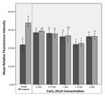
Relative fluorescent intensity in the vacuole (light grey) and cytosol (dark grey) of fresh and calcium chloride-treated tobacco microspores after incubation at 33ºC for 1 day using Fluo 3-AM staining. Different letter notations indicate significant difference at p-value: 0.05 based on Dunn's test.
3.3. Metabolite profile of stress-induced tobacco microspores under calcium chloride treatment
Untargeted GC-MS based metabolite profiling identified a total of 34 metabolites, including 29 metabolites from fresh microspore extracts, 18 metabolites from the extract of microspores cultured on medium B without the addition of calcium chloride, 17 metabolites from the treatment of 0.5 mM calcium chloride, 15 metabolites from the treatment of 1 mM calcium chloride, 12 metabolites from the treatment of 1.5 mM calcium chloride, and 3 metabolites from the treatment of 2 mM calcium chloride (as shown in Table 3). The majority of the identified compounds were fatty acids (20 compounds) and some organic acids (5 compounds), sugars (4 compounds), amino acids (3 compounds), and sugar alcohols (2 compounds). The dominant compounds identified in all treatments were oleic acid, palmitic acid and stearic acid. The higher the Ca2+ concentration, the lower the relative abundance of the three compounds.
List of compounds identified from samples of fresh tobacco microspores and microspores cultured under calcium chloride treatment in B medium at temperature of 33ºC for 6 days.
Fourteen metabolites identified only on fresh microspore extract, including L-alanine, glycine, L-proline, 3,4-dimethylbenzoic acid, L-(+)-tartaric acid, glycerol, D-fructose, D-glucose, sucrose, octanoic acid, nonanoic acid, decanoic acid, dodecanoic acid, and galactaric acid. Some compounds were only identified in certain treatments, including -(+)-galactose identified in the treatment of 0.5 mM calcium chloride, 6-oxoheptanoic identified in the treatment without the addition of calcium chloride, and behenic acid in the treatment of 1 mM calcium chloride. Metabolites identified in all samples had a tendency to decrease in relative abundance in treatments with higher calcium chloride concentrations, except for azelaic acid and sebacic acid. However, both compounds were not identified in the treatment with addition of 2 mM calcium chloride.
4. Discussion
Although the microspore embryogenesis system has proven effective for producing double haploid plants, its successful application in several important crops is still not well established. The induction phase represents a pivotal stage in the success of microspore embryogenesis, as it initiates the activation of altered developmental pathways. Changes in calcium concentration inside microspores provide a signal in initiating a series of embryogenesis processes. It has been demonstrated that the concentration of exogenous calcium in the culture medium can influence the efficiency of microspore embryogenesis induction. However, the cellular changes that occur under various calcium concentrations have not yet been described. Here, we utilised tobacco microspore to evaluate the optimum calcium chloride concentration for embyogenic microspore formation and also to observe the dynamics of intracellular calcium along with the changes of metabolite profile and cytological development during early stage of microspore embryogenesis.
Our study revealed calcium chloride concentration in the B medium influenced cytological development of tobacco microspore. After six days of incubation at 33ºC, microspores developed star-like structures and underwent symmetric first division, both of which increased with increasing calcium chloride concentration up to 2 mM. Star-like structure microspore serves as a marker for the formation of embryogenic microspores in tobacco and other species such as wheat (Indrianto et al., 2001), barley (Maraschin et al., 2005), rice (Nurbaiti et al., 2019), and eggplant (Pagalla et al., 2020).
In the process of androgenic plant formation, microspores that have been induced by stress treatment will pass through certain developmental pathways to form multicellular structures within the microspore wall (Bhojwani and Dantu, 2013). In tobacco microspore culture, the first division is reported to occur through both asymmetric and symmetric division pathways (Touraev et al., 1996a; Luo et al., 2022). In later development, nuclei similar to vegetative nuclei will divide again to form multicellular structures (Touraev et al., 1996a). The formation of symmetrical binucleate microspores, which does not naturally occur in the gametophytic developmental pathway, shows clear evidence that the induction process has successfully changed the microspore developmental pathway towards sporophytic development. Therefore, our results underscored the addition of 2 mM calcium chloride was optimum for embryogenic microspore formation.
Following the study of cytological development, we evaluated the calcium dynamics in microspore to observe the changes of intracellular calcium concentrations during early microspore embryogenesis. In line with the report of Rivas-Sendra et al. (2017), intracellular calcium signal was also found to be significantly increased in tobacco microspores after heat stress treatment for one day. The analysis also revealed that there was a rise in calcium signal within the cytosol and a corresponding decline in calcium signal within the vacuole compared to fresh microspores. This suggests a transfer of calcium ions from the vacuole to the cytosol during heat stress.
Luo et al. (2022) reported the involvement of phospholipase C (PLC) in tobacco microspore embryogenesis via lipid metabolism, suggesting its role in calcium ion release from intracellular compartments, particularly the vacuole. Phosphatidylinositol-specific PLC (PI-PLC) is implicated in the hydrolysis of phosphatidylinositol 4,5-bisphosphate (PIP2) into inositol 1,4,5-trisphosphate (IP3) and diacylglycerol (DAG) in response to stimuli such as heat stress, according to previous studies (Pauls et al., 2006; Singh et al., 2015; Canonne et al., 2011).
During the sixth day of the incubation period, calcium signals were detected across all experimental conditions, which showed consistent distribution patterns. However, there were significant differences in the average fluorescent intensity among the various treatments. This result suggests that the extracellular calcium concentration affects the intracellular Ca2+ concentration after six days of incubation under heat stress conditions.
The decrease in the intracellular Ca2+ signal in the 2 mM CaCl2 treatment after six days may be due to the reduced permeability of the plasma membrane from prolonged exposure to high extracellular Ca2+. Qiu and Su (1998) found that high extracellular calcium levels can reduce membrane fluidity. Hepler (2005) also found that increasing extracellular Ca2+ to 10 mM significantly decreased intracellular Ca2+ in lily pollen tubes. This possible mechanism is beneficial for microspore development because high cytosolic Ca2+ levels are toxic (Sanders et al., 1999).
Based on the metabolite profile, the relative abundance of oleic acid, palmitic acid and stearic acid were dominant in all samples (see Figure 6). The three compounds belong to the fatty acid class and are constituent components of the lipid bilayer membrane (Reszczyńska and Hanaka, 2020). Its relative abundance were decreased in induced microspores compared to fresh microspore, where the higher the Ca2+ concentration, the lower the relative abundance.

Chromatogram of GC-MS analysis results of tobacco microspores. A. Fresh microspores; B-F. Microspores after cultured on medium B with incubation temperature of 33ºC for 6 days; B. 0 mM CaCl2; C. 0.5 mM CaCl2; D. 1 mM CaCl2; E. 1.5 mM CaCl2; F. 2 mM CaCl2. 1: palmitic acid; 2: oleic acid; 3: stearic acid.
The fatty acid composition of membrane lipids, especially palmitic acid and stearic acid, plays a role in regulating the functional state of cells by altering membrane fluidity in response to stress (Sidorov et al., 2014). The higher the content of saturated fatty acids (palmitic acid and stearic acid) in lipids, the lower the membrane fluidity (Zhukov, 2015), while the presence of unsaturated fatty acids can increase their fluidity (Hąc-Wydro and Wydro 2007).
Oleic acid (C18:1) is a monounsaturated fatty acid that has one single double bond in its carbon chain (Reszczyńska and Hanaka, 2020). The results of this study showed a decrease in the relative abundance of oleic acid along with an increase in calcium chloride concentration. Based on Yu et al. (1998), calcium affects changes in plasma membrane lipid composition by reducing the content of unsaturated fatty acids. Therefore, we suggest that high extracellular calcium concentration affects intracellular calcium concentration by reducing membrane fluidity via altering fatty acid composition after six days of incubation. Further investigation is required to elucidate the mechanism of calcium action that causes these changes.
As a model plant for microspore embryogenesis, tobacco has been studied well in this area of research. However, our study used tobacco (local variety: Nicotiana tabacum L. var Manilo) to evaluate the effect of extracellular concentration in early microspore embryogenesis, which has not been widely explored. Our results provide novel evidence that high extracellular calcium moderates the undesirable effects of prolonged heat stress by altering the relative abundance of plasma membrane constituents, thereby increasing embryogenic microspores. These findings improve our understanding of the role of extracellular calcium in microspore embryogenesis and provide new strategies to improve the success of microspore embryogenesis in other important plants.
5. Conclusion
Overall, our study showed that the addition of 2 mM calcium chloride increased the percentage of microspores that developed into embryogenic microspores. This increase coincided with changes in intracellular calcium concentration and metabolite profile. We confirmed that an increase in calcium ions in the cytosol is required to activate microspore embryogenesis. In addition, exposure to heat stress for six days caused a prolonged increase in calcium ions in the cytosol which may be cytotoxic. Changes in the relative abundance of plasma membrane constituents may alter membrane fluidity, providing indirect evidence that high extracellular calcium mitigates the adverse effects of prolonged heat stress. These findings may lead to a better understanding of the role of extracellular calcium in microspore embryogenesis and provide new strategies to improve the success of microspore embryogenesis in important plants.
Acknowledgements
We would like to acknowledge Prof. Ir. Djajadi, M.Sc., Ph.D. and Supriyadi, SP. from the National Research and Innovation Agency Indonesia for providing the tobacco seeds used in this study. This work was supported by Universitas Airlangga under Grant number 1705/UN3.LPPM/PT.01.03/2023.
References
-
BHOJWANI, S.S. and DANTU, P.K., 2013. Plant tissue culture: an introductory text New Delhi: Springer India. http://doi.org/10.1007/978-81-322-1026-9
» http://doi.org/10.1007/978-81-322-1026-9 -
CANONNE, J., FROIDURE-NICOLAS, S. and RIVAS, S., 2011. Phospholipases in action during plant defense signaling. Plant Signaling & Behavior, vol. 6, no. 1, pp. 13-18. http://doi.org/10.4161/psb.6.1.14037 PMid:21248491.
» http://doi.org/10.4161/psb.6.1.14037 -
DUBAS, E., JANOWIAK, F., KRZEWSKA, M., HURA, T. and ŻUR, I., 2013. Endogenous ABA concentration and cytoplasmic membrane fluidity in microspores of oilseed rape (Brassica napus L.) genotypes differing in responsiveness to androgenesis induction. Plant Cell Reports, vol. 32, no. 9, pp. 1465-1475. http://doi.org/10.1007/s00299-013-1458-6 PMid:23736307.
» http://doi.org/10.1007/s00299-013-1458-6 -
HĄC-WYDRO, K. and WYDRO, P., 2007. The influence of fatty acids on model cholesterol/phospholipid membranes. Chemistry and Physics of Lipids, vol. 150, no. 1, pp. 66-81. http://doi.org/10.1016/j.chemphyslip.2007.06.213 PMid:17651712.
» http://doi.org/10.1016/j.chemphyslip.2007.06.213 -
HEPLER, P.K., 2005. Calcium: a central regulator of plant growth and development. The Plant Cell, vol. 17, no. 8, pp. 2142-2155. http://doi.org/10.1105/tpc.105.032508 PMid:16061961.
» http://doi.org/10.1105/tpc.105.032508 -
HOSP, J., TASHPULATOV, A., ROESSNER, U., BARSOVA, E., KATHOLNIGG, H., STEINBORN, R., MELIKANT, B., LUKYANOV, S., HEBERLE-BORS, E. and TOURAEV, A., 2006. Transcriptional and metabolic profiles of stress-induced, embryogenic tobacco microspores. Plant Molecular Biology, vol. 63, no. 1, pp. 137-149. http://doi.org/10.1007/s11103-006-9078-y PMid:17016740.
» http://doi.org/10.1007/s11103-006-9078-y -
INDRIANTO, A., BARINOVA, I., TOURAEV, A. and HEBERLE-BORS, E., 2001. Tracking individual wheat microspores in vitro: identification of embryogenic microspores and body axis formation in the embryo. Planta, vol. 212, no. 2, pp. 163-174. http://doi.org/10.1007/s004250000375 PMid:11216836.
» http://doi.org/10.1007/s004250000375 -
KYO, M. and HARADA, H., 1986. Control of the developmental pathway of tobacco pollen in vitro. Planta, vol. 168, no. 4, pp. 427-432. http://doi.org/10.1007/BF00392260 PMid:24232317.
» http://doi.org/10.1007/BF00392260 -
LUO, P., JIANG, A., ZHOU, Y., YANG, M., ZHOU, X., YANG, Y., YU, J. and TANG, X., 2022. Phospholipase C is a novel regulator at the early stages of microspore embryogenesis in Nicotiana tabacum. Plant Signaling & Behavior, vol. 17, no. 1, pp. 2094618. http://doi.org/10.1080/15592324.2022.2094618 PMid:35786356.
» http://doi.org/10.1080/15592324.2022.2094618 -
MARASCHIN, S.F., VENNIK, M., LAMERS, G.E.M., SPAINK, H.P. and WANG, M., 2005. Time-lapse tracking of barley androgenesis reveals position-determined cell death within pro-embryos. Planta, vol. 220, no. 4, pp. 531-540. http://doi.org/10.1007/s00425-004-1371-x PMid:15449059.
» http://doi.org/10.1007/s00425-004-1371-x -
NURBAITI, S., PURWESTRI, Y.A., DARYONO, B.S., SEMIARTI, E. and INDRIANTO, A., 2019. Developmental the pattern of embryogenic microspore of rice (Oryza sativa L.) based on morphological characteristic. Berkala Penelitian Hayati, vol. 25, no. 1, pp. 7-11. http://doi.org/10.23869/bphjbr.25.1.20192
» http://doi.org/10.23869/bphjbr.25.1.20192 -
PAGALLA, D.B., INDRIANTO, A., MARYANI, M. and SEMIARTI, E., 2020. Induction of microspore embryogenesis of eggplant (Solanum melongena L.) ‘Gelatik.’. Journal of Tropical Biodiversity and Biotechnology, vol. 5, no. 2, pp. 124-131. http://doi.org/10.22146/jtbb.53677
» http://doi.org/10.22146/jtbb.53677 -
PARRA-VEGA, V., CORRAL-MARTÍNEZ, P., RIVAS-SENDRA, A. and SEGUÍ-SIMARRO, J.M., 2015. Induction of embryogenesis in brassica napus microspores produces a callosic subintinal layer and abnormal cell walls with altered levels of callose and cellulose. Frontiers in Plant Science, vol. 6, pp. 1018. http://doi.org/10.3389/fpls.2015.01018 PMid:26635844.
» http://doi.org/10.3389/fpls.2015.01018 -
PAULS, K.P., CHAN, J., WORONUK, G., SCHULZE, D. and BRAZOLOT, J., 2006. When microspores decide to become embryos: cellular and molecular changesThis review is one of a selection of papers published in the Special Issue on Plant Cell Biology. Canadian Journal of Botany, vol. 84, no. 4, pp. 668-678. http://doi.org/10.1139/b06-064
» http://doi.org/10.1139/b06-064 -
QIU, Q.-S. and SU, X.-F., 1998. The influence of extracellular-side Ca2+ on the activity of the plasma membrane H+-ATPase from wheat roots. Functional Plant Biology, vol. 25, no. 8, pp. 923-928. http://doi.org/10.1071/PP98036
» http://doi.org/10.1071/PP98036 -
QU, H., JIANG, X., SHI, Z., LIU, L. and ZHANG, S., 2012. Fast loading ester fluorescent Ca2+ and pH indicators into pollen of Pyrus pyrifolia. Journal of Plant Research, vol. 125, no. 1, pp. 185-195. http://doi.org/10.1007/s10265-011-0440-z PMid:21789557.
» http://doi.org/10.1007/s10265-011-0440-z -
RESZCZYŃSKA, E. and HANAKA, A., 2020. Lipids composition in plant membranes. Cell Biochemistry and Biophysics, vol. 78, no. 4, pp. 401-414. http://doi.org/10.1007/s12013-020-00947-w PMid:33034870.
» http://doi.org/10.1007/s12013-020-00947-w -
REYNOLDS, T.L., 2000. Effects of calcium on embryogenic induction and the accumulation of abscisic acid, and an early cysteine-labeled metallothionein gene in androgenic microspores of Triticum aestivum. Plant Science, vol. 150, no. 2, pp. 201-207. http://doi.org/10.1016/S0168-9452(99)00187-9
» http://doi.org/10.1016/S0168-9452(99)00187-9 -
RIVAS-SENDRA, A., CALABUIG-SERNA, A. and SEGUÍ-SIMARRO, J.M., 2017. Dynamics of calcium during In vitro microspore embryogenesis and In vivo microspore development in Brassica napus and Solanum melongena. Frontiers in Plant Science, vol. 8, pp. 1177. http://doi.org/10.3389/fpls.2017.01177 PMid:28736567.
» http://doi.org/10.3389/fpls.2017.01177 -
SANDERS, D., BROWNLEE, C. and HARPER, J.F., 1999. Communicating with calcium. The Plant Cell, vol. 11, no. 4, pp. 691-706. http://doi.org/10.1105/tpc.11.4.691 PMid:10213787.
» http://doi.org/10.1105/tpc.11.4.691 -
SEGUÍ-SIMARRO, J.M., TESTILLANO, P.S., JOUANNIC, S., HENRY, Y. and RISUEÑO, M.C., 2005. Mitogen-activated protein kinases are developmentally regulated during stress-induced microspore embryogenesis in Brassica napus L. Histochemistry and Cell Biology, vol. 123, no. 4-5, pp. 541-551. http://doi.org/10.1007/s00418-004-0749-y PMid:15895239.
» http://doi.org/10.1007/s00418-004-0749-y - SIDOROV, R.A., ZHUKOV, A.V., PCHELKIN, V.P. and TSYDENDAMBAEV, V.D., 2014. Palmitic acid in higher plant lipids. In: L.F. PORTO, ed. Palmitic acid: occurrence, biochemistry and health effects New York: Nova Science Publishers, Inc., pp. 125-144.
-
SINGH, A., BHATNAGAR, N., PANDEY, A. and PANDEY, K., 2015. Plant phospholipase C family: regulation and functional role in lipid signaling. Cell Calcium, vol. 58, no. 2, pp. 139-146. http://doi.org/10.1016/j.ceca.2015.04.003 PMid:25933832.
» http://doi.org/10.1016/j.ceca.2015.04.003 -
TOURAEV, A. and HEBERLE-BORS, E., 2003. Anther and microspore culture in tobacco. In: M. MALUSZYNSKI, K.J. KASHA, B.P. FORSTER and I. SZAREJKO, eds. Doubled haploid production in crop plants Dordrecht: Springer Netherlands, pp. 223-228. http://doi.org/10.1007/978-94-017-1293-4_33
» http://doi.org/10.1007/978-94-017-1293-4_33 -
TOURAEV, A., ILHAM, A., VICENTE, O. and HEBERLE-BORS, E., 1996b. Stress-induced microspore embryogenesis in tobacco: an optimized system for molecular studies. Plant Cell Reports, vol. 15, no. 8, pp. 561-565. http://doi.org/10.1007/BF00232453 PMid:24178518.
» http://doi.org/10.1007/BF00232453 -
TOURAEV, A., PFOSSER, M., VICENTE, O. and HEBERLE-BORS, E., 1996a. Stress as the major signal controlling the developmental fate of tobacco microspores: towards a unified model of induction of microspore/pollen embryogenesis. Planta, vol. 200, no. 1, pp. 144-152. http://doi.org/10.1007/BF00196662
» http://doi.org/10.1007/BF00196662 -
TSUWAMOTO, R. and TAKAHATA, Y., 2008. Identification of genes specifically expressed in androgenesis-derived embryo in rapeseed (Brassica napus L.). Breeding Science, vol. 58, no. 3, pp. 251-259. http://doi.org/10.1270/jsbbs.58.251
» http://doi.org/10.1270/jsbbs.58.251 -
YU, B., GONG, H. and LIU, Y., 1998. Effects of calcium on lipid composition and function of plasma membrane and tonoplast vesicles isolated from roots of barley seedlings under salt stress. Journal of Plant Nutrition, vol. 21, no. 8, pp. 1589-1600. http://doi.org/10.1080/01904169809365506
» http://doi.org/10.1080/01904169809365506 -
ZHUKOV, A.V., 2015. Palmitic acid and its role in the structure and functions of plant cell membranes. Russian Journal of Plant Physiology, vol. 62, no. 5, pp. 706-713. http://doi.org/10.1134/S1021443715050192
» http://doi.org/10.1134/S1021443715050192