
 Virulent Beauveria bassiana OR500626 strain effectively control Tetranychus urticae
Abstract
The entomopathogenic fungus Beauveria bassiana OR500626 plays a pivotal role as a biological control agent for various agricultural pests, including the two-spotted red spider mite, Tetranychus urticae Koch. In the current study, we tested the ability of a promising strain of the fungus isolated from the tissues of the red palm weevil, Rhynchophorus ferrugineus in the eastern region of the Kingdom of Saudi Arabia. B. bassiana virulence was estimated based on LC50 values calculated by Probit analysis of T. urticae, which were treated with a series of concentrations (1 × 10², 1 × 10⁴, 1 × 10⁶, and 1 × 10⁸ conidia/ml). The mortality percentages of immature stages were 15.98-100%, males 15.94%-100%, and females (28.92%-100%) with an estimated LC50 value of 24.09×10³, 35.43×10³, and 11.03×10³ conidia/ml, respectively. Furthermore, electron microscopy confirmed the ability of B. bassiana to infect T. urticae. The results indicated that B. bassiana spores attached to the T. urticae spider body within about 12 hours. The germination of spores increased within 24-36 and penetrated the T. urticae cuticle after 96 hours of exposure.
Keywords:
Tetranychus urticae; Beauveria bassiana; biological control; scanning electron microscopy; LC50, entomopathogenic fungus
Resumo
O fungo entomopatogênico Beauveria bassiana OR500626 desempenha papel fundamental como agente de controle biológico para diversas pragas agrícolas, incluindo o ácaro-vermelho-de-duas-manchas, Tetranychus urticae Koch. No presente estudo, testamos a capacidade de uma cepa promissora do fungo isolada dos tecidos do gorgulho-vermelho-das-palmeiras, Rhynchophorus ferrugineus, na região leste do Reino da Arábia Saudita. A virulência de B. bassiana foi estimada com base nos valores de CL50 calculados pela análise Probit de T. urticae, que foram tratados com uma série de concentrações (1 × 10², 1 × 10⁴, 1 × 10⁶ e 1 × 10⁸ conídios/ml). As porcentagens de mortalidade dos estágios imaturos foram de 15,98% a 100%, dos machos de 15,94% a 100% e das fêmeas de 28,92% a 100%, com um valor estimado de CL50 de 24,09 × 10³, 35,43 × 10³ e 11,03 × 10³ conídios/ml, respectivamente. Além disso, a microscopia eletrônica confirmou a capacidade de B. bassiana de infectar T. urticae. Os resultados indicaram que os esporos de B. bassiana aderiram ao corpo da aranha T. urticae em cerca de 12 horas. A germinação dos esporos aumentou em 24-36 horas e penetrou na cutícula de T. urticae após 96 horas de exposição.
Palavras-chave:
Tetranychus urticae; Beauveria bassiana; controle biológico; microscopia eletrônica de varredura; CL50; fungo entomopatogênico
1. Introduction
The largest agricultural family of plant-feeding mites is red spider mite (Acari: Prostigmata: Tetranychidae). Small members (200-900 μm) of this family employ mobile styles to penetrate host plant cells (Van Leeuwen et al., 2009). They reduce agricultural productivity by 18–26% annually, damaging various field crops, both open and greenhouse, including vegetables (e.g., cucumber, tomatoes, peppers, eggplants, beans), fruits, maize, cotton, roses, medicinal, and decorative plants (Elhakim et al., 2020; Farahani et al., 2020). With over 1200 red spider mite species, over 10% are agricultural pests (Lugo-Sánchez et al., 2019). The most important pest is the two-spotted red spider mite (TSSM) Tetranychus urticae Koch, which has been found on more than 3,877 host plant species and is economically relevant to at least 150 plants (Elhakim et al., 2020; Farahani et al., 2020; Jakubowska et al., 2022).
Tetranychus urticae's primary concern in open fields and greenhouses is mobile stages that feed on leaves by putting their stylets on the underside and sucking their contents. Plant cells suffer direct and indirect damage. Yellowish-white, gray, or silvery patches called tiny stippling indicate damaged cells, which may become blackish if there are more damaged cells (Farahani et al., 2020). The ongoing feeding process lowers nitrogen, phosphate, and protein concentrations, disrupts cell shape, and inhibits photosynthesis and transpiration. As feeding continues, leaves necrotize and die (Riahi et al., 2013; Agut et al., 2018; Osman et al., 2019). Only 18 red spider mite species from 10 genera of Tetranychidae were found in Saudi Arabia (Al Riyadh, Al Baha, Qassim, Al Medina, Makkah, Tabuk, Wadi Al Dawasir, and Al Ahsa) (Osman et al., 2019).
The most frequent way to reduce T. urticae is through chemical control; however, long-term pesticide usage has increased its resistance. Due to its short life cycle, high fertility, and prolific offspring, T. urticae is the most resistant species to acaricides that may be created quickly (Zhang et al., 2018; Wu et al., 2019). Thus, an alternate T. urticae control strategy was needed. In most biological control research, predatory mites and entomopathogenic fungi (E.P.F.) are used (Attia et al., 2013; Gámez-Guzmán et al., 2019). Phytoseiidae: predatory mites, Neoseiulus californicus and Phytoseiulus persimilis Athias-Henriot have effectively reduced T. urticae populations in numerous crops (Greco et al., 2005). They're less efficient at managing pest mites because predatory mites eat eggs and larvae (Zhang et al., 2018).
Recently, entomopathogenic fungi have reduced mite infestations without harming non-target species, the environment, or people, making them suitable for biological control programs (Sholla and Kottb, 2017). Beauveria bassiana is the most well-known and successful species in lowering the population of several mite species, and its usage in bio-preparations has grown popular worldwide (Faria and Wraight, 2007; Yucel, 2021). Entomopathogenic fungi develop conidia that germinate, penetrate the cuticle, and rapidly colonize the hemocoel before appearing and sporulating on the mite cadaver, causing death from mechanical damage, nutrient reduction, and toxicities (Dogan et al., 2017). Additionally, entomopathogenic fungi synthesize several metabolic substances with varied roles (Yun et al., 2017).
The current study aims to apply the insect-pathogenic fungus B. bassiana, isolated locally from fields in Saudi Arabia, to combat the T. urticae on sweet pepper leaves in the lab, calculating the mortality of different stages and proving the ability of B. bassiana to penetrate the T. urticae cuticle using a scanning electron microscope.
2. Materials and Methods
2.1. Collection and rearing of T. urticae
Samples were obtained from the Agricultural Research and Training Station in King Faisal University in Al-Ahsa city (25°15'57.6”N 49°41'43.9” E) and a palm farm in Anak city (26°31'02.7”N 50°01'08.6” E) in the Eastern Province of Saudi Arabia. These samples of living T. urticae were collected from beans, eggplants, and cucumber leaves growing in the greenhouses. Samples were transported at room temperature in plastic bags to the Basic and Applied Scientific Research Center (BASRC) in Imam Abdulrahman bin Faisal University, Dammam city.
Immediately once received, triplicate breeding experiments were conducted with mature mites to generate a population of T. urticae encompassing both developmental stages of immature and mature mites. A T. urticae male was mated with three females in a 3 cm sweet pepper leaf disc. Once eggs had been laid, six leaf discs were then placed in a petri dish on a moist cotton wool pad for 24-48 hours at 16L: 8D photoperiod, 28 ± 1ºC, and 65 ± 5% R.H until hatching. Newly hatched eggs were placed individually on fresh sweet pepper leaf discs and used at this study as the immature and mature (female and male) T. urticae (Osman et al., 2019).
2.2. Source of B. bassiana OR500626 inoculum
The Beauveria bassiana OR500626 fungus was obtained from a previous study (Al-Zahrani et al., 2023) at the Basic and Applied Scientific Research Center (BASRC) in Imam Abdulrahman bin Faisal University, Dammam city.
These isolates were isolated from the tissues of Rhynchophorus ferrugineus, also known as red palm weevil, in the eastern region of the Kingdom of Saudi Arabia. To cultivate B. bassiana, the isolate was placed on an autoclaved Potato Dextrose Agar prepared as follows: 1L of distilled water, dissolved potato infusion (200 g/L), dextrose (20 g/L), yeast extract (20 g/L), and agar (20 g/L). The fungal culture was incubated at 25±1 °C for 10-14 days (Örtücü and Albayrak, 2017) to produce conidia. Following the incubation, conidia were scraped from the culture and suspended in 10 ml sterile distilled water with 0.02% Tween 80 in preparation for spore suspensions. Using a Neubauer-improved hemocytometer (Germany), spore concentrations of 1 × 10², 1 × 10⁴, 1 × 10⁶, and 1 × 10⁸ (conidia/ml) were prepared (Sewify et al., 2015).
2.3. In vitro acaricidal of B. bassiana against T. urticae
Fifteen T. urticae were individually transplanted on a pepper leaf disc (3 cm in diameter) at each stage of immature, mature female, and mature male. Each isolate has six leaf discs and is maintained on a moist cotton wool pad (15-cm petri dish). Direct spraying of B. bassiana spore suspensions were administered with a hand sprayer at 25–30 cm, while control replicates were sprayed with water. Live and dead T. urticae were counted daily using a dissecting microscope for 7 days after application.
After three days on wet Petri plates under the same laboratory settings, cadavers were declared “mycotised” if fungal outgrowths were visible, the treatment was replicated three times, with independent batches of fungal suspension used for each replication (Sewify et al., 2015).
2.4. Scanning Electron Microscope (S.E.M.)
In the scanning electron microscopy (SEM) study, infected T. urticae were selected based on the density of fungal mycelium growth on their instrument. Mites' slow movement and color change were also compared to the negative control. Infected individuals (females only) were fixed and prepared according to standard protocols before SEM imaging. A sample size of 10 adult females was selected from multiple replicates to ensure an accurate representation of the studied condition. Infected T. urticae were fixed in 3% glutaraldehyde at 25±3°C for three hours. Next, the samples were dried in ethanol at 30, 50, 70, 90, and 100% for five minutes at each concentration. After installing the samples on scanning electron microscopy, ions were sprayed to cover them with a thin layer of gold (Emam and Mohamed, 2021).
2.5. Statistical analyses
All experiments were conducted in triplicates. Data was analyzed using ANOVA and compared using Tukey HSD (SPSS 22.0) (IBM Corp, 2014). Corrected Mortality percentages were calculated using the Abbott Formula (Abbott, 1925).
Since lethal doses produced 50% mortality (LC50), the statistical software LdP line computed confidence limits and probit line slope values (Bakr, 2007).
Where: n = insect population, T = treated, Co = control.
Since lethal doses produced 50% mortality (LC50), the statictical software LdP line computed confidence limits and probit line slope values (Bakr, 2007).
3. Results and Discussion
3.1. Acaricidal activity of B. bassiana against T. urticae
Under laboratory conditions at 25±1ºC, 65±5% R.H., and 16:8 h photoperiod, the survivability of the developing stages of T. urticae (immature, adult male, and adult female) of the entomopathogenic fungus B. bassiana was assessed. Four concentrations (1×10², 1×10⁴, 1×10⁶, and 1×10⁸ conidia/ml) of B. bassiana were used for each stage. Adult females and males were more vulnerable to fungal infection than immature stages at all doses. While B. bassiana demonstrates effectiveness in controlling T. urticae, its efficacy varies across life stages, with adults exhibiting higher susceptibility; the reduced susceptibility of immature stages, attributed to conidia release during molting—particularly pronounced in species with short molting periods—is partially mitigated by the fungal concentration, which remains a critical determinant of mortality across all stages. Irigaray et al. (2003); Al-Alawi (2019) found comparable findings in T. urticae and other arthropods such as Tetranychus evansi (Wekesa et al., 2006) and tick species (Samish et al., 2001; Gindin et al., 2002). The release of a large number of conidia from the mites' outer layer during molting may reduce their susceptibility to B. bassiana, especially in species with short molting periods. Nevertheless, the concentration level affected the mortality rate of T. urticae stage.
3.1.1. Treatment of immature T. urticae
In Table 1, the mean number of dead immature stages of T. urticae after 1 day of spraying B. bassiana at various concentrations (1×10², 1×10⁴, 1×10⁶, and 1×10⁸ conidia/ml) was 5.38, 5.66, 9.33, and 11.50 respectively. (F4,25 = 28.59, p = 0.00). At 1×10², 1×10⁴, 1×10⁶, and 1×10⁸ conidia/ml concentrations, immature stages decreased by 15.98, 32.53, 59.04, and 74.70% respectively. Moreover, after 2 days of spraying B. bassiana at various doses (1×10², 1×10⁴, 1×10⁶, and 1×10⁸ conidia/ml), the mean dead immature T. urticae was 9.50, 10.66, 13.00, and 14.66 (F4,25 = 99.97, p = 0.00). At 1×10², 1×10⁴, 1×10⁶, and 1×10⁸ conidia/ml concentrations, immature T. urticae was reduced by 58.75, 67.50, 85.00, and 97.50%. After three days of spraying B. bassiana at various concentrations (1×10², 1×10⁴, 1×10⁶, and 1×10⁸ conidia/ml), dead immature T. urticae population decreased by 84.42, 97.40, 98.70, and 100.00%, respectively (F4,25 = 125.28, p = 0.00). The study demonstrates the efficacy of B. bassiana in controlling the immature stages of T. urticae, where mortality rates increase with higher fungal concentrations and longer exposure times. This is accompanied by variations in results when compared to previous studies in this field, attributed to factors such as fungal strains and experimental conditions, which were observed when comparing the obtained results with prior research in this area, including Wekesa et al. (2005) reported lower mortality in T. urticae compared to our results from our investigation. On T. evansi, death rates were highest in larvae (20.0, 48%), protonymph (22.0, 51.0%), and deutonymph (41.0, 78.0%) at concentrations of 3.0 ×10⁶ and 1 ×10⁸ conidia/ml. Elsherbeni (2021) tested B. bassiana against T. urticae larvae. Death rates were from 16.67% to 43.33% at concentration 1x10³ and 36.67% to 66.67% at concentration 1x10⁴ in a lab setting of 25 ± 2 ºC and 75 ± 5% R.H. Moreover, B. bassiana fungal sprays at 1×10⁷ conidia/ml concentration resulted in 17.5-25.25% immature T. urticae mortality after 7 days. Topuz et al. (2016) compared four B. bassiana isolates to effect on Tetranychus cinnabarinus at the deutonymph stage, observing a death rate of 11.68% and 8.33% after 10 days of spraying at 1×10⁷ conidia/ml. A combination of Trichoderma harzianum (2x10⁶ spores/ml) and Cladosporium herbarium (1x10⁶ spores/ml) caused mite mortality rate of immatures from 58.4% to 65.7% at 30 oC and 90% R.H., and 57.1%–67.8% at 35 oC and 90% R.H. (Afifi et al., 2013). B. bassiana showed 67.00% and 90.00% efficiency against T. urticae nymphs at doses of 1 × 10⁶ and 1 × 10⁸, respectively. The mortality rate of six Jordanian B. bassiana isolates and the commercially available GHA (BotaniGard®, Bio Works, U.S.A.) on T. urticae deutonymphs was 27-55% at a concentration of 1×10⁷ conidia/ml (Al-Alawi, 2019).
The mean mortality, ± standard error (SE), and the percentage decrease of T. urticae immatures treated with B. bassiana.
Means in each column followed by the same letter is not significantly different using Tukey HSD test.
3.1.2. Treatment of adult females of T. urticae
In Table 2, the mean number of dead adult females after 1 day of spraying B. bassiana at different concentrations (1 × 10², 1 × 10⁴, 1 × 10⁶, and 1 × 10⁸ conidia/ml) were 5.33, 7.50, 11.16, and 12.83 respectively, (F4,25 =31.759, p= 0.000). At the same concentrations, adult females decreased 28.92, 34.78, 66.67, and 81.16% respectively. Dead adult females had a mean of 10.33, 12.50, 14.33, and 14.66 (F4,25 = 28.793, p= 0.000) after 2 days of spraying B. bassiana at various concentrations (1 × 10², 1 × 10⁴, 1 × 10⁶, and 1 × 10 conidia/ml). At the same concentrations, adult females decreased 52.54, 74.58, 93.22, and 96.61% respectively. After 3 days of spraying B. bassiana at different concentrations (1 × 10², 1 × 10⁴, 1 × 10⁶, and 1 × 10⁸ conidia/ml), the mean number of dead adult females was 14.666, 15.00, 15.00, and 15.000 (F4,25 = 17.505, p= 0.000), resulting in a 95.35, 100.00, 100.00, and 100.00% decrease (Table 2), Elsherbeni (2021) also tested B. bassiana against T. urticae females. The death rate was 26.67-43.33 at 1x10⁴ concentration in a lab setting of 25 ± 2°C, 75 ± 5% R.H. After the first, second, and third days, the mortality of T. urticae female at 106 and 108 conidia/ml was 34.5, 51.8, 61.0%, and 60.4, 72.7, 77.2% respectively, which was greater than Sewify et al. (2015). Another investigation found that 5 B. bassiana isolates at 106 conidia/ml of the conidial solution were deadly 68.49 to 83.78%) on the first day and 100% on the fourth day (Draganova and Simova, 2010). Research by Yanar et al. (2018) found that various isolates had a 46.2-72.2% impact on T. urticae at a 1x10⁸ effect rate. At concentration 2×10⁴, T. urticae death rates by Gatarayiha (2012) were 35.1-42.6%. Treatment at 1×10⁶ and 1×10⁸ (conidia/ml) concentrations resulted in B. bassiana mycosis in T.urticae individuals within 3 days, with rapid and fatal effects. Ullah and Lim (2015) found that T. urticae cadavers grown on bean leaves 3 days after B. bassiana treatment displayed fungal overgrowth. The effectiveness of B. bassiana against T. urticae may depend on spore suspension dosage, isolation identity, and experimental settings such as humidity, temperature, host type, plant variety, and treatment interval. The enzymes generated by B. bassiana isolates may also affect their virulence against T. urticae. Secondary compounds generated by pathogenic fungi, such as beauvericin poisons in B. bassiana, may potentially explain the observed virulence differential (Roberts and Leger, 2004).
The mean mortality, ± standard error (SE), and the percentage decrease of T. urticae females treated with B. bassiana.
3.1.3. Treatment of adult males of T. urticae
After spraying B. bassiana at the same concentrations mentioned above on T. urticae male, mortality increased with higher concentrations. The number of deaths was directly proportional to the concentration of the suspension and also increased with longer exposure periods, from one to three days (Table 3). Seiedy et al. (2010) reported lower mortality rates for T. urticae using two B. bassiana isolates: 4.06-38.75 and 4.37-35.00% at 1 × 10⁴ conidia/ml, 13.43-70.31% and 10.00-59.37% at 1 × 10⁶ conidia/ml, and 15.93-82.81% and 11.87-74.68% at 1 × 10⁸ conidia/ml.
The mean mortality, shown by the standard error (S.E.), and the percentage decrease of male T. urticae when treated with B. bassiana.
3.2. Observation Scanning Electron Microscopic (S.E.M.) on infected T. urticae by B. bassiana
When we examined T. urticae by Scanning electron microscopic, we found many conidia attached to the setae base and body folds of the T. urticaeFigures 1A, B, C, and D. B. bassiana conidia adhered to T. urticae within about 12 hours. After that, these conidia germinate and form germ tubes about 3 mm long, some of them produced near the T. urticae cuticle, the appressoria of fungi secrete extracellular main epidermal degrading enzymes (lipases, proteases, and chitinases) to enhance penetration of the T. urticae integument within 24-36 hours Figure. 2A. Within 48 hours, germ tube length and profuse germination rates increased. Fungi were significantly formed on T. urticae integument Figures 2B, C, and D. After 72 h, these mycelia formed a mat-like structure and released conidiophores and new conidia (Figure 3A). After 96 h, numerous hyphae entered the cuticle from the setae base or stomata via the body (Figures 3B and C). After 120 h, the T. urticae integument was covered with white hyphae with many conidia (Figure 3D). Conidia on the cuticle and setae base germinated well. Results showed conidia germination was linked to T. urticae cuticle. Germinal tubes entered T. urticae from all areas except the head capsule, where epidermal sclerosis was considerable. The development of mycosis, characterized by the proliferation of fungal hyphae and conidia on the mite's cadaver, signifies successful colonization and nutrient acquisition. This proliferation is a direct result of the fungus's ability to overcome the host's immune responses and establish itself within the hemocoel. The time-dependent increase in toxicity reflects the cumulative effect of these processes, including the secretion of toxins and enzymes, mechanical penetration, and ultimately, the disruption of vital physiological functions. The findings supported Sewify et al. (2015); Wu et al. (2016); Hassan et al. (2017) in the infection and germination of the entomopathogenic fungus B. bassiana against T. urticae.

Adhesion of Beauveria bassiana conidia to the body of the red spider mites Tetranychus urticae, a2 arrow; (A), especially in body folds, a2 arrow; (B & C) and setae base, a3 arrow; (D) within about 12 hours.

Germination of Beauveria bassiana conidia and production of appressoria, b1 arrow near the cuticle with germ tubes, b2 arrow; (A) within 24-36 hours. Germ tube length, b3 arrow; (B), germination rates increased (C), and fungi were significantly formed on the surface of TSSM (D) within 48 hours.

Emergence of conidiophores, c1 arrow and new conidia, d1 arrow from the mycelium; (A). After 96 hours, the hyphae invade the cuticle from the base of the mites' body stomata, e1 arrow; (B) or setae base and spread to the bristles, f1 arrow; (C). After 120 h, T. urticae integument was covered with white threads showing many conidia (D).
The strategy of the infection stage is represented by a combination of mechanical factors with pressure resulting from the germ tubes, which contribute to the breakdown of the cuticle layers and the secretion of enzymes produced by the fungus, such as lipase, protease, and chitinase. First, lipase, the first enzyme to be synthesized, degrades long-chain alkenes and fatty lipids that make up the cuticle surface layer (epicuticle) into nutritious raw materials. Then a protease enzyme is released, which breaks down the soluble proteins into amino acids and facilitates penetration before the chitin enzyme is secreted. Chitinase is produced in the final stage of enzymatic analysis, which breaks down chitin and ends the penetration process. Thus, the fungus penetrated all layers of the epidermis until reaching the hemolymph of the mite. After entering T. urticae hemolymph, the fungus develops from filamentous bodies to shorter hyphal bodies and creates blastospores that bud within the hemocoel (a yeast-like development phase), infiltrate interior tissues, and disrupt the host immune system (Khan et al., 2012). B. bassiana and hyphal development create several secondary metabolites, the most significant of which are poisons that confound the insect's immunological defense systems, speeding infection (Dannon et al., 2020). These toxins include beauvericin, beauverolides, bassianolides, and isarolides. The host dies from these occurrences (Singh et al., 2017; Dannon et al., 2020).
The saprophytic phase starts following host death (Sani et al., 2020). The fungus creates secondary compounds like “Oosporin,” a red antibiotic that colors cadavers and battles bacteria in insects' guts after an infected host dies. B. bassiana hyphae cross T. urticae integument intersegmentally and reverse (Figure 4) (Dannon et al., 2020; Sani et al., 2020). Finally, pathogenic fungi-infected cadavers passively transmit spores to new hosts (Sani et al., 2020).

The hyphae penetrate the cuticle of the T. urticae to the outside of the body, h1 arrow. And mycelium forming, i1 arrow, in addition to the sprouting conidia forming, j1 arrow.
3.3. Toxicity of tested B. bassiana against developmental stages of T. urticae
B. bassiana led to the death of T. urticae 72 hours after fungal treatment. The deadly toxicity of B. bassiana was time-dependent, meaning that the toxicity of this fungus increased with the progression of time, and it was noted that the body of T. urticae was covered with fungal conidia causing mycosis compared to the dead mite in the control (Figure 5). These agree with Islam et al. (2017) and Yucel (2021). They observed an increase in the toxicity of B. bassiana with time, in addition to the development of mycosis on the dead groups of T. urticae after three days of application.

Infection caused by B. bassiana on T. urticae, (A) the appearance of symptoms of infestation in T. urticae female front legs after 3 days, (B) Mycosis develops after 5 days, (C) Mycosis develops after 7 days, and (D) Dead T. urticae under control without symptoms of infection.
3.3.1. Toxicity of tested B. bassiana against different stages of T. urticae after 24 h from exposure
According to Table 4, At 24 hours post-treatment, the LC50 values for immature, adult male, and adult female T. urticae were 5.98×10³, 6.40×10³, and 2.50×10³ (conidia/ml) for B. bassiana delivered by T. urticae. In addition, the lowest toxicity values for immature, adult male, and adult female T. urticae were 2.24×10³, 2.80×10³, and 1.84×10³ (mg L-1) for B. bassiana. Maximum toxicity values for B. bassiana were 10.49×10³, 12.50×10³, and 4.68×10³ (mg L-1) for the same three species. In addition, T. urticae's adult male, female, and immature stages had toxicity slope values of 0.28, 0.31, and 0.32 (Figure 6). Our B. bassiana isolate, from the red palm weevil in Anak, Saudi Arabia, was more virulent than isolates used in previous studies on T. urticae development. B. bassiana direct spraying bioassays against T. urticae life stages (Geroh et al., 2015). The LC50 values for eggs, larvae, nymphs, and adults were 3.0 × 10⁵, 1.4 × 10⁶, 0.4 × 10⁷, and 0.3 × 10⁸ conidia/ml, respectively. He also noted that the low LC50 values of the direct spray Bioassay technique make B. bassiana concentrations in all stages of T. urticae far more hazardous than those from the processed meal approach. Both the slope (Slope ± SE) and 95% Confidence limits (Lower and Upper) intervals for the LC50 value of B. bassiana against the immature and adult males and females of T. urticae after 24 h from exposure have already been calculated and are reported in Tables 4 and 5. The observed variability in B. bassiana toxicity against T. urticae, as reflected in LC50 values and toxicity slopes, is attributable to a confluence of factors including fungal strain genetics, host physiological differences, environmental conditions, and experimental methodologies. Sewify et al. (2015) found that the entomopathogenic fungus B. bassiana might affect T. urticae embryos and adult stages. The first-day LC50 of 7.59 x 10⁷ (conidia/ml) showed that B. bassiana was more pathogenic to adults. Elhakim et al. (2020) calculated LC50 values at 3.3 × 10⁶ conidia/ml. On day 53, LC50 was 1.1107 conidia/ml. Irigaray et al. (2003) found that a commercial isolate of B. bassiana (Naturalis L.) had an LC50 value of 3.1 × 10³ (conidia/ml) against larvae and juveniles of T. urticae, and 1.9 × 10³ against adult T. urticae.
Toxicity of tested B. bassiana against the immature male and female of T. urticae after 24 h from exposure.
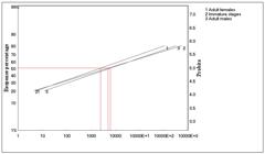
Ldp-lines of toxicity effect of B. bassiana against the immature male and female of T. urticae after 24 h from exposure.
Toxicity of tested B. bassiana against the immature male and female of T. urticae after 48 h from exposure.
3.3.2. Toxicity of tested B. bassiana against different stages of T. urticae after 48 h from exposure
The LC50 values for B. bassiana following direct spraying of T. urticae were 24.09×10³, 35.43×10³, and 11.03×10³ (conidia/ml) 48 hours after treatment for immature, adult male, and adult female T. urticae, respectively (Table 5 Additionally, B. bassiana toxicity values ranged from 9.04×10³ to 24.05×10³ (mg L-1) for immature, adult male, and adult female T. urticae, respectively. Additionally, juvenile, adult males, and adult females T. urticae had toxicity slope values of 0.27, 0.30, and 0.33, respectively (Figure 7). Compared to previous research, our local isolate, B. bassiana, demonstrated increased toxicity or virulence against T. urticae development with time. Hassan et al. (2017) found that B. bassiana strains were most effective against T. urticae, causing 88.5% mortality to adulthood at 108 (conidia/ml) concentration and a highest LC50 of 6.61 × 10⁶ (conidia/ml) on the seventh day. Additionally, the egg stage showed a hatchability of 25.2% and a highest LC50 of 1.14 × 10⁷ (conidia/ml) on the seventh day. Sewify et al. (2015) found that the entomopathogenic fungus B. bassiana harmed T. urticae eggs and adults. B. bassiana was more pathogenic against adulthood, with mortality rates increasing and LC50 values decreasing: 1.08 x 10⁷ (conidia/ml) on the second day, 5.61 x 10⁶ on the third day, 5.19 x 10⁶ on the fourth day, 3.16 x 10⁶ on the fifth day, and 1.22 x 10⁶ on the sixth day. Yucel (2021) found that BGF14 and BCA32, B. bassiana isolates, killed T. urticae adults quickly and increased with time. The BGF14 isolate had an LC50 of 1.1×10⁷ (conidia/ml) on the third day, 0.9×10⁷ on the fifth day, and 2.6×10⁶ on the seventh day. The BCA32 isolate was more toxic, with an LC50 of 1.3×10⁶ on the third day, 2.7×10⁵ on the fifth day, and 6.3×10⁴ on the seventh day.

Ldp-lines of toxicity effect of B. bassiana against the immature male and female of T. urticae after 48 h from exposure.
4. Conclusion
This study advocates for the application of B. bassiana as a viable and environmentally benign alternative to chemical pesticides for the management of T. urticae in Saudi Arabian greenhouses. The strategic application of this entomopathogenic fungus should prioritize targeting adult T. urticae female, utilizing concentrations aligned with the determined LC50 values. Furthermore, the integration of this biological control agent with sound agricultural practices and rigorous post-application monitoring is crucial to optimize efficacy and ensure sustainable pest management.
Data Availability Statement
The entire data set that supports the results of this study was published in the article itself.
Acknowledgements
The authors would like to acknowledge the authorities of the Basic and Applied Scientific Research Center (BASRC) of Imam Abdulrahman bin Faisal University (IAU) for providing the required equipment and infrastructure to carry out this research. We are also grateful to the engineers of the Pests and Plant Diseases Unit of the Ministry of Environment, Water and Agriculture for their assistance in providing samples of plants infected with T. urticae.
References
-
ABBOTT, W.S., 1925. A method of computing the effectiveness of an insecticide. Journal of Economic Entomology, vol. 18, no. 2, pp. 265-266. http://doi.org/10.1093/jee/18.2.265a
» http://doi.org/10.1093/jee/18.2.265a - AFIFI, A., EL-BISHLAWY, S. and MAHMOUD, A., 2013. Resistance of two eggplant cultivars against the two-spotted red spider mite; Tetranychus urticue koch infestation, with notes on its biology. ACARTNES, vol. 7, pp. 23-27. http://doi.org/10.21608/AJESA.2013.163690.
-
AGUT, B., PASTOR, V., JAQUES, J. and FLORS, V., 2018. Can plant defense mechanisms provide new approaches for the sustainable control of the two-spotted red spider mite Tetranychus urticae. International Journal of Molecular Sciences, vol. 19, no. 2, pp. 614. http://doi.org/10.3390/ijms19020614 PMid:29466295.
» http://doi.org/10.3390/ijms19020614 - AL-ALAWI, M., 2019. Evaluation of Jordanian isolates of Beauveria bassiana (Balsamo) Vuillemin and their interaction with essential plant oils when combined for the two-spotted red spider mite, Tetranychus urticae Koch control. Advances in Environmental Biology, vol. 13, pp. 4-10. http://doi.org/10.22587/aeb.2019.13.9.2.
-
AL-ZAHRANI, J.K., AL-ABDALALL, A.H., OSMAN, M.A., ALDAKHEEL, L.A., ALAHMADY, N.F., ALDAKEEL, S.A., ABDULAZEEZ, S., BORGIO, J.F., ELNAGGAR, M.A., ALABDALLAH, N.M. and ALMUSTAFA, M.M., 2023. Entomopathogenic fungi and their biological control of Tetranychus urticae: two-spotted red spider mite s. Journal of King Saud University. Science, vol. 35, no. 8, pp. 102910. http://doi.org/10.1016/j.jksus.2023.102910
» http://doi.org/10.1016/j.jksus.2023.102910 -
ATTIA, S., GRISSA, K., LOGNAY, G., BITUME, E., HANCE, T. and MAILLEUX, A., 2013. A review of the major biological approaches to control the worldwide pest Tetranychus urticae (Acari: Tetranychidae) with special reference to natural pesticides. Journal of Pest Science, vol. 86, no. 3, pp. 361-386. http://doi.org/10.1007/s10340-013-0503-0
» http://doi.org/10.1007/s10340-013-0503-0 -
BAKR, E. 2007 [accessed 26 January 2025]. LdP Line [software]. Available from: http://embakr.tripod.com/ldpline/ index.htm
» http://embakr.tripod.com/ldpline/ -
DANNON, H., DANNON, A., DOURO-KPINDOU, O., ZINSOU, A., HOUNDETE, A., TOFFA-MEHINTO, J., ELEGBEDE, I., OLOU, B. and TAMÒ, M., 2020. Toward the efficient use of Beauveria bassiana in integrated cotton insect pest management. Journal of Cotton Research, vol. 3, no. 1, pp. 24. http://doi.org/10.1186/s42397-020-00061-5
» http://doi.org/10.1186/s42397-020-00061-5 -
DOGAN, Y., HAZIR, S., YILDIZ, A., BUTT, T. and CAKMAK, I., 2017. Evaluation of entomopathogenic fungi for the control of Tetranychus urticae (Acari: Tetranychidae) and the effect of Metarhizium brunneum on the predatory mites (Acari: Phytoseiidae). Biological Control, vol. 111, pp. 6-12. http://doi.org/10.1016/j.biocontrol.2017.05.001
» http://doi.org/10.1016/j.biocontrol.2017.05.001 -
DRAGANOVA, S. and SIMOVA, S., 2010. Susceptibility of Tetranychus urticae koch. (Acari: Tetranychidae) to isolates of entomopathogenic fungus Beauveria bassiana. Pesticidi i Fitomedicina, vol. 25, no. 1, pp. 51-57. http://doi.org/10.2298/PIF1001051D
» http://doi.org/10.2298/PIF1001051D -
ELHAKIM, E., MOHAMED, O. and ELAZOUNI, I., 2020. Virulence and proteolytic activity of entomopathogenic fungi against the two-spotted red spider mite, Tetranychus urticae Koch (Acari: tetranychidae). Egyptian Journal of Biological Pest Control, vol. 30, no. 1, pp. 30. http://doi.org/10.1186/s41938-020-00227-y
» http://doi.org/10.1186/s41938-020-00227-y -
ELSHERBENI, M., 2021. Efficacy of Cladosporium cladosporioides, as a biocontrol agent for controlling Tetranychus urticae Koch (Acari: Tetranychidae) under laboratory conditions Cladosporium cladosporioides Tetranychus urticae Journal of Plant Protection and Pathology, vol. 12, pp. 319-321. http://doi.org/10.21608/jppp.2021.171278
» http://doi.org/10.21608/jppp.2021.171278 -
EMAM, H. and MOHAMED, S., 2021. Isolation and identification of the fungal pathogen aspergillus aflatoxiformans strain and its role in controlling the two-spotted red spider mite Tetranychus urticae (Koch). Journal of Plant Protection and Pathology, vol. 12, pp. 695-702. http://doi.org/10.21608/jppp.2021.99994.1044
» http://doi.org/10.21608/jppp.2021.99994.1044 -
FARAHANI, S., BANDANI, A. and AMIRI, A., 2020 [accessed 26 January 2025]. Toxicity and repellency effects of three essential oils on two populations of Tetranychus urticae (Acari: tetranychidae). Persian Journal of Acarology [online], vol. 9, pp. 67-81. Available from: https://www.biotaxa.org/pja/article/view/202016
» https://www.biotaxa.org/pja/article/view/202016 -
FARIA, M. and WRAIGHT, S., 2007. Mycoinsecticides and Mycoacaricides: a comprehensive list with worldwide coverage and international classification of formulation types. Biological Control, vol. 43, no. 3, pp. 237-256. http://doi.org/10.1016/j.biocontrol.2007.08.001
» http://doi.org/10.1016/j.biocontrol.2007.08.001 -
GÁMEZ-GUZMÁN, A., TORRES-ROJAS, E. and GAIGL, A., 2019. Potential of a Cladosporium cladosporioides strain for the control of Tetranychus urticae koch (Acari: Tetranychidae) under laboratory conditions. Agronomia Colombiana, vol. 37, no. 1, pp. 21-26. http://doi.org/10.15446/agron.colomb.v37n1.73353
» http://doi.org/10.15446/agron.colomb.v37n1.73353 -
GATARAYIHA, M., 2012. Selection of Beauveria bassiana strains against the two-spotted red spider mite, Tetranychus urticae Koch, in laboratory and greenhouse trials. African Journal of Microbiological Research, vol. 6, no. 11, pp. 2694-2703. http://doi.org/10.5897/AJMR11.1184
» http://doi.org/10.5897/AJMR11.1184 - GEROH, M., GULATI, R. and TEHRI, K., 2015. Determination of lethal concentration and lethal time of entomopathogen Beauveria bassiana (Balsamo) Vuillemin against Tetranychus urticae Koch. International Journal of Agricultural Sciences, vol. 7, pp. 523-528.
-
GINDIN, G., SAMISH, M., ZANGI, G., MISHOUTCHENKO, A. and GLAZER, I., 2002. The susceptibility of different species and stages of ticks to entomopathogenic fungi. Experimental & Applied Acarology, vol. 28, no. 1-4, pp. 283-288. http://doi.org/10.1023/A:1025379307255 PMid:14570142.
» http://doi.org/10.1023/A:1025379307255 -
GRECO, N.M., SÁNCHEZ, N.E. and LILJESTHRÖM, G.G., 2005. Neoseiulus californicus (Acari: Phytoseiidae) as a potential control agent of Tetranychus urticae (Acari: Tetranychidae. Effect of pest/predator ratio on pest abundance on strawberry. Experimental & Applied Acarology, vol. 37, no. 1-2, pp. 57-66. http://doi.org/10.1007/s10493-005-0067-7 PMid:16180072.
» http://doi.org/10.1007/s10493-005-0067-7 - HASSAN, D., RIZK, M., SOBHY, H., MIKHAIL, W. and NADA, M., 2017. Virulent entomopathogenic fungi against the two-spotted red spider mite Tetranychus urticae and some associated predator mites as non-target organisms. Egyptian Academic Journal of Biological Sciences, vol. 10, pp. 37-56. http://doi.org/10.21608/EAJB.2017.12124.
- IBM CORP, 2014. IBM SPSS Statistics for Windows, Version 22.00 [software]. Armonk, NY: IBM Corp.
-
IRIGARAY, F., MARCO-MANCEBÓN, V. and PEREZ-MORENO, I., 2003. The entomopathogenic fungus Beauveria bassiana and its compatibility with triflumuron: effects on the two-spotted red spider mite Tetranychus urticae. Biological Control, vol. 26, no. 2, pp. 168-173. http://doi.org/10.1016/S1049-9644(02)00123-8
» http://doi.org/10.1016/S1049-9644(02)00123-8 -
ISLAM, M., JAHAN, M., GOTOH, T. and ULLAH, M., 2017. Host-dependent life history and life table parameters of Tetranychus truncatus (Acari: tetranychidae). Systematic and Applied Acarology, vol. 22, no. 12, pp. 2068-2082. http://doi.org/10.11158/saa.22.12.4
» http://doi.org/10.11158/saa.22.12.4 - JAKUBOWSKA, M., DOBOSZ, R., ZAWADA, D. and KOWALSKA, J., 2022. A review of crop protection methods against the twospotted red spider mite—Tetranychus urticae Koch (Acari: Tetranychidae)—with special reference to alternative methods. Agriculture, vol. 12, pp. 898. http://doi.org/10.3390/agriculture12070898.
-
KHAN, S., GUO, L., SHI, H., MIJIT, M. and QIU, D., 2012. Bioassay and enzymatic comparison of six entomopathogenic fungal isolates for virulence or toxicity against green peach aphids Myzus persicae. African Journal of Biotechnology, vol. 11, no. 77, pp. 14193-14203. http://doi.org/10.5897/AJB12.1592
» http://doi.org/10.5897/AJB12.1592 - LUGO-SÁNCHEZ, M., FLORES-CANALES, R., ISIORDIA-AQUINO, N., LUGO-GARCÍA, G. and REYES-OLIVAS, Á., 2019. Tomato-associated phytophagous mites in northern Sinaloa, Mexico. Revista Mexicana de Ciencias Agrícolas, vol. 10, pp. 1541-1550. http://doi.org/10.29312/remexca.v10i7.1756.
- ÖRTÜCÜ, S. and ALBAYRAK, N., 2017. Determination of control potentials and enzyme activities of Beauveria bassiana (Bals.) vull. isolates against Tetranychus urticae Koch (Acari: tetranychidae). Trakya University Journal of Natural Sciences, vol. 18, pp. 33-38. http://doi.org/10.23902/trkjnat.285656.
-
OSMAN, M., AL DHAFAR, Z. and ALQAHTANI, A., 2019. Biological responses of the two-spotted red spider mite, Tetranychus urticae to different host plant. Archiv für Phytopathologie und Pflanzenschutz, vol. 52, no. 17-18, pp. 1229-1238. http://doi.org/10.1080/03235408.2019.1703299
» http://doi.org/10.1080/03235408.2019.1703299 - RIAHI, E., NEMATI, A., SHISHEHBOR, P. and SAEIDI, Z., 2013. Temperature effects on development and life table parameters of Tetranychus urticae (Acari: tetranychidae). Journal of Agricultural Science and Technology, vol. 15, pp. 661-672.
-
ROBERTS, D.W. and LEGER, R.J.S., 2004. Metarhizium spp., cosmopolitan insect-pathogenic fungi: mycological aspects. Advances in Applied Microbiology, vol. 54, pp. 1-70. http://doi.org/10.1016/S0065-2164(04)54001-7 PMid:15251275.
» http://doi.org/10.1016/S0065-2164(04)54001-7 -
SAMISH, M., GINDIN, G., ALEKSEEV, E. and GLAZER, I., 2001. Pathogenicity of entomopathogenic fungi on different developmental stages of Rhipicephalus sanguineus (Acari: ixodidae). The Journal of Parasitology, vol. 87, no. 6, pp. 1355-1359. http://doi.org/10.1645/0022-3395(2001)087[1355:POEFTD]2.0.CO;2 PMid:11780821.
» http://doi.org/10.1645/0022-3395(2001)087[1355:POEFTD]2.0.CO;2 -
SANI, I., ISMAIL, S., ABDULLAH, S., JALINAS, J., JAMIAN, S. and SAAD, N., 2020. A review of the biology and control of whitefly, Bemisia tabaci (Hemiptera: Aleyrodidae), with special reference to biological control using entomopathogenic fungi. Insects, vol. 11, no. 9, pp. 619. http://doi.org/10.3390/insects11090619 PMid:32927701.
» http://doi.org/10.3390/insects11090619 -
SEIEDY, M., SABOORI, A., ALLAHYARI, H., TALAEI-HASSANLOUI, R. and TORK, M., 2010. Laboratory investigation on the virulence of two isolates of the entomopathogenic fungus Beauveria bassiana against the two-spotted red spider mite Tetranychus urticae (Acari: tetranychidae). International Journal of Acarology, vol. 36, no. 6, pp. 527-532. http://doi.org/10.1080/01647954.2010.519718
» http://doi.org/10.1080/01647954.2010.519718 -
SEWIFY, G., MIKHAIL, W., RIZK, M. and HASSAN, D., 2015. Using a biological control method for controlling red red spider mite. Egyptian Academic Journal of Biological Sciences, vol. 7, no. 1, pp. 115-126. http://doi.org/10.21608/eajbsf.2015.17246
» http://doi.org/10.21608/eajbsf.2015.17246 - SHOLLA, S. and KOTTB, M., 2017. Bioactivity of Trichoderma (6-Pentyl α-pyrone) against Tetranychus urticae Koch (Acari: tetranychidae). The Journal of Biological Sciences, vol. 10, pp. 29-34. http://doi.org/10.21608/eajbsa.2017.12532.
- SINGH, D., RAINA, T. and SINGH, J., 2017. Entomopathogenic fungi: an effective biocontrol agent for management of insect populations naturally. Journal of Pharmaceutical Sciences, vol. 9, pp. 830-839.
-
TOPUZ, E., ERLER, F. and GUMRUKCU, E., 2016. Survey of indigenous entomopathogenic fungi and evaluation of their pathogenicity against the carmine red spider mite, Tetranychus cinnabarinus (Boisd.), and the whitefly, Bemisia tabaci (Genn.) biotype B. Pest Management Science, vol. 72, no. 12, pp. 2273-2279. http://doi.org/10.1002/ps.4266 PMid:26929052.
» http://doi.org/10.1002/ps.4266 -
ULLAH, M.S. and LIM, U., 2015. Laboratory bioassay of Beauveria bassiana against Tetranychus urticae (Acari: Tetranychidae) on leaf discs and potted bean plants. Experimental & Applied Acarology, vol. 65, no. 3, pp. 307-318. http://doi.org/10.1007/s10493-014-9871-2 PMid:25500970.
» http://doi.org/10.1007/s10493-014-9871-2 -
VAN LEEUWEN, T., VONTAS, J., TSAGKARAKOU, A. and TIRRY, L., 2009. Mechanisms of acaricide resistance in the two-spotted red spider mite Tetranychus urticae In: I. ISHAAYA and A. HOROWITZ, eds. Book biorational control of arthropod pests 1st ed. Dordrecht: Springer, pp. 347-393. http://doi.org/10.1007/978-90-481-2316-2_14
» http://doi.org/10.1007/978-90-481-2316-2_14 -
WEKESA, V., KNAPP, M., MANIANIA, N. and BOGA, H., 2006. Effects of Beauveria bassiana and Metarhizium anisopliae on mortality, fecundity and egg fertility of Tetranychus evansi. Journal of Applied Entomology, vol. 130, no. 3, pp. 155-159. http://doi.org/10.1111/j.1439-0418.2006.01043.x
» http://doi.org/10.1111/j.1439-0418.2006.01043.x -
WEKESA, V.W., MANIANIA, N., KNAPP, M. and BOGA, H., 2005. Pathogenicity of Beauveria bassiana and Metarhizium anisopliae to the tobacco red spider mite Tetranychus evansi. Experimental & Applied Acarology, vol. 36, no. 1-2, pp. 41-50. http://doi.org/10.1007/s10493-005-0508-3 PMid:16082922.
» http://doi.org/10.1007/s10493-005-0508-3 - WU, M., ADESANYA, A., MORALES, M., WALSH, D., LAVINE, L., LAVINE, M. and ZHU, F., 2019. Multiple acaricide resistance and underlying mechanisms in Tetranychus urticae on hops. Journal of Pesticide Science, vol. 92, pp. 543-555. http://doi.org/10.1007/s10340-018-1050-5.
-
WU, S., XIE, H., LI, M., XU, X. and LEI, Z., 2016. Highly virulent Beauveria bassiana strains against the two-spotted red spider mite, Tetranychus urticae, show no pathogenicity against five phytoseiid mite species. Experimental & Applied Acarology, vol. 70, no. 4, pp. 421-435. http://doi.org/10.1007/s10493-016-0090-x PMid:27783179.
» http://doi.org/10.1007/s10493-016-0090-x -
YANAR, D., YANAR, Y., BELGÜZAR, S., ESER, İ. and ÜNALAN, K.H., 2018. Efficacy of entomopathogenic fungus Beauveria bassiana isolates against the two-spotted red spider mite, Tetranychus urticae Koch (Acari: tetranychidae). Applied Ecology and Environmental Research, vol. 16, no. 6, pp. 7903-7911. http://doi.org/10.15666/aeer/1606_79037911
» http://doi.org/10.15666/aeer/1606_79037911 -
YUCEL, C., 2021. Effects of local isolates of Beauveria bassiana (Balsamo) Vuillemin on the two-spotted red spider mite, Tetranychus urticae (Koch) (Acari: tetranychidae). Egyptian Journal of Biological Pest Control, vol. 31, no. 1, pp. 63. http://doi.org/10.1186/s41938-021-00409-2
» http://doi.org/10.1186/s41938-021-00409-2 - YUN, H., KIM, D., LEE, J., MA, J., GWAK, W. and WOO, S., 2017. Comparative evaluation of conidia, blastospores and culture filtrates from entomopathogenic fungi against Tetranychus urticae. International Journal of Industrial Entomology, vol. 35, pp. 58-62. http://doi.org/10.7852/ijie.2017.35.1.58.
-
ZHANG, X.N., GUO, J., ZOU, X. and JIN, D., 2018. Pathogenic differences of the entomopathogenic fungus Isaria cateniannulata to the red spider mite Tetranychus urticae (Trombidiformes: Tetranychidae) and its predator Euseius nicholsi (Mesostigmata: Phytoseiidae). Experimental & Applied Acarology, vol. 75, no. 1, pp. 69-84. http://doi.org/10.1007/s10493-018-0247-x PMid:29611070.
» http://doi.org/10.1007/s10493-018-0247-x
Edited by
-
Editor:
Takako Matsumura Tundisi


