
 Mycosynthesis of silver nanoparticles from Pleurotus ostreatus extracts: antibacterial and catalytic activity evaluation
Abstract
Biosynthesized silver nanoparticles (AgNPs) are attracting increasing attention due to their unique properties, including simplicity, cost-effectiveness, and eco-friendliness. In this study, AgNPs were successfully synthesized at room temperature using extracts from Pleurotus ostreatus (P. ostreatus). The resulting POS-AgNPs were characterized using UV–Vis spectroscopy, FTIR, TEM, and Zetasizer analysis. The UV–Vis spectra displayed a characteristic peak for AgNPs within the wavelength range of 250–800 nm, confirming successful nanoparticle formation. TEM analysis revealed uniform, spherical POS-AgNPs with an average size of approximately 19.9 nm and a zeta potential of −10.8 mV. FTIR spectroscopy confirmed the presence of functional groups involved in the reduction and stabilization of the nanoparticles. The green-synthesized PO-AgNPs exhibited potent antibacterial activity against the tested bacterial strains, showing greater efficacy compared to both the plant extract and AgNO3 alone. Among the tested bacteria, Pseudomonas aeruginosa was the most sensitive, showing the highest inhibition zone at a concentration of 100 µg/mL (15.6 ± 1.2 mm), followed by Bacillus cereus and Escherichia coli (13.2 ± 0.3 mm and 12.5 ± 0.5 mm, respectively). The concentration-dependent inhibition of bacterial growth suggests that PO-AgNPs are a promising antimicrobial agent. Furthermore, the catalytic activity of POS-AgNPs was evaluated by monitoring the reduction of 4-nitroaniline (4-NA) and 4-nitrophenol (4-NP). The nanoparticles demonstrated efficient catalytic activity, leading to the rapid degradation of these compounds. Thus, this study highlights P. ostreatus as a green and sustainable source for the synthesis of AgNPs with notable antibacterial and catalytic properties, holding significant promise for applications in healthcare and various industries.
Keywords:
Pleurotus ostreatus; antibacterial activity; catalytic; silver nitrate
Resumo
Nanopartículas de prata biossintetizadas (AgNPs) estão atraindo cada vez mais a atenção devido às suas propriedades únicas, incluindo simplicidade, custo-benefício e respeito ao meio ambiente. Neste estudo, AgNPs foram sintetizadas com sucesso, à temperatura ambiente, utilizando extratos de Pleurotus ostreatus (P. ostreatus). As POS-AgNPs resultantes foram caracterizadas por espectroscopia UV-Vis, FTIR, MET e análise Zetasizer. Os espectros UV-Vis apresentaram um pico característico para AgNPs na faixa de comprimento de onda de 250 a 800 nm, confirmando o sucesso da formação das nanopartículas. A análise por MET revelou POS-AgNPs uniformes e esféricas, com tamanho médio de aproximadamente 19,9 nm e potencial zeta de -10,8 mV. A espectroscopia FTIR confirmou a presença de grupos funcionais envolvidos na redução e na estabilização das nanopartículas. As PO-AgNPs sintetizadas em verde exibiram potente atividade antibacteriana contra as cepas bacterianas testadas, mostrando maior eficácia em comparação ao extrato vegetal e ao AgNO3 isoladamente. Dentre as bactérias testadas, Pseudomonas aeruginosa foi a mais sensível, apresentando a maior zona de inibição na concentração de 100 µg/mL (15,6 ± 1,2 mm), seguida por Bacillus cereus e Escherichia coli (13,2 ± 0,3 mm e 12,5 ± 0,5 mm, respectivamente). A inibição do crescimento bacteriano dependente da concentração sugere que as PO-AgNPs são um agente antimicrobiano promissor. Além disso, a atividade catalítica das POS-AgNPs foi avaliada monitorando a redução de 4-nitroanilina (4-NA) e 4-nitrofenol (4-NP). As nanopartículas demonstraram atividade catalítica eficiente, levando à rápida degradação destes compostos. Assim, este estudo destaca Pleurotus ostreatus como uma fonte verde e sustentável para a síntese de AgNPs com notáveis propriedades antibacterianas e catalíticas, sendo uma promessa significativa para aplicações na área da saúde e em diversas indústrias.
Palavras-chave:
Pleurotus ostreatus; atividade antibacteriana; catalítico; nitrato de prata
1. Introduction
The increasing prevalence of antibiotic-resistant bacteria has become a significant global health threat, leading to higher morbidity and mortality rates, longer hospital stays, and escalating healthcare costs (Theuretzbacher, 2017). The rapid emergence of resistance in several bacterial strains to existing antibiotics, coupled with the slow pace of new drug development, has exacerbated the need for alternative antimicrobial agents (Adil et al., 2019; Coriolano et al., 2021). Alarmingly, bacteria are acquiring resistance at a rate that outpaces the development of new antimicrobial agents (Lopez-Carrizales et al., 2018). This growing gap highlights the urgent need for innovative research and the development of novel, effective antimicrobial compounds.
Silver nanoparticles (AgNPs) have attracted significant attention in recent years due to their unique physicochemical properties, including a high surface area-to-volume ratio, enhanced reactivity, and potent antimicrobial activity (Keshari et al., 2020). These attributes make AgNPs highly effective in a variety of applications, such as in medicine, environmental remediation, food packaging, surgical tools, textiles, cosmetics, dental care products, catheters, and wound dressings (Bruna et al., 2021). Notably, AgNPs exhibit selective toxicity towards prokaryotic cells, including bacteria and viruses, while remaining relatively non-toxic to eukaryotic cells, such as those found in humans (Hussain et al., 2016). Their ability to interact with bacterial cell membranes and disrupt key cellular processes, such as DNA replication and enzyme function, enhances their effectiveness against a broad spectrum of pathogens. Moreover, the small size and high surface area of AgNPs facilitate their penetration into microbial cells, further amplifying their antibacterial effects (Smekalova et al., 2016).
Nanotechnology is playing an increasingly important role in addressing environmental issues caused by the chemical industry, particularly with respect to the release of pollutants into wastewater and accidental spillages. Pollutants such as organic dyes, oils, mercury, and arsenic have become widespread contaminants (Anand et al., 2015). Among these, benzene derivatives, including nitro aromatic compounds, represent a significant threat to aquatic ecosystems and the organisms that depend on them. As a result, there is growing research focused on catalytic reduction methods for nitroaromatics to reduce their toxicity, particularly in the context of industrial wastewater effluents. Traditional catalytic reduction methods carry certain risks; therefore, modern nanotechnologists are investigating innovative solutions using metal nanoparticles, which offer reduced toxicity to aquatic environments while still effectively reducing nitro compounds (Anand et al., 2015).
Nanotechnology plays a pivotal role in developing nanoparticles with enhanced antibacterial properties through an interdisciplinary approach to biochemical applications (Reddy et al., 2014). Noble metal nanoparticles, known for their remarkable biological, chemical, and physical characteristics, have gained widespread attention (Lalegani and Ebrahimi, 2020). The green synthesis of noble metal nanoparticles using plant extracts or microorganisms has become increasingly popular due to its cost-effectiveness and environmentally friendly nature (Saravanakumar et al., 2015). Among these, silver nanoparticles (AgNPs) stand out in nanotechnology due to their stability, conductivity, catalytic, and antibacterial properties (Saravanakumar et al., 2015). Silver has long been recognized for its potent antibacterial effects, with silver-containing products serving as traditional remedies for various infections (Ravichandran et al., 2019). Notably, plant extract-mediated nanoparticle synthesis leads to more stable nanoparticles compared to those synthesized with organic solvents (Abdelhakim et al., 2020). This eco-friendly approach has made plant extract-assisted synthesis a preferred method for nanomaterial production (Sivasankarapillai et al., 2023). Additionally, medicinal plants are valuable sources of antimicrobial compounds, and secondary metabolites derived from these plants are extensively studied for their natural antimicrobial properties (Dridi et al., 2022).
The genus Pleurotus, commonly known as oyster mushrooms, comprises edible fungi from the phylum Basidiomycota (Akyüz et al., 2023). These mushrooms are highly valued for their nutritional richness and medicinal properties, particularly their high protein content (Manimaran et al., 2023). Among them, Pleurotus ostreatus is one of the most widely cultivated species and is known to contain bioactive compounds such as flavonoids, polyphenols, and polysaccharides, which contribute to its strong antioxidant activity (Zhang et al., 2020; Kocak et al., 2023). Studies have demonstrated that P. ostreatus exhibits a range of biological activities, including antibacterial, antifungal, immunomodulatory, anticancer, hepatoprotective, antiglycemic, and antioxidant effects (Martínez-Flores et al., 2021; Raman et al., 2015). In the present study, P. ostreatus was utilized for the green synthesis of silver nanoparticles, with its extract serving as both a reducing and stabilizing agent. The primary objective was to evaluate the antimicrobial efficacy of the synthesized silver nanoparticles against both Gram-positive and Gram-negative bacteria, as well as to assess their catalytic properties.
2. Experimental
2.1. Materials
Pleurotus ostreatus, also known as oyster mushrooms, were purchased from Food Lover's Market in Durban, KwaZulu-Natal, South Africa, and stored in a cold environment at -4°C until required.
2.2. Methods
2.2.1. Preparation of mushroom extract
The mushrooms were thoroughly washed several times with distilled water to remove any adhering debris. The cleaned caps were then sliced and oven-dried at 45 °C for 24 hours. Once dried, the mushroom slices were ground into a fine powder. For extract preparation, 10 g of the mushroom powder was mixed with distilled water in a 1:10 (w/v) ratio in a conical flask. The mixture was heated at 60 °C for 30 minutes on a hotplate equipped with a magnetic stirrer. After heating, the flask was covered and allowed to cool to room temperature. The cooled mixture was subsequently centrifuged for 30 minutes, and the resulting supernatant was collected and filtered through Whatman No. 1 filter paper into a clean conical flask. The final filtrate was transferred into 50 mL centrifuge tubes and stored at 4 °C for further use (Al-Bahrani et al., 2017).
2.2.3. Green synthesis of silver nanoparticles
Silver nanoparticles (AgNPs) were synthesized using Pleurotus ostreatus (Pe) aqueous extract, following a modified protocol adapted from Oves et al. (2022). A 250 mL solution of 1 mM silver nitrate (AgNO3) was mixed with 50 mL of the mushroom extract. The mixture was incubated at 30 °C in the dark for 60 minutes under continuous stirring to promote the biological reduction of silver ions (Ag+ to Ag0). A visible color change from colorless to yellow-brown indicated the successful formation of silver nanoparticles.
2.3. Characterization of silver nanoparticles
2.3.1. UV/Vis spectroscopy
The formation of silver nanoparticles was confirmed using a Thermo Scientific GENESYS UV-Vis Spectrophotometer. A 1 mL aliquot was taken from the 50 mL centrifuge tube containing the reduced solution. The reduction of silver ions was monitored at room temperature over a wavelength range of 250–800 nm, with a resolution of 1 nm, at 24-hour intervals. Absorbance measurements were carried out using a quartz cuvette with a 10 mm optical path length (Owaid et al., 2015).
2.3.2. Fourier-Transform Infrared Spectroscopy (FTIR)
Fourier-transform infrared spectroscopy (FTIR) was employed to identify the chemical bonds present in both the mushroom extract and the POS-AgNPs. The analysis was conducted using a Shimadzu 8400S FTIR spectrometer, operating in the mid-infrared range of 400–4000 cm−1 with a resolution of 2 cm−1 (Mirunalini et al., 2012).
2.3.3. Scanning Electron Microscopy (SEM) coupled with Energy Dispersive X-Ray Spectroscopy (EDX)
Scanning electron microscopy (SEM) was used to analyze the elemental composition, particle distribution, microstructure, and morphological characteristics of the synthesized silver nanoparticles. The AgNP solution was first freeze-dried using an SP Scientific FM35EL-85° freeze-dryer at −55 °C for 48 hours. The resulting freeze-dried sample was then examined using a ZEISS LEO 1450 SEM, operated at an accelerating voltage of 20 kV and a working distance of 8.3 mm. Additionally, energy-dispersive X-ray spectroscopy (EDS) was employed in conjunction with SEM to determine the elemental composition of the nanoparticles (Sanguiñedo et al., 2018).
2.3.4. Transmission Electron Microscopy (TEM)
Transmission electron microscopy (TEM) was employed to examine the particle size, shape, distribution, agglomeration, and surface morphology of the synthesized silver nanoparticles. A small amount of the freeze-dried sample was deposited onto a copper grid coated with a carbon film. TEM analysis was conducted using a JEOL JEM-1010 Transmission Electron Microscope, operating at an accelerating voltage of 200 kV, located at the University of KwaZulu-Natal’s Westville Campus. Particle size measurements were performed using an automatic image analyzer, which evaluated multiple micrographs to ensure accuracy. High-resolution images were captured with a high-resolution charge-coupled device (CCD) camera, providing detailed visualization of the silver nanoparticles.
2.3.5. Zeta potential
The surface charge and zeta potential of the synthesized AgNPs were measured at 25 °C using a Litesizer 500 Dynamic Light Scattering (DLS) Particle Analyzer (Anton Paar).
2.3.6. Dynamic light scattering
The mean hydrodynamic size of the synthesized AgNPs was determined using a particle analyzer based on Dynamic Light Scattering (DLS) technology. A 1 mL sample of the nanoparticle suspension was placed in a 10 mm optical path quartz cuvette for analysis. The DLS method measures fluctuations in the intensity of scattered light resulting from the Brownian motion of particles, allowing for the calculation of particle size (Haider and Kang, 2015). The hydrodynamic diameter of the AgNPs was derived from these measurements.
2.4. Antibacterial activity
The antibacterial activity of the biosynthesized silver nanoparticles (AgNPs) was evaluated using the standard disc diffusion method, as described by Kasumbwe et al. (2014), against Bacillus cereus, Staphylococcus aureus, Escherichia coli, and Pseudomonas aeruginosa. Each bacterial strain was cultured in Muller Hinton broth (MHB) at 37°C for 18–24 hours. The bacterial inoculum was prepared by suspending at least five young colonies of the respective bacteria in normal saline and adjusting the turbidity to match the 0.5 McFarland standard, resulting in a bacterial concentration of 1.5 × 10⁸ CFU/mL. This suspension was then diluted with MHB medium to a final concentration of 1 × 10⁶ CFU/mL.
Next, 100 μL of the diluted suspension was spread on Muller Hinton Agar (MHA) plates using sterile cotton swabs and allowed to dry. Sterile 5 mm discs, loaded with 10 μL of AgNO3, green-synthesized AgNPs, or P. ostreatus aqueous extract (at varying concentrations of 12.5, 25, 50, and 100 µg/mL), were placed on the agar surface. After 24 hours of incubation at 37°C, the diameters of the inhibition zones were measured. The experiment was conducted in triplicate to ensure reproducibility, and the average antibacterial activity was calculated from the results of the three independent trials. Gentamicin and ciprofloxacin served as positive controls, while distilled water was used as a negative control.
2.5. Catalytic reduction of 4-nitrophenol (4-NP) and 4-nitroaniline (4-NA)
In the catalytic reduction experiments, an ice-cold aqueous solution of sodium borohydride (1 ml, 15 mM) was combined with 4-nitrophenol (1.7 ml, 0.2 mM) in a standard quartz cuvette. Initially, the 4-nitrophenol solution exhibited a light yellow color, which shifted to yellowish green after the addition of sodium borohydride. This color change indicated the formation of the 4-nitrophenolate ion, which is the reduced form of 4-nitrophenol. Subsequently, gold nanoparticles (0.3 ml) that were prepared at room temperature were added to the reaction mixture. The time-dependent absorbance spectra of the solution were recorded every 0.5 minutes using a UV-Vis spectrophotometer, scanning from 200 to 700 nm, at room temperature. The absorption spectrum was monitored to track the progress of the reduction reaction, with the peak shifts and changes in absorbance providing insights into the reaction kinetics and efficiency. A similar experimental approach was employed for the catalytic study with 4-nitroaniline. The method followed for 4-nitrophenol was applied to 4-nitroaniline, where the nitro group (-NO2) was reduced to the amine group (-NH2), and the resulting product was monitored via UV-Vis absorption spectra (Anand et al., 2015).
3. Results and Discussion
3.1. UV analysis of POS AgNPs
The study aimed to synthesize silver nanoparticles (AgNPs) using aqueous extracts of P. ostreatus mushrooms. The formation of AgNPs was confirmed by a visible color change from colorless to yellow or brown, indicating the presence of silver nanoparticles. Optical measurements using a UV-visible spectrophotometer were employed to monitor the reduction of silver ions to metallic silver nanoparticles within the 250-800 nm wavelength range. The UV-Vis spectra of the reaction mixture containing POS AgNPs exhibited an increase in the intensity of the silver surface plasmon resonance band between 280-300 nm over time (Figure 1). Although the exact mechanism of biosynthesis of AgNPs using mushroom extract remains not fully understood, it is hypothesized that the NADH-dependent nitrate reductase enzyme may play a key role in reducing silver ions to their nanoparticulate form (Mukherjee et al., 2018). The results suggest that spherical-shaped nanoparticles are formed, likely due to the bioactive components in the mushroom extract, which influence both the reduction and stabilization processes. The observed color change and the corresponding absorbance values highlight the effectiveness of the P. ostreatus extract in facilitating nanoparticle formation.

UV-Vis absorption spectra of silver nanoparticles synthesized using Pleurotus ostreatus aqueous extract. The spectra show the characteristic surface plasmon resonance (SPR) peak, indicating the formation of silver nanoparticles.
3.2. FTIR analysis of POS AgNPs
The FTIR analysis of both the mushroom extract and the synthesized nanoparticles provides valuable insights into the bioreduction process and the functional groups involved in the synthesis of POS AgNPs. The FTIR spectrum of the extract shows peaks at 3737.1, 3265.1, 2352, 2109, and 1632.6 cm−1, which correspond to various functional groups, including N-H stretching, water, alcohol, phenolic groups, alkyne or C-N groups, and carbonyl or primary amide bands (Figure 2). These functional groups likely play a significant role in the reduction of silver ions and the stabilization of the nanoparticles. The presence of proteins and carbohydrates in the extract further supports their role as effective reducing agents in the bioreduction process (Al-Bahrani et al., 2017).

FT-IR spectrum of P. ostreatus extract (red) and the silver nanoparticle complex (blue). The spectrum highlights the functional groups in the extract and their involvement in the synthesis of silver nanoparticles.
The superimposed FTIR spectra of freeze-dried POS AgNPs and the filtrate of POS AgNPs (Figure 3) demonstrate that the freeze-drying process does not significantly alter the chemical composition of the nanoparticles. This suggests that the freeze-dried POS AgNPs maintain their bioactive components and functional groups, which are crucial for their stability and potential applications (Owaid, 2019). Overall, the FTIR analysis provides essential information about the chemical composition and functional groups present in both the mushroom extract and the synthesized nanoparticles, enhancing our understanding of the bioreduction process and highlighting the potential applications of POS AgNPs.

FT-IR spectrum of freeze-dried P. ostreatus silver nanoparticle complex (red) and the filtrate of the P. ostreatus silver nanoparticle complex (blue). The comparison highlights the retention of key bioactive components and functional groups after freeze-drying.
3.3. SEM and EDX analysis of POS AgNPs
Spectroscopy is a powerful analytical technique widely used in various scientific fields to study the interaction of matter with electromagnetic radiation. In the context of silver nanoparticle (AgNP) synthesis via mycosynthesis, spectroscopy plays a pivotal role in characterizing the properties of the nanoparticles. The scanning electron microscopy (SEM) images presented in Figures 4a and 4b provide valuable insights into the morphology and size distribution of the AgNPs synthesized through mycosynthesis. The analysis revealed that the AgNPs were spherical in shape, with an average size ranging from 10 to 45 nm, indicating a polydisperse nature (Mohanta et al., 2018). This information is crucial for understanding the physical characteristics of the nanoparticles and their potential applications.

SEM images of POS AgNPs at (a) 10× magnification and (b) 20× magnification. EDX images of (c) Pleurotus ostreatus synthesized silver nanoparticle extract and (d) freeze-dried POS AgNPs.
Energy-dispersive X-ray spectroscopy (EDX) was employed to determine the elemental composition of the AgNPs. The EDX spectrum displayed peaks corresponding to potassium, carbon, oxygen, and silver, confirming the elemental composition of the nanoparticles. The carbon, oxygen, and potassium signals are likely attributed to biomolecules present on the nanoparticle surface, which highlights the role of biological entities in the synthesis process. The characteristic absorption peak around 3 keV further confirmed the presence of metallic silver nanoparticles. The analysis revealed that silver nanoparticles accounted for 4.6% of the total composition, along with carbon and oxygen, providing additional evidence of the successful synthesis of AgNPs via mycosynthesis (Raman et al., 2015).
The spectroscopic techniques such as SEM and EDX are essential for characterizing nanoparticles synthesized through mycosynthesis. These analytical tools offer valuable information about the morphology, size distribution, and elemental composition of the nanoparticles, thereby enabling researchers to better understand their physical and chemical properties.
3.4. TEM analysis of POS AgNPs
The spherical shape of the silver nanoparticles is a common feature observed in many studies and is attributed to the reduction and stabilization mechanisms involved in their synthesis. The size range of 5-20 nm (Figure 5d) is typical for silver nanoparticles and is considered ideal for various applications due to their unique properties at the nanoscale. The well-dispersed and uniform size distribution of the nanoparticles is essential for their stability and performance in diverse applications. The presence of lattice fringes in the TEM images indicates the crystalline nature of the nanoparticles, which is crucial for their structural and functional properties. The crystalline structure can significantly influence their optical, electronic, and catalytic properties, making them suitable for a wide range of uses. Thus, the TEM analysis offers valuable insights into the morphology, size, and structure of the silver nanoparticles synthesized using Pleurotus ostreatus extract. Understanding these characteristics is critical for optimizing the synthesis process and tailoring the nanoparticles for specific applications.

(a) TEM image of P. ostreatus synthesized silver nanoparticles (POS AgNPs) at 200 nm, showing the overall morphology, (b) TEM image of POS AgNPs at 100 nm, providing a closer view of the nanoparticles, (c-d) TEM images of POS AgNPs at 50 nm, highlighting the uniform size and spherical shape of the nanoparticles.
3.5. Zeta potential
The stability of the AgNPs was confirmed by the zeta potential value (Figure 6a). Nanoparticles typically carry a surface charge, leading to the formation of a thin layer of counter ions, known as the Stern layer, around their surface. This double layer remains attached to the nanoparticle as it moves through the solution. The electric potential at the boundary of this double layer is referred to as the zeta potential, which usually ranges from +100 to -100 mV. The magnitude of the zeta potential is a crucial indicator of the colloidal stability of the solution (Kumar and Dixit, 2017). The measured zeta potential value of -10.8 mV indicates that the AgNPs are negatively charged in the dispersed medium, resulting in repulsion between particles. Nanoparticles with a zeta potential between -10 and +10 mV are considered nearly neutral. Silver nanoparticles typically exhibit a negative charge due to the presence of hydroxyl and carboxyl groups on the surface, leading to electrostatic repulsion (Mohanraj and Chen, 2006). The zeta potential value of -10.8 mV suggests strong repulsion between the silver nanoparticles, contributing to their stability.
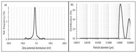
(a) DLS analysis showing the surface charge (zeta potential) of AgNPs synthesized using P. ostreatus extract, (b) Size distribution analysis of AgNPs by dynamic light scattering (DLS), illustrating the particle size distribution.
The hydrodynamic diameter of the nanoparticles was determined to be 10.527 µm (Figure 6b), which indicates some aggregation of the particles, leading to larger sizes. The polydispersity index (PDI) was used to measure the distribution of particle sizes. The PDI is calculated by dividing the standard deviation of the particle size distribution by the mean particle size (Raj et al., 2018). A PDI below 0.3 indicates a monodisperse sample with uniform particle sizes, while a PDI above 0.3 suggests polydispersity with particles of varying sizes. The calculated PDI value of 0.3309 for the sample indicates polydispersity, which may be due to factors such as incomplete homogenization or the presence of particles of different sizes during sample preparation.
3.6. Antibacterial activity
The antibacterial potential of green-synthesized PO AgNO3 nanoparticles was compared to plant extract, chemically synthesized AgNO3 nanoparticles, and a standard drug against four bacterial strains: Bacillus cereus (ATCC10876), Staphylococcus aureus (ATCC29213), Escherichia coli (ATCC25922), and Pseudomonas aeruginosa (ATCC27853) at varying concentrations (12.5, 25, 50, and 100 µg/mL). The results, presented as the mean zone of inhibition (mm) ± standard deviation, indicated a concentration-dependent antibacterial activity for all samples, with the maximum inhibition observed at 100 µg/mL (Table 1)/ Figure 7. The plant extract exhibited increasing antimicrobial activity with higher concentrations, showing the highest zone of inhibition at 100 µg/mL for all organisms. Comparatively, the plant extract showed antimicrobial activity similar to or slightly lower than silver nitrate (AgNO3) at the same concentration, while PO AgNO3 nanoparticles demonstrated significantly greater inhibition of bacterial growth. P. aeruginosa showed the highest sensitivity to PO AgNO3 nanoparticles, with the highest inhibition at 100 µg/mL (15.6±1.2 mm), followed by B. cereus and E. coli with 13.2±0.3 mm and 12.5±0.5 mm, respectively. Staphylococcus aureus showed moderate sensitivity, with increasing inhibition at higher concentrations of PO AgNO3 nanoparticles (9.5±0.5 mm). The synthesized nanoparticles consistently showed greater antimicrobial activity than both the plant extract and AgNO3 across all bacteria and concentrations.

Antibacterial activity of P. Ostreatus synthesized silver nanoparticles against (A) B. cereus (50 µg/ml), (B), E. coli (12.5 µg/ml), (C), P. aeruginosa and (D) S. aureus (100 µg/ml) [-: negative control, +: positive control, P: plant extract, N: green-synthesized silver nanoparticles)].
Previous research has suggested that the interaction of Ag+ with the cell membranes of E. coli can disrupt their function due to the cells' negatively charged surface (Gogoi et al., 2006). Exposure to AgNPs has also been found to inhibit the replication ability of S. aureus DNA after 6 hours (Kim et al., 2011; Li et al., 2011). The effectiveness of silver nanoparticles (AgNPs) in killing bacteria depends on the concentration of AgNPs and the specific type of bacteria being targeted (Patil et al., 2016). Studies have reported varying results regarding which type of bacteria is more sensitive to AgNPs. Some suggest that Gram-negative bacteria are more sensitive than Gram-positive bacteria (Kuppusamy et al., 2016; Verma et al., 2016; Choudhary et al., 2016; Chandrasekaran et al., 2016; Kim et al., 2007), while others show contradictory findings (Banerjee et al., 2014; Kayalvizhi et al., 2016; Paul et al., 2016).
The difference in sensitivity between Gram-positive and Gram-negative bacteria to AgNPs is attributed to the differences in the thickness and composition of their membrane structures (Timotina et al., 2022), as well as factors such as the final inoculated concentration of bacteria, and the size, shape, and concentration of AgNPs (Rai et al., 2012; Pal et al., 2007). Gram-negative bacteria, such as E. coli and P. aeruginosa, have an outer layer of lipopolysaccharides and a thinner peptidoglycan layer, while Gram-positive bacteria, like B. cereus and S. aureus, have a thicker peptidoglycan layer. Despite these differences, both types of bacteria carry a negative charge on their surfaces (Ramalingam et al., 2016). The antimicrobial effectiveness of the plant extract may also depend on the permeability of the microbial membranes and their metabolic processes. Green-synthesized AgNPs are thought to possess antibacterial properties due to compounds like polysaccharides, alkaloids, steroids, and terpenoids (Karaman et al., 2010). Notably, P. aeruginosa, a pathogenic bacterium known for its resistance to conventional synthetic antibiotics, exhibited susceptibility to POS AgNPs. This susceptibility may be linked to the mushroom's natural defense mechanisms, which include the production of various antiviral and antifungal compounds (Fakoya et al., 2020).
The suggested mechanisms responsible for the inhibitory effects of nanoparticles on bacteria include: the attachment of nanoparticles to the bacterial cell surface, causing damage to the cell membrane and disrupting transport systems; the penetration of nanoparticles into the cells, where they interact with different biomolecules and organelles, potentially affecting inhibitory activity; the participation of nanoparticles in generating reactive oxygen species (ROS), leading to cellular damage; and the internalization of nanoparticles into bacterial cells, which can trigger mutations and genotoxic effects (Kisimba et al., 2023).
The antibacterial mechanism of silver ions is not fully understood, but research suggests that the positive charge on Ag ions plays a key role in their antimicrobial activity. Silver nanoparticles likely interact with the negatively charged cell membrane of microorganisms through electrostatic attraction (Sondi and Salopek-Sondi, 2004), disrupting their respiratory function and membrane permeability, ultimately leading to bacterial cell death (Kvítek et al., 2008). Gram-negative bacteria are more vulnerable to silver ions due to their cell wall structure, which allows easier access to the cytoplasmic membrane (Okafor et al., 2013). The bactericidal activity of silver ions involves multiple mechanisms, including membrane disruption and interference with DNA (Ahmed et al., 2015).
3.7. Catalytic activity
In this study, we synthesized silver nanoparticles (AgNPs) using a green synthesis approach with P. ostreatus extract as both a reducing and stabilizing agent. Green synthesis methods using natural extracts have gained considerable attention due to their eco-friendliness and antioxidant properties, as supported by Flieger et al. (2021) and Sudha et al. (2017). The catalytic activity of the resulting P. ostreatus-silver nanoparticle complexes was evaluated in the reduction of 4-nitroaniline and 4-nitrophenol using sodium borohydride (NaBH₄) as the reducing agent. The catalytic reduction of both nitroaromatic compounds was highly efficient, demonstrating the excellent catalytic performance of the P. ostreatus-silver nanoparticle complexes. UV-Vis absorption spectra, shown in Figure 8, confirmed the successful reduction of 4-nitroaniline and 4-nitrophenol. In the case of 4-nitroaniline (Figure 8A), the absorbance peak decreased over time after the addition of NaBH₄, indicating the conversion of the nitro group (–NO₂) into an amine group, yielding 4-phenylenediamine. This reduction process occurred rapidly, with high conversion rates, consistent with prior reports on similar nitroaromatic compound behavior (Dexlin et al., 2023). For 4-nitrophenol (Figure 8B), the reduction process resulted in a significant shift in the absorption peak from 420 nm to 480 nm, corresponding to the reduction of 4-nitrophenol to 4-aminophenol. The P. ostreatus-silver nanoparticle complexes facilitated this transformation effectively, confirming their capability to catalyze the reduction of nitroaromatic compounds. Previous studies have also shown that biogenic metal nanoparticles, such as gold and silver, can catalyze such reductions effectively due to their nanoscale properties (Rajan et al., 2015).

(A) Time-dependent UV–Vis absorption spectra illustrating the catalytic reduction of 4-nitroaniline (4-NA). (B) Time- dependent UV–Vis absorption spectra illustrating the catalytic reduction of 4-nitrophenol (4-NP).
The observed catalytic efficiency can be attributed to the large surface area, high reactivity, and abundance of active sites on the P. ostreatus-silver nanoparticle surface, which promote the adsorption of the nitro compounds and facilitate electron transfer during the reduction process. Moreover, the use of P. ostreatus, a mushroom known for its antioxidant and bioactive peptide content (Chakrabarti et al., 2018; (Wongaem et al., 2021), may further enhance nanoparticle stability and activity. This study highlights the potential of P. ostreatus-silver nanoparticle complexes as efficient and environmentally friendly catalysts for the reduction of nitroaromatic compounds, with possible applications in environmental remediation and synthetic chemistry.
4. Conclusion
In conclusion, the green synthesis of silver nanoparticles using Pleurotus ostreatus extract offers a sustainable and environmentally friendly approach for the production of nanoparticles with diverse applications. The synthesized nanoparticles exhibited potent antibacterial activity against various bacterial strains and demonstrated excellent catalytic performance in the reduction of nitroaromatic compounds. These findings underscore the potential of P. ostreatus-silver nanoparticle complexes as versatile materials for biomedical, environmental, and catalytic applications. Further research is warranted to explore the full range of their properties and applications, paving the way for the development of innovative and sustainable nanotechnologies.
Acknowledgements
The authors thank the Durban University of Technology for its support.
Data Availability Statement
All data generated or analyzed during this study are included in this published article.
References
-
ABDELHAKIM, H.K., EL‐SAYED, E. and RASHIDI, F., 2020. Biosynthesis of zinc oxide nanoparticles with antimicrobial, anticancer, antioxidant and photocatalytic activities by the endophytic Alternaria tenuissima. Journal of Applied Microbiology, vol. 128, no. 6, pp. 1634-1646. http://doi.org/10.1111/jam.14581 PMid:31954094.
» http://doi.org/10.1111/jam.14581 -
ADIL, M., KHAN, T., AASIM, M., KHAN, A.A. and ASHRAF, M., 2019. Evaluation of the antibacterial potential of silver nanoparticles synthesized through the interaction of antibiotic and aqueous callus extract of Fagonia indica. AMB Express, vol. 9, no. 1, pp. 75. http://doi.org/10.1186/s13568-019-0797-2 PMid:31134363.
» http://doi.org/10.1186/s13568-019-0797-2 -
AHMED, M.J., MURTAZA, G., MEHMOOD, A. and BHATTI, T.M., 2015. Green synthesis of silver nanoparticles using leaves extract of Skimmia laureola: characterization and antibacterial activity. Materials Letters, vol. 153, pp. 10-13. http://doi.org/10.1016/j.matlet.2015.03.143
» http://doi.org/10.1016/j.matlet.2015.03.143 -
AKYÜZ, M., İNCI, Ş. and KIRBAĞ, S., 2023. Evaluation of antimicrobial, antioxidant, cytotoxic and DNA protective effects of oyster mushroom: Pleurotus pulmonarius (Fr.) Quel. Arabian Journal for Science and Engineering, vol. 48, no. 6, pp. 7273-7283. http://doi.org/10.1007/s13369-022-07418-9
» http://doi.org/10.1007/s13369-022-07418-9 -
AL-BAHRANI, R., RAMAN, J., LAKSHMANAN, H., HASSAN, A.A. and SABARATNAM, V., 2017. Green synthesis of silver nanoparticles using tree oyster mushroom Pleurotus ostreatus and its inhibitory activity against pathogenic bacteria. Materials Letters, vol. 186, pp. 21-25. http://doi.org/10.1016/j.matlet.2016.09.069
» http://doi.org/10.1016/j.matlet.2016.09.069 -
ANAND, K., GENGAN, R.M., PHULUKDAREE, A. and CHUTURGOON, A., 2015. Agroforestry waste Moringa oleifera petals mediated green synthesis of gold nanoparticles and their anti-cancer and catalytic activity. Journal of Industrial and Engineering Chemistry, vol. 21, pp. 1105-1111. http://doi.org/10.1016/j.jiec.2014.05.021
» http://doi.org/10.1016/j.jiec.2014.05.021 -
BANERJEE, P., SATAPATHY, M., MUKHOPAHAYAY, A. and DAS, P., 2014. Leaf extract mediated green synthesis of silver nanoparticles from widely available Indian plants: synthesis, characterization, antimicrobial property and toxicity analysis. Bioresources and Bioprocessing, vol. 1, no. 1, pp. 3. http://doi.org/10.1186/s40643-014-0003-y
» http://doi.org/10.1186/s40643-014-0003-y -
BRUNA, T., MALDONADO-BRAVO, F., JARA, P. and CARO, N., 2021. Silver nanoparticles and their antibacterial applications. International Journal of Molecular Sciences, vol. 22, no. 13, pp. 7202. http://doi.org/10.3390/ijms22137202 PMid:34281254.
» http://doi.org/10.3390/ijms22137202 -
CHAKRABARTI, S., GUHA, S. and MAJUMDER, K., 2018. Food-derived bioactive peptides in human health: challenges and opportunities. Nutrients, vol. 10, no. 11, pp. 1738. http://doi.org/10.3390/nu10111738 PMid:30424533.
» http://doi.org/10.3390/nu10111738 -
CHANDRASEKARAN, R., GNANASEKAR, S., SEETHARAMAN, P., KEPPANAN, R., AROCKIASWAMY, W. and SIVAPERUMAL, S., 2016. Formulation of Carica papaya latex-functionalized silver nanoparticles for its improved antibacterial and anticancer applications. Journal of Molecular Liquids, vol. 219, pp. 232-238. http://doi.org/10.1016/j.molliq.2016.03.038
» http://doi.org/10.1016/j.molliq.2016.03.038 -
CHOUDHARY, M.K., KATARIA, J., CAMEOTRA, S.S. and SINGH, J., 2016. A facile biomimetic preparation of highly stabilized silver nanoparticles derived from seed extract of Vigna radiata and evaluation of their antibacterial activity. Applied Nanoscience, vol. 6, no. 1, pp. 105-111. http://doi.org/10.1007/s13204-015-0418-6
» http://doi.org/10.1007/s13204-015-0418-6 -
CORIOLANO, D.L., SOUZA, J.B., BUENO, E.V., MEDEIROS, S.M.F.R.S., CAVALCANTI, I.D.L. and CAVALCANTI, I.M.F., 2021. Antibacterial and antibiofilm potential of silver nanoparticles against antibiotic-sensitive and multidrug-resistant Pseudomonas aeruginosa strains. Brazilian Journal of Microbiology, vol. 52, no. 1, pp. 267-278. http://doi.org/10.1007/s42770-020-00406-x PMid:33231865.
» http://doi.org/10.1007/s42770-020-00406-x - DEXLIN, X.D., TARIKA, J.D., KUMAR, S.M., RATHIKA, A. and BEAULA, T.J., 2023. Insights into spectral elucidations, reactivity sites, invitro assay, molecular docking and pharmacokinetic studies of non-covalently bonded 4-Aminobenzoic acid 4-Nitroaniline. Polycyclic Aromatic Compounds, vol. 44, no. 3, pp. 1722-1744. http://doi.org/10.1080/10406638.2023.2205152.
-
DRIDI, R., ESSGHAIER, B., HANNACHI, H., KHEDHER, G.B., CHAFFEI, C. and ZID, M.F., 2022. Biosynthesized silver nanoparticles using Anagallis monelli: evaluation of antioxidant activity, antibacterial and antifungal effects. Journal of Molecular Structure, vol. 1251, pp. 132076. http://doi.org/10.1016/j.molstruc.2021.132076
» http://doi.org/10.1016/j.molstruc.2021.132076 -
FAKOYA, S., ADEGBEHINGBE, K. and ADEMAKINWA, I., 2020. Bio-therapeutic, phytochemical screening and antioxidant efficacies of oyster mushroom (Pleurotus ostreatus) obtained from the wild. Open Journal of Medical Microbiology, vol. 10, no. 2, pp. 58-70. http://doi.org/10.4236/ojmm.2020.102006
» http://doi.org/10.4236/ojmm.2020.102006 -
FLIEGER, J., FRANUS, W., PANEK, R., SZYMAŃSKA-CHARGOT, M., FLIEGER, W., FLIEGER, M. and KOŁODZIEJ, P., 2021. Green synthesis of silver nanoparticles using natural extracts with proven antioxidant activity. Molecules, vol. 26, no. 16, pp. 4986. http://doi.org/10.3390/molecules26164986 PMid:34443574.
» http://doi.org/10.3390/molecules26164986 -
GOGOI, S.K., GOPINATH, P., PAUL, A., RAMESH, A., GHOSH, S.S. and CHATTOPADHYAY, A., 2006. Green fluorescent protein-expressing escherichia c oli as a model system for investigating the antimicrobial activities of silver nanoparticles. Langmuir, vol. 22, no. 22, pp. 9322-9328. http://doi.org/10.1021/la060661v PMid:17042548.
» http://doi.org/10.1021/la060661v -
HAIDER, A. and KANG, I.-K., 2015. Preparation of silver nanoparticles and their industrial and biomedical applications: a comprehensive review. Advances in Materials Science and Engineering, vol. 2015, pp. 165257. http://doi.org/10.1155/2015/165257
» http://doi.org/10.1155/2015/165257 -
HUSSAIN, I., SINGH, N., SINGH, A., SINGH, H. and SINGH, S., 2016. Green synthesis of nanoparticles and its potential application. Biotechnology Letters, vol. 38, no. 4, pp. 545-560. http://doi.org/10.1007/s10529-015-2026-7 PMid:26721237.
» http://doi.org/10.1007/s10529-015-2026-7 -
KARAMAN, Ş., TÜTEM, E., BAŞKAN, K.S. and APAK, R., 2010. Comparison of total antioxidant capacity and phenolic composition of some apple juices with combined HPLC–CUPRAC assay. Food Chemistry, vol. 120, no. 4, pp. 1201-1209. http://doi.org/10.1016/j.foodchem.2009.11.065
» http://doi.org/10.1016/j.foodchem.2009.11.065 -
KASUMBWE, K., VENUGOPALA, K.N., MOHANLALL, V. and ODHAV, B., 2014. Antimicrobial and antioxidant activities of substituted halogenated coumarins. Journal of Medicinal Plants Research, vol. 8, no. 5, pp. 274-281. http://doi.org/10.5897/JMPR2013.4419
» http://doi.org/10.5897/JMPR2013.4419 -
KAYALVIZHI, T., RAVIKUMAR, S. and VENKATACHALAM, P., 2016. Green synthesis of metallic silver nanoparticles using Curculigo orchioides rhizome extracts and evaluation of its antibacterial, larvicidal, and anticancer activity. Journal of Environmental Engineering, vol. 142, no. 9, pp. C4016002. http://doi.org/10.1061/(ASCE)EE.1943-7870.0001098
» http://doi.org/10.1061/(ASCE)EE.1943-7870.0001098 -
KESHARI, A.K., SRIVASTAVA, R., SINGH, P., YADAV, V.B. and NATH, G., 2020. Antioxidant and antibacterial activity of silver nanoparticles synthesized by Cestrum nocturnum. Journal of Ayurveda and Integrative Medicine, vol. 11, no. 1, pp. 37-44. http://doi.org/10.1016/j.jaim.2017.11.003 PMid:30120058.
» http://doi.org/10.1016/j.jaim.2017.11.003 -
KIM, J.S., KUK, E., YU, K.N., KIM, J.H., PARK, S.J., LEE, H.J., KIM, S.H., PARK, Y.K., PARK, Y.H., HWANG, C.Y., KIM, Y.K., LEE, Y.S., JEONG, D.H. and CHO, M.H., 2007. Antimicrobial effects of silver nanoparticles. Nanomedicine; Nanotechnology, Biology, and Medicine, vol. 3, no. 1, pp. 95-101. http://doi.org/10.1016/j.nano.2006.12.001 PMid:17379174.
» http://doi.org/10.1016/j.nano.2006.12.001 - KIM, S.H., LEE, H.S., RYU, D.S., CHOI, S.J. and LEE, D.S., 2011. Antibacterial activity of silver-nanoparticles against Staphylococcus aureus and Escherichia coli. Microbiology and Biotechnology Letters, vol. 39, no. 1, pp. 77-85.
-
KISIMBA, K., KRISHNAN, A., FAYA, M., BYANGA, K., KASUMBWE, K., VIJAYAKUMAR, K. and PRASAD, R., 2023. Synthesis of metallic nanoparticles based on green chemistry and their medical biochemical applications: synthesis of metallic nanoparticles. Journal of Renewable Materials, vol. 11, no. 6, pp. 2575-2591. http://doi.org/10.32604/jrm.2023.026159
» http://doi.org/10.32604/jrm.2023.026159 -
KOCAK, Y., MEYDAN, I., GUR KARAHAN, T. and SEN, F., 2023. Investigation of mycosynthesized silver nanoparticles by the mushroom Pleurotus eryngii in biomedical applications. International Journal of Environmental Science and Technology, vol. 20, no. 5, pp. 4861-4872. http://doi.org/10.1007/s13762-023-04786-z
» http://doi.org/10.1007/s13762-023-04786-z -
KUMAR, A. and DIXIT, C.K., 2017. Methods for characterization of nanoparticles. In: S. NIMESH, R. CHANDRA and N. GUPTA, eds. Advances in nanomedicine for the delivery of therapeutic nucleic acids Sawston: Woodhead Publishing, pp. 43-58. http://doi.org/10.1016/B978-0-08-100557-6.00003-1
» http://doi.org/10.1016/B978-0-08-100557-6.00003-1 -
KUPPUSAMY, P., YUSOFF, M.M., MANIAM, G.P. and GOVINDAN, N., 2016. Biosynthesis of metallic nanoparticles using plant derivatives and their new avenues in pharmacological applications: an updated report. Saudi Pharmaceutical Journal, vol. 24, no. 4, pp. 473-484. http://doi.org/10.1016/j.jsps.2014.11.013 PMid:27330378.
» http://doi.org/10.1016/j.jsps.2014.11.013 -
KVÍTEK, L., PANÁČEK, A., SOUKUPOVÁ, J., KOLÁŘ, M., VEČEŘOVÁ, R., PRUCEK, R., HOLECOVÁ, M. and ZBOŘIL, R., 2008. Effect of surfactants and polymers on stability and antibacterial activity of silver nanoparticles (NPs). The Journal of Physical Chemistry C, vol. 112, no. 15, pp. 5825-5834. http://doi.org/10.1021/jp711616v
» http://doi.org/10.1021/jp711616v -
LALEGANI, Z. and EBRAHIMI, S.S., 2020. Optimization of synthesis for shape and size controlled silver nanoparticles using response surface methodology. Colloids and Surfaces. A, Physicochemical and Engineering Aspects, vol. 595, pp. 124647. http://doi.org/10.1016/j.colsurfa.2020.124647
» http://doi.org/10.1016/j.colsurfa.2020.124647 -
LI, W.R., XIE, X.B., SHI, Q.S., DUAN, S.S., OUYANG, Y.S. and CHEN, Y.B., 2011. Antibacterial effect of silver nanoparticles on Staphylococcus aureus. Biometals, vol. 24, no. 1, pp. 135-141. http://doi.org/10.1007/s10534-010-9381-6 PMid:20938718.
» http://doi.org/10.1007/s10534-010-9381-6 -
LOPEZ-CARRIZALES, M., VELASCO, K.I., CASTILLO, C., FLORES, A., MAGAÑA, M., MARTINEZ-CASTANON, G.A. and MARTINEZ-GUTIERREZ, F., 2018. In vitro synergism of silver nanoparticles with antibiotics as an alternative treatment in multiresistant uropathogens. Antibiotics, vol. 7, no. 2, pp. 50. http://doi.org/10.3390/antibiotics7020050 PMid:29921822.
» http://doi.org/10.3390/antibiotics7020050 -
MANIMARAN, K., YANTO, D.H.Y., ARDIATI, F.C., OKTAVIANI, M., NATARAJAN, D., RAGAVENDRAN, C., KAMARAJ, C., KARUNANITHI, B. and LOGANATHAN, S., 2023. Enhanced catalytic degradation, antimicrobial and anticancer efficiency of mycosynthesized TiO2 nanoparticles using Pleurotus ostreatus mushroom extract: an eco-friendly approach. Journal of Environmental Chemical Engineering, vol. 11, no. 6, pp. 111512. http://doi.org/10.1016/j.jece.2023.111512
» http://doi.org/10.1016/j.jece.2023.111512 -
MARTÍNEZ-FLORES, H.E., CONTRERAS-CHÁVEZ, R. and GARNICA-ROMO, M.G., 2021. Effect of extraction processes on bioactive compounds from Pleurotus ostreatus and Pleurotus djamor: their applications in the synthesis of silver nanoparticles. Journal of Inorganic and Organometallic Polymers and Materials, vol. 31, no. 3, pp. 1406-1418. http://doi.org/10.1007/s10904-020-01820-2
» http://doi.org/10.1007/s10904-020-01820-2 -
MIRUNALINI, S., ARULMOZHI, V., DEEPALAKSHMI, K. and KRISHNAVENI, M., 2012. Intracellular biosynthesis and antibacterial activity of silver nanoparticles using edible mushrooms. Notulae Scientia Biologicae, vol. 4, no. 4, pp. 55-61. http://doi.org/10.15835/nsb448051
» http://doi.org/10.15835/nsb448051 - MOHANRAJ, V. and CHEN, Y., 2006. Nanoparticles: a review. Tropical Journal of Pharmaceutical Research, vol. 5, pp. 561-573. http://doi.org/10.4314/tjpr.v5i1.14634.
-
MOHANTA, Y.K., NAYAK, D., BISWAS, K., SINGDEVSACHAN, S.K., ABDALLAH, E.F., HASHEM, A., ALQARAWI, A.A., YADAV, D. and MOHANTA, T.K., 2018. Silver nanoparticles synthesized using wild mushroom show potential antimicrobial activities against food borne pathogens. Molecules, vol. 23, no. 3, pp. 655. http://doi.org/10.3390/molecules23030655 PMid:29538308.
» http://doi.org/10.3390/molecules23030655 -
MUKHERJEE, K., GUPTA, R., KUMAR, G., KUMARI, S., BISWAS, S. and PADMANABHAN, P., 2018. Synthesis of silver nanoparticles by Bacillus clausii and computational profiling of nitrate reductase enzyme involved in production. Journal of Genetic Engineering and Biotechnology, vol. 16, no. 2, pp. 527-536. http://doi.org/10.1016/j.jgeb.2018.04.004 PMid:30733770.
» http://doi.org/10.1016/j.jgeb.2018.04.004 -
OKAFOR, F., JANEN, A., KUKHTAREVA, T., EDWARDS, V. and CURLEY, M., 2013. Green synthesis of silver nanoparticles, their characterization, application and antibacterial activity. International Journal of Environmental Research and Public Health, vol. 10, no. 10, pp. 5221-5238. http://doi.org/10.3390/ijerph10105221 PMid:24157517.
» http://doi.org/10.3390/ijerph10105221 -
OVES, M., RAUF, M.A., ASLAM, M., QARI, H.A., SONBOL, H., AHMAD, I., ZAMAN, G.S. and SAEED, M., 2022. Green synthesis of silver nanoparticles by Conocarpus Lancifolius plant extract and their antimicrobial and anticancer activities. Saudi Journal of Biological Sciences, vol. 29, no. 1, pp. 460-471. http://doi.org/10.1016/j.sjbs.2021.09.007 PMid:35002442.
» http://doi.org/10.1016/j.sjbs.2021.09.007 -
OWAID, M.N., 2019. Green synthesis of silver nanoparticles by Pleurotus (oyster mushroom) and their bioactivity. Environmental Nanotechnology, Monitoring & Management, vol. 12, pp. 100256. http://doi.org/10.1016/j.enmm.2019.100256
» http://doi.org/10.1016/j.enmm.2019.100256 -
OWAID, M.N., RAMAN, J., LAKSHMANAN, H., AL-SAEEDI, S.S.S., SABARATNAM, V. and ABED, I.A., 2015. Mycosynthesis of silver nanoparticles by Pleurotus cornucopiae var. citrinopileatus and its inhibitory effects against Candida sp. Materials Letters, vol. 153, pp. 186-190. http://doi.org/10.1016/j.matlet.2015.04.023
» http://doi.org/10.1016/j.matlet.2015.04.023 -
PAL, S., TAK, Y.K. and SONG, J.M., 2007. Does the antibacterial activity of silver nanoparticles depend on the shape of the nanoparticle? A study of the gram-negative bacterium Escherichia coli. Applied and Environmental Microbiology, vol. 73, no. 6, pp. 1712-1720. http://doi.org/10.1128/AEM.02218-06 PMid:17261510.
» http://doi.org/10.1128/AEM.02218-06 -
PATIL, M.P., ROKADE, A.A., NGABIRE, D. and KIM, G.D., 2016. Green synthesis of silver nanoparticles using water extract from galls of Rhus chinensis and its antibacterial activity. Journal of Cluster Science, vol. 27, no. 5, pp. 1737-1750. http://doi.org/10.1007/s10876-016-1037-4
» http://doi.org/10.1007/s10876-016-1037-4 -
PAUL, B., BHUYAN, B., PURKAYASTHA, D.D. and DHAR, S.S., 2016. Photocatalytic and antibacterial activities of gold and silver nanoparticles synthesized using biomass of Parkia roxburghii leaf. Journal of Photochemistry and Photobiology. B, Biology, vol. 154, pp. 1-7. http://doi.org/10.1016/j.jphotobiol.2015.11.004 PMid:26590801.
» http://doi.org/10.1016/j.jphotobiol.2015.11.004 -
RAI, M.K., DESHMUKH, S.D., INGLE, A.P. and GADE, A.K., 2012. Silver nanoparticles: the powerful nanoweapon against multidrug‐resistant bacteria. Journal of Applied Microbiology, vol. 112, no. 5, pp. 841-852. http://doi.org/10.1111/j.1365-2672.2012.05253.x PMid:22324439.
» http://doi.org/10.1111/j.1365-2672.2012.05253.x -
RAJ, S., MALI, S.C. and TRIVEDI, R., 2018. Green synthesis and characterization of silver nanoparticles using Enicostemma axillare (Lam.) leaf extract. Biochemical and Biophysical Research Communications, vol. 503, no. 4, pp. 2814-2819. http://doi.org/10.1016/j.bbrc.2018.08.045 PMid:30100057.
» http://doi.org/10.1016/j.bbrc.2018.08.045 -
RAJAN, A., VILAS, V. and PHILIP, D., 2015. Studies on catalytic, antioxidant, antibacterial and anticancer activities of biogenic gold nanoparticles. Journal of Molecular Liquids, vol. 212, pp. 331-339. http://doi.org/10.1016/j.molliq.2015.09.013
» http://doi.org/10.1016/j.molliq.2015.09.013 -
RAMALINGAM, B., PARANDHAMAN, T. and DAS, S.K., 2016. Antibacterial effects of biosynthesized silver nanoparticles on surface ultrastructure and nanomechanical properties of gram-negative bacteria viz. Escherichia coli and Pseudomonas aeruginosa. ACS Applied Materials & Interfaces, vol. 8, no. 7, pp. 4963-4976. http://doi.org/10.1021/acsami.6b00161 PMid:26829373.
» http://doi.org/10.1021/acsami.6b00161 -
RAMAN, J., REDDY, G.R., LAKSHMANAN, H., SELVARAJ, V., GAJENDRAN, B., NANJIAN, R., CHINNASAMY, A. and SABARATNAM, V., 2015. Mycosynthesis and characterization of silver nanoparticles from Pleurotus djamor var. roseus and their in vitro cytotoxicity effect on PC3 cells. Process Biochemistry, vol. 50, no. 1, pp. 140-147. http://doi.org/10.1016/j.procbio.2014.11.003
» http://doi.org/10.1016/j.procbio.2014.11.003 -
RAVICHANDRAN, V., VASANTHI, S., SHALINI, S., SHAH, S.A.A., TRIPATHY, M. and PALIWAL, N., 2019. Green synthesis, characterization, antibacterial, antioxidant and catalytic activity of Parkia speciosa leaves extract mediated silver nanoparticles. Results in Physics, vol. 15, pp. 102565. http://doi.org/10.1016/j.rinp.2019.102565
» http://doi.org/10.1016/j.rinp.2019.102565 -
REDDY, N.J., VALI, D.N., RANI, M. and RANI, S.S., 2014. Evaluation of antioxidant, antibacterial and cytotoxic effects of green synthesized silver nanoparticles by Piper longum fruit. Materials Science and Engineering C, vol. 34, pp. 115-122. http://doi.org/10.1016/j.msec.2013.08.039 PMid:24268240.
» http://doi.org/10.1016/j.msec.2013.08.039 -
SANGUIÑEDO, P., FRATILA, R.M., ESTEVEZ, M.B., FUENTE, J.M., GRAZÚ, V. and ALBORÉS, S., 2018. Extracellular biosynthesis of silver nanoparticles using fungi and their antibacterial activity. Nano Biomedicine and Engineering, vol. 10, no. 2, pp. 156-164. http://doi.org/10.5101/nbe.v10i2.p165-173
» http://doi.org/10.5101/nbe.v10i2.p165-173 -
SARAVANAKUMAR, A., GANESH, M., JAYAPRAKASH, J. and JANG, H.T., 2015. Biosynthesis of silver nanoparticles using Cassia tora leaf extract and its antioxidant and antibacterial activities. Journal of Industrial and Engineering Chemistry, vol. 28, pp. 277-281. http://doi.org/10.1016/j.jiec.2015.03.003
» http://doi.org/10.1016/j.jiec.2015.03.003 -
SIVASANKARAPILLAI, V.S., KRISHNAMOORTHY, N., ELDESOKY, G.E., WABAIDUR, S.M., ISLAM, M.A., DHANUSURAMAN, R. and PONNUSAMY, V.K., 2023. One-pot green synthesis of ZnO nanoparticles using Scoparia Dulcis plant extract for antimicrobial and antioxidant activities. Applied Nanoscience, vol. 13, no. 9, pp. 6093-6103. http://doi.org/10.1007/s13204-022-02610-7 PMid:36120603.
» http://doi.org/10.1007/s13204-022-02610-7 -
SMEKALOVA, M., ARAGON, V., PANACEK, A., PRUCEK, R., ZBORIL, R. and KVITEK, L., 2016. Enhanced antibacterial effect of antibiotics in combination with silver nanoparticles against animal pathogens. Veterinary Journal, vol. 209, pp. 174-179. http://doi.org/10.1016/j.tvjl.2015.10.032 PMid:26832810.
» http://doi.org/10.1016/j.tvjl.2015.10.032 -
SONDI, I. and SALOPEK-SONDI, B., 2004. Silver nanoparticles as antimicrobial agent: a case study on E. coli as a model for Gram-negative bacteria. Journal of Colloid and Interface Science, vol. 275, no. 1, pp. 177-182. http://doi.org/10.1016/j.jcis.2004.02.012 PMid:15158396.
» http://doi.org/10.1016/j.jcis.2004.02.012 -
SUDHA, A., JEYAKANTHAN, J. and SRINIVASAN, P., 2017. Green synthesis of silver nanoparticles using Lippia nodiflora aerial extract and evaluation of their antioxidant, antibacterial and cytotoxic effects. Resource-Efficient Technologies, vol. 3, no. 4, pp. 506-515. http://doi.org/10.1016/j.reffit.2017.07.002
» http://doi.org/10.1016/j.reffit.2017.07.002 -
THEURETZBACHER, U., 2017. Antibiotic innovation for future public health needs. Clinical Microbiology and Infection, vol. 23, no. 10, pp. 713-717. http://doi.org/10.1016/j.cmi.2017.06.020 PMid:28652114.
» http://doi.org/10.1016/j.cmi.2017.06.020 -
TIMOTINA, M., AGHAJANYAN, A., SCHUBERT, R., TRCHOUNIAN, K. and GABRIELYAN, L., 2022. Biosynthesis of silver nanoparticles using extracts of Stevia rebaudiana and evaluation of antibacterial activity. World Journal of Microbiology & Biotechnology, vol. 38, no. 11, pp. 196. http://doi.org/10.1007/s11274-022-03393-3 PMid:35989355.
» http://doi.org/10.1007/s11274-022-03393-3 -
VERMA, D.K., HASAN, S.H. and BANIK, R.M., 2016. Photo-catalyzed and phyto-mediated rapid green synthesis of silver nanoparticles using herbal extract of Salvinia molesta and its antimicrobial efficacy. Journal of Photochemistry and Photobiology. B, Biology, vol. 155, pp. 51-59. http://doi.org/10.1016/j.jphotobiol.2015.12.008 PMid:26735000.
» http://doi.org/10.1016/j.jphotobiol.2015.12.008 -
WONGAEM, A., REAMTONG, O., SRIMONGKOL, P., SANGTANOO, P., SAISAVOEY, T. and KARNCHANATAT, A., 2021. Antioxidant properties of peptides obtained from the split gill mushroom (Schizophyllum commune). Journal of Food Science and Technology, vol. 58, no. 2, pp. 680-691. http://doi.org/10.1007/s13197-020-04582-4 PMid:33568862.
» http://doi.org/10.1007/s13197-020-04582-4 -
ZHANG, L., LU, Y., FENG, X., LIU, Q., LI, Y., HAO, J., WANG, Y., DONG, Y. and WANG, H.D., 2020. Hepatoprotective effects of Pleurotus ostreatus protein hydrolysates yielded by pepsin hydrolysis. Catalysts, vol. 10, no. 6, pp. 595. http://doi.org/10.3390/catal10060595
» http://doi.org/10.3390/catal10060595
Edited by
-
Editor:
Takako Matsumura Tundisi


