The impact of anaerobic exercise on nephro-hazards of androgenic-anabolic steroids in male albino rats
Abstract
Although the misuse of androgenic-anabolic steroids is increasing, not much research has been done to determine how it affects renal function. Our research sought to ascertain how Nandrolone decanoate (ND) treatment affected kidney function and histological changes in adult male albino rats. Additionally, we sought to evaluate the impact of combining ND therapy with anaerobic exercise. A total of 39 male rats were divided into three groups (n = 13 each): control, ND treated (15mg/kg/day for 8 weeks; SC), and trained-ND treated (8-12 ladder climbs, three times per week for eight weeks). Blood and kidney samples were taken after eight weeks of anaerobic exercise and therapy. The study assessed organ weight, serum urea, and creatinine levels, inflammation (TNF-α, IL-6), oxidative stress markers (MDA, SOD, TAC), kidney histology, and collagen content. Caspase3 and BAX immunohistochemistry were performed, whereas Sirt1, Beclin-1, and microtubule-associated protein light chain 3 (LC3) mRNA were analyzed using RT-PCR. The current study illustrated that exercise significantly improved kidney function, histological results, and damage score while reducing collagen deposition, inflammation, and oxidative stress. Apoptosis proteins (CASPASE3 and BAX) were dramatically reduced, whereas Sirt1 and autophagy proteins (Beclin-1 and LC3) mRNA levels were increased. In our conclusion, because of its anti-inflammatory, antioxidative, and antiapoptotic properties as well as its modification of autophagy protein mRNA, anaerobic exercise in conjunction with ND administration in rats led to a reduction in renal damage and kidney fibrosis.
Keywords:
nandrolone decanoate; exercise; kidney injury; apoptosis; autophagy
Resumo
Embora o uso indevido de esteroides anabolizantes androgênicos esteja aumentando, poucas pesquisas foram realizadas para determinar como ele afeta a função renal. Nossa pesquisa buscou verificar como o tratamento com decanoato de nandrolona (ND) afetou a função renal e as alterações histológicas em ratos albinos machos adultos. Além disso, buscamos avaliar o impacto da combinação da terapia de ND com exercício anaeróbico. Um total de 39 ratos machos foram divididos em três grupos (n = 13 cada): controle, tratado com ND (15 mg/kg/dia por 8 semanas; SC) e tratado com ND treinado (8 a 12 subidas em escada, três vezes por semana, durante oito semanas). Amostras de sangue e rim foram coletadas após oito semanas de exercício anaeróbico e terapia. O estudo avaliou o peso dos órgãos, os níveis séricos de ureia e creatinina, a inflamação (TNF-α, IL-6), os marcadores de estresse oxidativo (MDA, SOD, TAC), a histologia renal e o conteúdo de colágeno. Foram realizadas imuno-histoquímicas para caspase-3 e BAX, enquanto o mRNA de Sirt1, Beclin-1 e da cadeia leve 3 da proteína associada a microtúbulos (LC3) foi analisado por RT-PCR. O presente estudo demonstrou que o exercício físico melhorou significativamente a função renal, os resultados histológicos e o escore de dano, reduzindo a deposição de colágeno, a inflamação e o estresse oxidativo. As proteínas de apoptose (Caspase-3 e BAX) foram drasticamente reduzidas, enquanto os níveis de mRNA de Sirt1 e das proteínas de autofagia (Beclin-1 e LC3) aumentaram. Em nossa conclusão, devido às suas propriedades anti-inflamatórias, antioxidantes e antiapoptóticas, bem como à sua modificação do mRNA da proteína de autofagia, o exercício anaeróbico, em conjunto com a administração de ND em ratos, levou à redução do dano renal e da fibrose renal.
Palavras-chave:
decanoato de nandrolona; exercício; lesão renal; apoptose; autofagia
1. Introduction
The use of androgenic-anabolic steroids (AAS) by professional and non-competitive bodybuilders, as well as its adverse effects, has emerged as a major community health issue (Khalili et al., 2023). The abuse of AAS is not confined to athletes; it has also expanded to teenagers and young adults in the general population (Perry et al., 2020).
AASs are testosterone analogs with functional and structural hormonal components (Saddick 2021). Because of its potent anabolic properties but lackluster androgenic effects, nandrolone decanoate (ND) is one of the most often misused AAS (Patanè et al., 2020).
Despite being used to treat a variety of medical conditions, including osteoporosis (Weber et al., 2022), the administration of AAS in supraphysiological doses may cause a variety of cardiovascular complications, including arterial stiffness, endothelial dysfunction, which can lead to atherosclerosis (Melsom et al., 2022), and renal tissue degeneration (Abbas et al., 2024).
ND increases inflammatory markers such as TNF-α and IL-1β, as well as oxidative stress (El Sawy et al., 2021) as shown that persistently administered AAS can affect cellular redox equilibrium, resulting in oxidative stress in renal and hepatic organs (Frankenfeld et al., 2014).
Additionally, a previous study shown that giving ND to Rattus norvegicus significantly affected renal histology, underscoring the possible health risks associated with AAS use. These results contribute to the growing body of information regarding the nephrotoxic effects of steroids, emphasizing the need for caution and further research to develop safer therapeutic alternatives (Abbas et al., 2024).
Regular exercise has been shown to be crucial in reducing oxidative stress, inflammation, vascular dysfunction, and immunological response in patients with chronic renal disease, in contrast to the detrimental health effects of alcohol consumption (Kanbay et al., 2024). Furthermore, exercise may effectively promote autophagy (Rocchi and He, 2017), which may be involved in maintaining tissue integrity, limiting the inflammatory response, managing tissue damage, or activating direct signaling pathways to respond to physiological changes in the body. Exercise-induced alterations in autophagy capacity, such as increased autophagy flux and activation of critical autophagy gene transcription, have been linked to increased autophagy activity (Wu et al., 2024).
In contrast to earlier studies, Heidari et al. (2020) found that ND treatment with and without exercise lowered antioxidant defense activity in kidney tissue, however, they suggested that additional research is needed in this area.
In order to elucidate some of the likely intervening mechanisms by which exercise can mitigate the nephron-toxic effects of ND, this study sought to examine the effects of ND treatment both alone and in conjunction with exercise training on renal structure and function.
2. Material and Methods
2.1. Animals
Thirty-nine male Sprague Dawley rats (~250 gm., 8 weeks old) were imported from King Faisal University - College of Medicine - Pharmaceutical Laboratory in Saudi Arabia. To prepare for the lab environment at Pharmaceutical Laboratory. The animals were housed in conventional plastic cages (3 per cage) with sanitary conditions (~25 °C, 12-hour light/dark cycle, free access to water, and 60% humidity) following animal house practices.
The Institutional Review Board (IRB) of Imam Abdulrahman Bin Faisal University (IAU) (Dammam, Saudi Arabia) carefully reviewed and approved all experimental protocols pertaining to animal experimentation, which were conducted in the King Faisal University - College of Medicine - Pharmaceutical Laboratory in Saudi Arabia. All protocols were in compliance with the European Union Directive 2010 /63/ EU, the U.K-Animals (Scientific Procedures) Act, 1986, and the National Research Council's Guide for the Care and Use of Laboratory Animals; approval number: IRB-A-2025-10-0239.
2.2. Experimental design
The study's rats were classified into three main groups (13 rats each) as follows as Group I (Control group, n=13): In which the rats were vehicle-treated (sesame oil 1ml/kg, subcutaneously (S.C.)) for the whole 8 weeks of study, Group II (ND-treated group, n=13): In which the rats received the ND (Belugas Pharmaceuticals, Brussels, Belgium; 50mg/1ml injection) in a dose of 15mg/kg/day- 5 times a week for 8 weeks S.C. (Magalhães et al., 2022).
Animals in group I and II were left at the top of the vertical ladder for 15 min/ 3 times per week to expose animals in this group to the same environment (vertical ladder apparatus) as the exercise group (group III) and Group III (T+ND group n=13): After two weeks of familiarization, rats were trained for 8 weeks on vertical ladder apparatus with housing chamber at the top of the ladder with application of maximal load test (Miguel-dos-Santos et al., 2021), with concomitant administration of the ND (15mg/kg/day; SC); five times per week for 8 weeks (Magalhães et al., 2022).
2.3. The familiarization protocol
By “climbing a vertical ladder (110 cm high*18 cm wide, 2 cm grid, with 80° incline), with a housing chamber located at the top of the ladder that served as a shelter during the resting period (1 minute),” all rats were acclimated to the training procedure. For two weeks in a row, rats were permitted to ascend the ladder with the load apparatus but without the weight three times a week (every other day), for a total of six adaption sessions.
2.4. Anaerobic resistance exercise protocol
Using “a load apparatus” that was attached to the tail by wrapping the proximal portion with self-adhesive foam. Each animal completed a test to determine its maximum voluntary carrying capacity (MVCC), which included climbs with increasingly greater loads. The initial climb was performed with 75% of the animal's body mass, followed by an additional 30g weight until the rat was unable to climb the complete length of the ladder. The maximum load that the animal could successfully carry through the ladder was termed the MVCC for that training session. For each climb, the animals had to undertake 8-12 repeating dynamic movements to reach the housing chamber, three times a week for 8 weeks (Miguel-dos-Santos et al., 2021).
The rats carried 50%, 75%, 90%, and 100% of their previously determined MVCC for the first four ladder climbs. An extra 30g burden was progressively added during each ladder climb until the rat was unable to make nine climbs or ascend the entire ladder “(Silvestre et al., 2017).
2.5. Samples collection and analysis at the end of the experiment for all groups
2.5.1. Blood sample
Blood was collected in non-heparinized tubes and centrifugated at 3500 rpm for 15 minutes at 4 °C, then the serum was collected and frozen at -20 °C until analysis for:
Kidney function tests: Urea and Creatinine using the commercially available assay Kits (Elabscience, USA; Cat. E-BC-K183-S, and E-BC-K188-M; respectively), following the method described by Kaplan (1984) and Murray (1984) respectively.
Inflammatory mediators (TNF-a, IL-6): they were analyzed using Rat ELISA kits (Sigma-Aldrich, USA). as described by Fernando et al. (1998) and Li et al. (2013).
Oxidative stress parameters: lipid peroxidation was assessed by measuring Malondialdehyde (MDA) according to Chi et al. (2002), however, antioxidant activities were determined by measuring superoxide dismutase (SOD) content and total antioxidant capacity (TAC) activity that were determined according to the method described by Erel (2004) using the corresponding rat ELISA Kit (Abcam Limited; USA).
2.5.2. Macro-morphological examination
Rats were secured in a ventro-dorsal position, and the kidneys were exposed by opening the abdomens using sterile scissors after rubbing the skin with Betadine, a skin disinfectant. They were separated from the surrounding fat and eliminated. The right kidneys were evaluated for any macro-morphological abnormalities and photographed, while the left kidneys were frozen at -80 °C and used for PCR analysis.
2.5.3. Tissue preparation for histopathological examination
Following morphological evaluation, right kidneys were extracted and promptly fixed in “10% buffered neutral formalin solution” for 24 hours, then dehydrated in gradually increasing ethanol concentrations (70, 80, 95, 95, and 100%), cleaned in xylene, and lastly embedded in paraffin. Suvarna et al. (2018) described the use of a microtome (Leica RM 2155, England) to slice 5µm thick paraffin slices. The sections were produced and normally stained with hematoxylin and eosin (H and E), as well as with the Masson Trichrome special stain, to detect collagen deposition. All section photographs were taken with a “Swift microscope associated with Swift digital camera”. The histopathological score was evaluated using a semiquantitative method as follows: “0= no, 1= mild, 2= moderate, and 3= severe alterations” (Table 1; Gibson-Corley et al., 2013). Furthermore, Image-J analysis software was used for morphometric analysis of glomerular diameter, Bowman space diameter, and renal tubular diameter. This technique took place in Department of Biology, College of Science, Imam Abdulrahman Bin Faisal University, Saudi Arabia.
2.5.4. Immunohistochemistry (IHC) analysis
Paraffin sections of kidney tissues from various groups were stained for IHC examination according to the manufacturer protocol and (Hsu, Raine, and Fanger, 1981) instructions, using Anti-Caspase-3 and Anti-Bax antibodies (Cambridge, UK, Abcam). Tissue sections from each experimental group were dewaxed and hydrated. Staining was subsequently done with “the DAB chromogenic agent” (Expose mouse and rabbit specific HRP/DAB detection kit, Abcam; ready-to-use; Cat. #: ab80436). The counterstaining with hematoxylin was subsequently performed. “A Swift microscope associated with Swift digital camera” was used to photograph all IHC-stained tissue sections. Quantitative analysis was done by measuring the percentage of positively stained cells relative to the total cell population within selected fields. For each experimental group, five randomly selected, non-overlapping fields at ×400 magnification were analyzed. Both positive and negative regions were included to avoid selection bias. The mean percentage of positive staining across the five fields was calculated for each sample and used for statistical analysis using the Image J program. This technique took place in Department of Biology, College of Science, Imam Abdulrahman Bin Faisal University, Saudi Arabia.
2.5.5. Quantitative qRT-PCR analysis
The expression of SIRT-1, Beclin-1, and microtubule-associated protein light chain 3 (LC3) mRNA in the kidneys of various groups of rats was examined in left renal tissues that were homogenized to extract total RNA using the RNeasy Mini Kit from Qiagen, following the manufacturer's protocol. Mx3005P (Stratagene, CA, USA) performed the RT-qPCR. “Thermal cycling was performed with a denaturation step at 94 °C for 5 min, followed by 40 cycles of 94 °C for 30 s, annealing at specific temperatures for 30 s, and elongation at 72 °C for 30 s,” with a final extension step at 72 °C for 10 minutes. Data was standardized to GADPH transcript levels and relative gene expression was calculated using the 2-ΔΔCt technique (Livak and Schmittgen, 2001). The sequences of the used primers are shown in Table 2. This technique took place in the King Faisal University - College of Medicine - Pharmaceutical Laboratory in Saudi Arabia.
2.5.6. Statistical analysis
The data gathered in the current study were calculated using the “Graphpad prism program” version 5. The “Shapiro-Wilk test” was performed to assess the normality of the data distribution. Normally distributed data were described as mean ± SD. The ANOVA test was performed to determine the significance differences between all research groups, followed by the “Tukey post Hoc test” to identify significant pairs. The Kruskal-Wallis Test was employed to investigate variations in the Semi-quantitative technique of histopathological scoring since the data was heterogeneously distributed, and Dunn's Multiple Comparison test was utilized to identify significant pairs.
3. Results
According to the load lifted by the trained animals (T+ND) group at the end of weeks 1, 4, and 8. After 4 weeks of training, loads increased compared to week 1 (p < 0.001). After week 8, animals in the T+ND group improved their physical ability by lifting larger loads (p<0.001) compared to those in week 4 (Figure 1).

Load carried by trained rats at the end of weeks 1, 4, and 8. Data are expressed as means ± SD. ***P <0.001 vs load in week 1, ###P<0.001 vs load in week 4, maximum voluntary carrying capacity (MVCC).
3.1. Serum biochemical assay
The study found that ND treatment significantly increased serum urea and creatinine levels compared to the control group (p<0.001), while the T+ND group had significantly lower levels than the ND group (p<0.001). However, compared to the control group, both previously indicated values were significantly higher (p<0.001 and p<0.01, respectively) (Figures 2A and 2B).

Vertical scattered plot histogram showing mean ± SD of serum urea (A), and creatinine (B) in all groups. asig vs control, bsig vs ND group, ***p<0.001, **p<0.01 (n=13).
Regarding inflammatory markers, the ND group had considerably higher levels of TNF-α and IL-6 (p<0.001) than the control group, however, exercise dramatically reduced these levels (p<0.001). The results were significantly greater than the control group (p<0.001) (Figures 3A and 3B).

Vertical scattered plot histogram showing mean ± SD of serum tumor necrosis-α (TNF-α) (A), interleukin-6 (IL-6) (B), Malonaldehyde (MDA) (C), superoxide dismutase (SOD) (D) and total antioxidant capacity (TAC) (E) in all groups. asig vs control, bsig vs ND group, ***p<0.001, **p<0.01 (n=13), *: p<0.05.
A significant decrease in SOD and TAC (p<0.001) was also linked to ND therapy's spectacular decrease in serum MDA (p<0.001) when compared to controls. In comparison to rats that were not trained, exercise dramatically decreased MDA (p<0.001) and raised SOD (p<0.05) and TAC (p<0.01). The training group had significantly higher MDA levels than the control group (p<0.001), along with lower SOD (p<0.001) and TAC (p<0.001) (Figures 3C-E).
3.2. Renal tissue morphological and histopathological examination findings
The morphological results showed normal structure and color of kidney in control group as smooth, bright reddish-brown but couldn't demonstrate any abnormal morphological changes other groups (Figure 4A), however, there was a significant increase in kidney weight in the ND group when compared to the control group (p<0.001). Interestingly, the weight was significantly lesser in the T+ND group (p<0.001) than that of the ND group, but still higher than that of the control group (p<0.01) (Figure 4B).

Photomicrograph of rats' kidneys showing: Macro-morphology of kidneys in all groups (A), comparison of mean ± SD of renal weight in all groups (B), histopathological analysis of H&E-stained sections (C-E): Normal histological structures of renal corpuscles (arrows), renal tubular epithelium (arrowheads) and interstitial tissues in control group (C). Necrosis in a large number of renal tubular epithelium (red arrowhead), hyaline casts in some renal tubular lumina (thick arrows), congested renal vasculature (curved arrow), and aneurysmal capillaries at some renal corpuscles in the ND group (D). Mild degenerative changes at some tubular epithelium, shrinkage glomerular tufts (red arrow), and mild dilated renal blood vessel (curved arrow) in T+ND group (E) (Scale bar 100 μm, 20 μm).
The histological analysis of renal tissues showed normal histological structures of renal corpuscles, renal tubular epithelium and interstitial tissues in control group (Figure 4C). While in ND-treated rats, renal tissue showed degenerative and necrotic changes in renal tubular epithelium, hyaline casts in some renal tubular lumina, congested renal vasculatures, and aneurysmal capillaries at some renal corpuscles, with shrank glomeruli (Figure 4D) that were statistically analyzed and showed a significant increase in the injury scoring items compared to the control group (Table 3). In addition, morphometric analysis of stained sections showed a significant decrease in glomerular diameter, with increased Bowman's space diameter and renal tubular diameter when compared to the control group (p<0.001) (Table 4). However, trained animals in group 3 showed improvement in the histological changes in their renal tubules and glomerular corpuscles, with some minor dilated renal blood vessels, some tubular epithelial degenerative changes, and some glomerular tufts shrinking (Figure 4E), that was supported by the statistics results of injury score shown in Table 3. Furthermore, there was a significant increase in glomerular diameter (p<0.001), but a decrease in Bowman's space diameter (p<0.001) and renal tubular diameter (p<0.01) when compared to ND untrained rats (Table 4).
Scoring system for evaluation of commonly observed lesions in kidneys among different groups.
Moreover, Masson’s trichrome-stained sections exhibited few amounts of blue color staining for collagen fibers within interstitial tissues and glomerular structures in control group (Figure 5A). However, ND group revealed intense areas of positive staining for collagen fibers within interstitial tissues and glomerular structures (Figure 5B) with significant increase in area % of collagen deposition in this group when relative to control group (p<0.001) (Figure 5D). While, a moderate amount of collagen deposition was demonstrated in trained rats of group 3 (Figure 5C), with a significantly lower area % of collagen deposition than that of the ND group (p<0.001) (Figure 5D). The area % of collagen deposition in the T+ND group was significantly higher than that of controls (p<0.05) (Figure 5D).

Photomicrographs of kidney sections stained with Masson’s Trichrome (fig. A-C) showing: a few amounts of blue color staining for collagen fibers within interstitial tissues & glomerular structures in control group (A). Marked collagen fibers deposition within interstitial tissues & glomerular structures in ND group (B). Moderate amount of collagen staining T+ND group (C), (red arrows indicate collagen threads). Comparison of mean ± SE of area % of collagen deposition in all groups (D). asig vs control, bsig vs ND group, ***p<0.001, *p<0.05, data presented as mean ± SD.
3.3. Immunohistochemical findings
The immunohistochemical expression of caspase-3 and BAX for kidney tissues showed negative immunoreaction in the control group (Figures 6A and 6D; respectively). However, moderate cytoplasmic immunoreaction of caspase-3 and BAX within renal tubular epithelium were demonstrated in the ND group (Figures 6B and 6E; respectively). In contrast, mild immunoreactivity for both caspase 3 and BAX within a few numbers of the renal epithelium was detected in the T+ND group (Figures 6C and 6F; respectively). Comparing the ND group to the control group, morphometric analysis revealed a substantial increase in the area percentage expression of BAX and Caspase 3 (p<0.001). Caspase 3 and BAX area percentage expression was substantially lower in the T+ND group than in the ND group (p<0.001). But compared to the control group, their expression was noticeably higher (p<0.001) (Figure 6G and 6H).

Photomicrographs of immune-stained sections of kidney for Caspase 3 (A-C) and BAX (D-F) showing: Negative immunoreaction in the control group (A). Moderated cytoplasmic immunoreaction at renal tubular epithelium in ND group (B). Mild immunoreactivity within a few numbers of renal epithelium cytoplasm in the T+ND group (C). Non-detectable expression of BAX protein in the control group (D). Strong cytoplasmic BAX immune-expressions within numerous renal tubular epithelia in ND group (E). Moderate expression of immuno-labeled cells containing BAX at T+ND group (F). IHC counterstaining with Mayer's hematoxylin. Arrowheads refer to positive stained cells. (The positive expressed cells revealed golden brown color), (Scale bar 20 μm). Area % of immune-expression of Caspase 3 (G), BAX (H). **: p<0.01 --***: p<0.001.
3.4. Quantitative real time-PCR analysis results
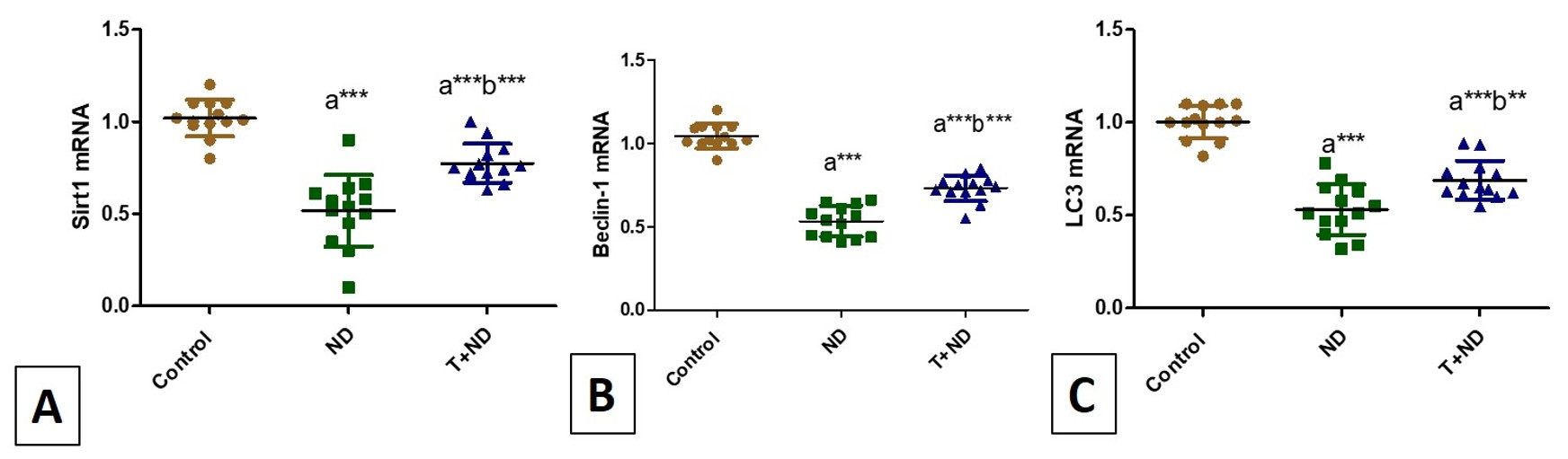
According to PCR analysis, the current study found that, in comparison to the controls, ND therapy significantly decreased the mRNA expression of Sirt1, Beclin-1, and LC3 (p<0.001). Anaerobic exercise for eight weeks, however, reduced the effects of ND on these parameters, as seen by the T+DN group's significantly higher mRNA expression compared to the ND group (p<0.001, p<0.001, and p<0.01, respectively). However, the T+ND group's mRNA expressions of Sirt1, Beclin-1, and LC3 were considerably lower than those of the control group (p<0.001) (Figures 7A-C).

Vertical scattered plot histogram showing mean ± SD of mRNA expressions of Sirt1 (A), Beclin-1 (B), and LC3 (C). asig vs control, bsig vs ND group, ***p<0.001, **p<0.01.
4. Discussion
The tremendous race for beauty standards, combined with the emergence of muscle dysmorphia, has increased AAS abuse among amateur bodybuilders. ND is one of the most regularly used AAS (Parente Filho et al., 2020), however, it is connected with several negative consequences and has become a public health concern. However, the protective effects of exercise on kidney structure and function are complex and not well known (Li and Lindholm, 2024), hence the purpose of this study is to investigate the effects of resistant anaerobic exercise combined with histopathology in ND-treated mice.
Consistent with previous studies, administration of ND at supra-pharmacological doses resulted in increased renal weight, indicating hypertrophic effects of ND on the kidney. This was accompanied by impaired renal function and altered histopathology, as evidenced by a significant elevation in serum urea and creatinine levels. Histological examination revealed pronounced degenerative and necrotic changes in many renal tubular epithelial cells, vascular congestion, shrunken glomeruli, and a marked reduction in glomerular diameter. These alterations were further associated with a significant increase in both Bowman’s space and renal tubule diameters in the ND-treated group (Abbas et al., 2024).
Interestingly, combining exercise with ND treatment in group T+ND significantly decreased the development of renal function impairment and histopathological abnormalities in comparison to untrained-ND-treated rats.
AAS abuse had developed proteinuria and renal failure (Herlitz et al., 2010). AAS could induce direct glomerular toxicity and glomerular hyperfiltration. Additionally, androgen receptor stimulation in podocytes induced renal damage, glomerulosclerosis, and apoptosis in female estrogen receptor knockout mice (Doublier et al., 2011).
Additionally, animals treated with AAS showed a disturbance in redox equilibrium in their liver, heart, and kidneys, which is in line with our findings because the current study demonstrates a significant rise in MDA and a fall in SOD and TAC in the NA-treated group relative to the control group (Frankenfeld et al., 2014).
In another experimental study on male rats, a protective effect of low-dose testosterone against ischemia-reperfusion injury (Patil et al., 2016). However, as in earlier studies, a high dose increased TNF-α and intrarenal T cells and failed to provoke a nephron-protective effect. Therefore, they concluded that TNF-α could be a potential key inflammatory cytokine involved in AAS-associated renal damage, which was in line with the results of the current study that showed a significant increase in TNF-α and IL-6 in this group (Magalhães et al., 2022; Shirpoor and Naderi, 2024).
Similarly, the reno-protective qualities of resistance exercise and anaerobic training were demonstrated by the considerable reduction of proteinuria and inflammation (cytokine-induced neutrophil chemoattractant 1, IL-1B, and IL-10) in diabetic rats. However, since they calculated their levels in rat kidney homogenate and we evaluated them in serum, and because their model had a diabetic kidney, they failed to identify a substantial decrease in TNF-α and IL-6 (Miguel-Dos-Santos et al., 2021).
In addition, anaerobic training provided renal protection that may be related to the increased vasodilator response, evidenced by the enhancement of renal angiotensin peptides 1-7 that seemed to counteract the Ang II-induced renal inflammatory effects in diabetic rats (Miguel-dos-Santos et al., 2021).
Interestingly, the protective effect of physical exercise on its anti-oxidative stress capabilities as well as strength training and treadmill training, respectively, were able to counteract renal oxidative damage produced by hypertension (Gu et al., 2015; Miguel-dos-Santos et al., 2021), Similar effects have been also shown in other strength training models in chronic kidney diseases (Souza et al., 2012).
The protective effect conferred by physical training appears crucial in preventing renal fibrosis. In the present study, this was demonstrated by the reduced collagen deposition observed in the renal tissues of the T+ND group compared with the ND-treated group. Conversely, collagen accumulation—and thus fibrosis—was more prominent in areas that had undergone oxidative damage (Miguel-Dos-Santos et al., 2021) that was mediated by ND treatment in high doses (Lima et al., 2020).
Apoptosis is another contributory factor in kidney fibrosis development. It can be induced by various inflammatory cytokines. Androgens play a crucial role in apoptosis of renal tubular cells through triggering a caspase-dependent apoptotic pathway (Davani-Davari et al., 2019; Verzola et al., 2009), as shown in the present study by increasing Caspase3 and BAX area% of IHC expression in ND-treated animals. In addition, ND-induced overproduction of the pro-inflammatory cytokines TNF-α, and IL-6 observed in the current study can enhance the activity of androgen receptors (Culig 2004) that can explain enhancing the pro-apoptotic, as well as pro-fibrotic signaling and consequently, the increased level of tubule-interstitial fibrosis and kidney dysfunction noticed in this group, as shown in Masson trichrome stained sections with increased collagen deposition.
Remarkably, earlier research on chronic kidney disease demonstrated that elevated cytochrome c release could activate caspase-9, which in turn activated caspase-3. At the same time, upregulation of Bax further triggered the release of mitochondrial cytochrome c from mitochondria to the cytosol, creating a positive feedback loop (Fussenegger, Bailey, and Varner, 2000), this effect was effectively suppressed by exercise training, consistent with the fact that exercise was capable to ameliorate renal cells apoptosis by modulation of caspase-3, reduction of ROS and prevention of subsequent loss of intra-mitochondrial cytochrome c. in chronic (Chen et al., 2013) and acute kidney disease (Costanti-Nascimento et al., 2023).
Remarkably, SIRT1, a NAD+-dependent deacetylase, exhibits anti-inflammatory effects through NF-κB deacetylation (Liu et al., 2019). Our results showed that exercise significantly upregulated Sirt1 mRNA expression in group III (T+ND) compared with ND-treated rats. This increase may be linked to the improvement in oxidative stress and inflammation observed in this group. These findings are in line with previous reports demonstrating the regulatory effects of exercise on SIRT1 and other sirtuins across various tissues, including skeletal muscle, brain, adipose tissue, and heart, thereby contributing to protection against metabolic diseases and aging-related disorders (Pucci et al., 2013; Suwa and Sakuma, 2013). Also, previous data supported our finding, as they reported that, resveratrol, a chemical SIRT1 activator has been shown to improve renal function in diabetic animal models (Hui et al., 2017; Liu et al., 2019).
Interestingly, our investigation showed that, compared to rats treated with ND, the T+ND group exhibited a considerable increase in the mRNA expression of autophagy proteins (Beclin and LC3) in renal tissues. By breaking down and recycling damaged proteins, macromolecules, and organelles, a low level of autophagy plays a critical function in cellular metabolism and organelle stability (Kaushal et al., 2020). The impact of autophagy on the progression of kidney disease is still entirely unstated (Ai et al., 2023). Autophagy-related proteins are involved in the implementation of autophagy. Beclin-1 participates in the early stages of autophagy. It promotes the nucleation of the autophagic vesicle and recruits proteins from the cytosol (Chifenti et al., 2013).
The gathering of protein aggregates and inclusion bodies has been observed in autophagy-deficient renal tubular cells. In addition, proximal tubule-specific autophagy-KO mice accumulated deformed or damaged mitochondria, endoplasmic stress, exhibited increased proximal tubular cell apoptosis, renal function loss, nuclear DNA damage, and fibrosis (Yamamoto et al., 2016), which ended by the development of mild albuminuria, podocyte loss, late-onset glomerulosclerosis (Hartleben et al., 2010).
Autophagy exerts diverse effects depending on the context, cell type, and pathological condition, and it may act as either a profibrotic or antifibrotic process. In renal tubular cells, activation of autophagy can provide protection against stresses such as proteinuria-induced apoptosis and ischemia-induced acute kidney injury. Conversely, impaired or lost autophagy in renal cells markedly increases the risk of developing various renal disorders (Ruby et al., 2023).
Studies have indicated that SIRT1-mediated autophagy is critical for cell differentiation, survival, and stress resistance (Suzuki and Bartlett, 2014). SIRT1 regulates autophagy by acetylating autophagy-related proteins (ATGs), fork head box proteins (FOXOs), and microtubule-associated protein light chain 3 (LC3) in a rat model of cardiac ischemia/ reperfusion injury (Ding et al., 2024) and Beclin1 in cancer bladder (Sun et al., 2023).
In agreement with our results, the beneficial effects of exercise on kidney changes in aged animal models to the maintenance of podocytes, via stimulating autophagy, and alleviation of oxidative damage and inflammation (Salem and Faried, 2021) . Podocytes autophagy is involved in renal protection and may be a therapeutic target (Zhou et al., 2019). Moreover, the exercise could induce autophagy, modulate cellular signaling, and promote metabolic adaptation (Brandt et al., 2018). Additionally, the high Beclin-1 function is protective against ischemia/reperfusion-induced acute kidney injury through increasing autophagy activity (Li et al., 2020).
However, Beclin-1-induced autophagy is an excessive form of autophagy in animal models of myocardial ischemia/reperfusion, which contradicts the findings of the current study. They discovered that while Beclin1 activation is required for the start of autophagy in early cardiac ischemia, persistent Beclin-1 elevation during the reperfusion phase caused autophagy to become overactivated, removing vital proteins or organelles from cells, which led to cell dysfunction and the induction of autophagic death (Valentim et al., 2006).
5. Conclusion
The present study demonstrated that anaerobic exercise training for 8 weeks improved structural and functional renal changes induced by ND in adult male rats. Thus, anaerobic exercise could be recommended for protection against kidney disease. Besides, relevant Sirt1/autophagy agonists or blockers can also be further explored to investigate in-depth the different underlying mechanisms. Targeting SIRT1, autophagy, and their interactions is likely to be a new strategy for the treatment of ND-abuse-related complications in the future.
Acknowledgements
The authors express their appreciation to the Imam Abdulrahman Bin Faisal University and King Faisal University, Saudi Arabia.
Data Availability Statement
Data will be made available on request.
References
-
ABBAS, J., AKRAM, A., NISA, M., LIAQAT, W.M.T., TARIQ, M.A., BAKAR, M. and SATTAR, M., 2024. Unveiling the impact of nandrolone decanoate on kidney and heart of Rattus norvegicus. Journal of Health and Rehabilitation Research, vol. 4, no. 1, pp. 1079-1083. http://doi.org/10.61919/jhrr.v4i1.533
» http://doi.org/10.61919/jhrr.v4i1.533 -
AI, S., LI, Y., ZHENG, H., WANG, Z., LIU, W., TAO, J., LI, Y. and WANG, Y., 2023. Global research trends and hot spots on autophagy and kidney diseases: a bibliometric analysis from 2000 to 2022. Frontiers in Pharmacology, vol. 14, pp. 1275792. http://doi.org/10.3389/fphar.2023.1275792 PMid:38099142.
» http://doi.org/10.3389/fphar.2023.1275792 -
BRANDT, N., GUNNARSSON, T.P., BANGSBO, J. and PILEGAARD, H., 2018. Exercise and exercise training-induced increase in autophagy markers in human skeletal muscle. Physiological Reports, vol. 6, no. 7, e13651. http://doi.org/10.14814/phy2.13651 PMid:29626392.
» http://doi.org/10.14814/phy2.13651 -
CHEN, K.C., PENG, C.C., HSIEH, C.L. and PENG, R.Y., 2013. Exercise ameliorates renal cell apoptosis in chronic kidney disease by intervening in the intrinsic and the extrinsic apoptotic pathways in a rat model. Evidence-Based Complementary and Alternative Medicine, vol. 2013, pp. 368450. http://doi.org/10.1155/2013/368450 PMid:24106522.
» http://doi.org/10.1155/2013/368450 -
CHI, C.H., SHIESH, S.C. and LIN, X.Z., 2002. Total antioxidant capacity and malondialdehyde in acute abdominal pain. The American Journal of Emergency Medicine, vol. 20, no. 2, pp. 79-82. http://doi.org/10.1053/ajem.2002.30102 PMid:11880867.
» http://doi.org/10.1053/ajem.2002.30102 -
CHIFENTI, B., LOCCI, M.T., LAZZERI, G., GUAGNOZZI, M., DINUCCI, D., CHIELLINI, F., FILICE, M.E., SALERNO, M.G. and BATTINI, L., 2013. Autophagy-related protein LC3 and Beclin-1 in the first trimester of pregnancy. Clinical and Experimental Reproductive Medicine, vol. 40, no. 1, pp. 33-37. http://doi.org/10.5653/cerm.2013.40.1.33 PMid:23614114.
» http://doi.org/10.5653/cerm.2013.40.1.33 -
COSTANTI-NASCIMENTO, A.C., BRELAZ-ABREU, L., BRAGANÇA-JARDIM, E., PEREIRA, W.O., CAMARA, N.O.S. and AMANO, M.T., 2023. Physical exercise as a friend not a foe in acute kidney diseases through immune system modulation. Frontiers in Immunology, vol. 14, pp. 1212163. http://doi.org/10.3389/fimmu.2023.1212163 PMid:37928533.
» http://doi.org/10.3389/fimmu.2023.1212163 -
CULIG, Z., 2004. Androgen receptor cross-talk with cell signalling pathways. Growth Factors, vol. 22, no. 3, pp. 179-184. http://doi.org/10.1080/08977190412331279908 PMid:15518241.
» http://doi.org/10.1080/08977190412331279908 -
DAVANI-DAVARI, D., KARIMZADEH, I. and KHALILI, H., 2019. The potential effects of anabolic-androgenic steroids and growth hormone as commonly used sport supplements on the kidney: a systematic review. BMC Nephrology, vol. 20, no. 1, pp. 198. http://doi.org/10.1186/s12882-019-1384-0 PMid:31151420.
» http://doi.org/10.1186/s12882-019-1384-0 -
DING, X., ZHU, C., WANG, W., LI, M., MA, C. and GAO, B., 2024. SIRT1 is a regulator of autophagy: implications for the progression and treatment of myocardial ischemia-reperfusion. Pharmacological Research, vol. 199, pp. 106957. http://doi.org/10.1016/j.phrs.2023.106957 PMid:37820856.
» http://doi.org/10.1016/j.phrs.2023.106957 -
DOUBLIER, S., LUPIA, E., CATANUTO, P., PERIERA-SIMON, S., XIA, X., KORACH, K., BERHO, M., ELLIOT, S.J. and KARL, M., 2011. Testosterone and 17β-estradiol have opposite effects on podocyte apoptosis that precedes glomerulosclerosis in female estrogen receptor knockout mice. Kidney International, vol. 79, no. 4, pp. 404-413. http://doi.org/10.1038/ki.2010.398 PMid:20962747.
» http://doi.org/10.1038/ki.2010.398 -
EL SAWY, M., TASH, R. and DESOUKY, A., 2021. Evaluation of nandrolone decanoate effects on sinoatrial node in experimental rat model (histological and immunohistochemical study). Journal of Medical Histology, vol. 5, no. 2, pp. 93-104. http://doi.org/10.21608/jmh.2022.170705.1105
» http://doi.org/10.21608/jmh.2022.170705.1105 -
EREL, O., 2004. A novel automated direct measurement method for total antioxidant capacity using a new generation, more stable ABTS radical cation. Clinical Biochemistry, vol. 37, no. 4, pp. 277-285. http://doi.org/10.1016/j.clinbiochem.2003.11.015 PMid:15003729.
» http://doi.org/10.1016/j.clinbiochem.2003.11.015 -
FERNANDO, B., MARLEY, R., HOLT, S., ANAND, R., HARRY, D., SANDERSON, P., SMITH, R., HAMILTON, G. and MOORE, K., 1998. N-acetylcysteine prevents development of the hyperdynamic circulation in the portal hypertensive rat. Hepatology, vol. 28, no. 3, pp. 689-694. http://doi.org/10.1002/hep.510280314 PMid:9731560.
» http://doi.org/10.1002/hep.510280314 -
FRANKENFELD, S.P., OLIVEIRA, L.P., ORTENZI, V.H., REGO-MONTEIRO, I.C.C., CHAVES, E.A., FERREIRA, A.C., LEITÃO, A.C., CARVALHO, D.P. and FORTUNATO, R.S., 2014. The anabolic androgenic steroid nandrolone decanoate disrupts redox homeostasis in liver, heart and kidney of male Wistar rats. PLoS One, vol. 9, no. 9, e102699. http://doi.org/10.1371/journal.pone.0102699 PMid:25225984.
» http://doi.org/10.1371/journal.pone.0102699 -
FUSSENEGGER, M., BAILEY, J.E. and VARNER, J., 2000. A mathematical model of caspase function in apoptosis. Nature Biotechnology, vol. 18, no. 7, pp. 768-774. http://doi.org/10.1038/77589 PMid:10888847.
» http://doi.org/10.1038/77589 -
GIBSON-CORLEY, K.N., OLIVIER, A.K. and MEYERHOLZ, D.K., 2013. Principles for valid histopathologic scoring in research. Veterinary Pathology, vol. 50, no. 6, pp. 1007-1015. http://doi.org/10.1177/0300985813485099 PMid:23558974.
» http://doi.org/10.1177/0300985813485099 -
GU, Q., ZHAO, L., MA, Y.P. and LIU, J.D., 2015. Contribution of mitochondrial function to exercise-induced attenuation of renal dysfunction in spontaneously hypertensive rats. Molecular and Cellular Biochemistry, vol. 406, no. 1-2, pp. 217-225. http://doi.org/10.1007/s11010-015-2439-6 PMid:25963667.
» http://doi.org/10.1007/s11010-015-2439-6 -
HARTLEBEN, B., GÖDEL, M., MEYER-SCHWESINGER, C., LIU, S., ULRICH, T., KÖBLER, S., WIECH, T., GRAHAMMER, F., ARNOLD, S.J., LINDENMEYER, M.T., COHEN, C.D., PAVENSTÄDT, H., KERJASCHKI, D., MIZUSHIMA, N., SHAW, A.S., WALZ, G. and HUBER, T.B., 2010. Autophagy influences glomerular disease susceptibility and maintains podocyte homeostasis in aging mice. The Journal of Clinical Investigation, vol. 120, no. 4, pp. 1084-1096. http://doi.org/10.1172/JCI39492 PMid:20200449.
» http://doi.org/10.1172/JCI39492 -
HEIDARI, E., HOSSEINI, S. and AZARBAYJANI, M., 2020. Effect of nandrolone treatment with and without resistance training on superoxide dismutase concentration and pathology of kidney tissue in rats. Journal of Clinical Research in Paramedical Sciences, vol. 9, no. 2, e102497. http://doi.org/10.5812/jcrps.102497
» http://doi.org/10.5812/jcrps.102497 -
HERLITZ, L.C., MARKOWITZ, G.S., FARRIS, A.B., SCHWIMMER, J.A., STOKES, M.B., KUNIS, C., COLVIN, R.B. and D’AGATI, V.D., 2010. Development of focal segmental glomerulosclerosis after anabolic steroid abuse. Journal of the American Society of Nephrology, vol. 21, no. 1, pp. 163-172. http://doi.org/10.1681/ASN.2009040450 PMid:19917783.
» http://doi.org/10.1681/ASN.2009040450 -
HSU, S.M., RAINE, L. and FANGER, H., 1981. Use of Avidin-Biotin-Peroxidase Complex (ABC) in Immunoperoxidase Techniques: a comparison between ABC and unlabeled antibody (PAP) procedures. The Journal of Histochemistry and Cytochemistry, vol. 29, no. 4, pp. 577-580. http://doi.org/10.1177/29.4.6166661 PMid:6166661.
» http://doi.org/10.1177/29.4.6166661 -
HUI, Y., LU, M., HAN, Y., ZHOU, H., LIU, W., LI, L. and JIN, R., 2017. Resveratrol improves mitochondrial function in the remnant kidney from 5/6 nephrectomized rats. Acta Histochemica, vol. 119, no. 4, pp. 392-399. http://doi.org/10.1016/j.acthis.2017.04.002 PMid:28434671.
» http://doi.org/10.1016/j.acthis.2017.04.002 -
KANBAY, M., COPUR, S., YILDIZ, A.B., TANRIOVER, C., MALLAMACI, F. and ZOCCALI, C., 2024. Physical exercise in kidney disease: a commonly undervalued treatment modality. European Journal of Clinical Investigation, vol. 54, no. 2, e14105. http://doi.org/10.1111/eci.14105 PMid:37814427.
» http://doi.org/10.1111/eci.14105 - KAPLAN, A., 1984. Urea St. Louis: Mosby.
-
KAUSHAL, G.P., CHANDRASHEKAR, K., JUNCOS, L.A. and SHAH, S.V., 2020. Autophagy function and regulation in kidney disease. Biomolecules, vol. 10, no. 1, pp. 100. http://doi.org/10.3390/biom10010100 PMid:31936109.
» http://doi.org/10.3390/biom10010100 -
KHALILI, S., KHOSHRAVESH, S., BARATI, M., MAHJOUB, H. and FARADMAL, J., 2023. Androgenic-anabolic steroids use among bodybuilders in western Iran: application of ridge logistic regression model. BMC Sports Science, Medicine and Rehabilitation, vol. 15, no. 1, pp. 7. http://doi.org/10.1186/s13102-023-00616-4 PMid:36631858.
» http://doi.org/10.1186/s13102-023-00616-4 -
LI, P., SHI, M., MAIQUE, J., SHAFFER, J., YAN, S., MOE, O.W. and HU, M.C., 2020. Beclin 1/Bcl-2 complex-dependent autophagy activity modulates renal susceptibility to ischemia-reperfusion injury and mediates renoprotection by Klotho. American Journal of Physiology. Renal Physiology, vol. 318, no. 3, pp. F772-F792. http://doi.org/10.1152/ajprenal.00504.2019 PMid:31984794.
» http://doi.org/10.1152/ajprenal.00504.2019 -
LI, X. and LINDHOLM, B., 2024. The role of irisin in kidney diseases. Clinica Chimica Acta, vol. 554, pp. 117756. http://doi.org/10.1016/j.cca.2023.117756 PMid:38218331.
» http://doi.org/10.1016/j.cca.2023.117756 -
LI, Y., GE, S., PENG, Y. and CHEN, X., 2013. Inflammation and cardiac dysfunction during sepsis, muscular dystrophy, and myocarditis. Burns and Trauma, vol. 1, no. 3, pp. 109-121. http://doi.org/10.4103/2321-3868.123072 PMid:27574633.
» http://doi.org/10.4103/2321-3868.123072 -
LIMA, E.M., CASSARO, K.O.S., SILVA, C.L., SILVA, M.A., POLTRONIERI, M.P., NASCIMENTO, A.M., ANDRADE, T.U., BISSOLI, N.S. and BRASIL, G.A., 2020. Eight weeks of treatment with nandrolone decanoate in female rats promotes disruption in the redox homeostasis and impaired renal function. Life Sciences, vol. 242, pp. 117227. http://doi.org/10.1016/j.lfs.2019.117227 PMid:31881226.
» http://doi.org/10.1016/j.lfs.2019.117227 -
LIU, H.W., KAO, H.H. and WU, C.H., 2019. Exercise training upregulates SIRT1 to attenuate inflammation and metabolic dysfunction in kidney and liver of diabetic db/db mice. Nutrition & Metabolism, vol. 16, no. 1, pp. 22. http://doi.org/10.1186/s12986-019-0349-4 PMid:30988688.
» http://doi.org/10.1186/s12986-019-0349-4 -
LIVAK, K. and SCHMITTGEN, T., 2001. Analysis of relative gene expression data using real-time quantitative PCR and the 2− ΔΔCT method. Methods, vol. 25, no. 4, pp. 402-408. http://doi.org/10.1006/meth.2001.1262 PMid:11846609.
» http://doi.org/10.1006/meth.2001.1262 -
MAGALHÃES, C., OLIVEIRA, L., SANTOS, S., MENDES, B., DIAS, I.R., CRUZ, P.P., SAMPAIO, K., PEIXOTO, M., SOUSA, R., CASSILHAS, R., 2022. Nandrolone decanoate reduces the positive effects of resistance training on cognition, anxious behavior, and hippocampal morphology in rats. Research, Society and Development, vol. 11, no. 8, e10511830600. http://doi.org/10.33448/rsd-v11i8.30600
» http://doi.org/10.33448/rsd-v11i8.30600 -
MELSOM, H.S., HEIESTAD, C.M., EFTESTØL, E., TORP, M.K., GUNDERSEN, K., BJØRNEBEKK, A.K., THORSBY, P.M., STENSLØKKEN, K.O. and HISDAL, J., 2022. Reduced arterial elasticity after anabolic-androgenic steroid use in young adult males and mice. Scientific Reports, vol. 12, no. 1, pp. 9707. http://doi.org/10.1038/s41598-022-14065-5 PMid:35690664.
» http://doi.org/10.1038/s41598-022-14065-5 -
MIGUEL-DOS-SANTOS, R., DOS SANTOS, J.F., MACEDO, F.N., MARÇAL, A.C., SANTANA-FILHO, V.J., WICHI, R.B. and LAUTON-SANTOS, S., 2021. Strength training reduces cardiac and renal oxidative stress in rats with renovascular hypertension. Arquivos Brasileiros de Cardiologia, vol. 116, no. 1, pp. 4-11. http://doi.org/10.36660/abc.20190391 PMid:33566958.
» http://doi.org/10.36660/abc.20190391 - MURRAY, R.L., 1984. Nonprotein compounds. In: L.A. KAPLAN and A.J. PESCE, eds. Clinical chemistry: theory, analysis and correlation. Toronto: Mosby.
-
PARENTE FILHO, S.L.A., GOMES, P.E.A., FORTE, G.A., LIMA, L.L.L., SILVA JÚNIOR, G.B.D., MENESES, G.C., MARTINS, A.M.C. and DAHER, E.F., 2020. Kidney disease associated with androgenic-anabolic steroids and vitamin supplements abuse: be aware! Nefrologia, vol. 40, no. 1, pp. 26-31. http://doi.org/10.1016/j.nefroe.2019.06.005 PMid:31585781.
» http://doi.org/10.1016/j.nefroe.2019.06.005 -
PATANÈ, F.G., LIBERTO, A., MAGLITTO, A.N.M., MALANDRINO, P., ESPOSITO, M., AMICO, F., COCIMANO, G., LI ROSI, G., CONDORELLI, D., NUNNO, N. and MONTANA, A., 2020. Nandrolone decanoate: use, abuse and side effects. Medicina, vol. 56, no. 11, pp. 1-24. http://doi.org/10.3390/medicina56110606 PMid:33187340.
» http://doi.org/10.3390/medicina56110606 -
PATIL, C.N., WALLACE, K., LA MARCA, B.D., MOULANA, M., LOPEZ-RUIZ, A., SOLJANCIC, A., JUNCOS, L., GRANDE, J. and RECKELHOFF, J.F., 2016. Low-dose testosterone protects against renal ischemia-reperfusion injury by increasing renal IL-10-to-TNF-α ratio and attenuating T-cell infiltration. American Journal of Physiology. Renal Physiology, vol. 311, no. 2, pp. F395-F403. http://doi.org/10.1152/ajprenal.00454.2015 PMid:27252490.
» http://doi.org/10.1152/ajprenal.00454.2015 -
PERRY, J.C., SCHUETZ, T.M., MEMON, M.D., FAIZ, S. and CANCAREVIC, I., 2020. Anabolic steroids and cardiovascular outcomes: the controversy. Cureus, vol. 12, no. 7, e9333. http://doi.org/10.7759/cureus.9333 PMid:32850208.
» http://doi.org/10.7759/cureus.9333 -
PUCCI, B., VILLANOVA, L., SANSONE, L., PELLEGRINI, L., TAFANI, M., CARPI, A., FINI, M. and RUSSO, M.A., 2013. Sirtuins: the molecular basis of beneficial effects of physical activity. Internal and Emergency Medicine, vol. 8, suppl. 1, pp. S23-S25. http://doi.org/10.1007/s11739-013-0920-3 PMid:23462891.
» http://doi.org/10.1007/s11739-013-0920-3 -
ROCCHI, A. and HE, C., 2017. Activating autophagy by aerobic exercise in mice. Journal of Visualized Experiments, vol. 2017, no. 120, pp. 55099. http://doi.org/10.3791/55099 PMid:28190073.
» http://doi.org/10.3791/55099 -
RUBY, M., GIFFORD, C.C., PANDEY, R.P., RAJ, V.S., SABBISETTI, V.S. and AJAY, A.K., 2023. Autophagy as a therapeutic target for chronic kidney disease and the roles of TGF-β1 in autophagy and kidney fibrosis. Cells, vol. 12, no. 3, pp. 412. http://doi.org/10.3390/cells12030412 PMid:36766754.
» http://doi.org/10.3390/cells12030412 -
SADDICK, S.Y., 2021. Effect of Nandrolone decanoate induced-oxidative stress on rat testes, prostate, and seminal vesicle: biochemical, morphometric and histopathological studies. Saudi Journal of Biological Sciences, vol. 28, no. 1, pp. 196-203. http://doi.org/10.1016/j.sjbs.2020.09.039 PMid:33424297.
» http://doi.org/10.1016/j.sjbs.2020.09.039 -
SALEM, H.R. and FARIED, M.A., 2021. Treadmill exercise training ameliorates functional and structural age-associated kidney changes in male albino rats. The Scientific World Journal, vol. 2021, pp. 1393372. http://doi.org/10.1155/2021/1393372 PMid:34887703.
» http://doi.org/10.1155/2021/1393372 -
SHIRPOOR, A. and NADERI, R., 2024. Nandrolone decanoate induced kidney injury through miRNA-146a targeting IRAK1 and TRAF6 via activation of the NF-κB pathway: the effect of moderate exercise. Steroids, vol. 211, pp. 109503. http://doi.org/10.1016/j.steroids.2024.109503 PMid:39208922.
» http://doi.org/10.1016/j.steroids.2024.109503 -
SILVESTRE, J.G., SPERETTA, G.F.F., FABRIZZI, F., MORAES, G. and OLIVEIRA DUARTE, A.C.G., 2017. Acute effects of resistance exercise performed on ladder on energy metabolism, stress, and muscle damage in rats. Motriz, vol. 23, no. spe, e101602. http://doi.org/10.1590/s1980-6574201700si0010
» http://doi.org/10.1590/s1980-6574201700si0010 -
SOUZA, P.S., ROCHA, L.G.C., TROMM, C.B., SCHEFFER, D.L., VICTOR, E.G., SILVEIRA, P., SOUZA, C., SILVA, L. and PINHO, R.A., 2012. Therapeutic action of physical exercise on markers of oxidative stress induced by chronic kidney disease. Life Sciences, vol. 91, no. 3-4, pp. 132-136. http://doi.org/10.1016/j.lfs.2012.06.028 PMid:22771699.
» http://doi.org/10.1016/j.lfs.2012.06.028 -
SUN, Y., LIU, X., TONG, H., YIN, H., LI, T., ZHU, J., CHEN, J., WU, L., ZHANG, X., GOU, X. and HE, W., 2023. SIRT1 promotes cisplatin resistance in bladder cancer via Beclin1 deacetylation-mediated autophagy. Cancers, vol. 16, no. 1, pp. 125. http://doi.org/10.3390/cancers16010125 PMid:38201552.
» http://doi.org/10.3390/cancers16010125 - SUVARNA, K., LAYTON, C. and BANCROFT, J., 2018. Bancroft’s theory and practice of histological techniques. New York: Churchil Livingstone.
-
SUWA, M. and SAKUMA, K., 2013. The potential role of sirtuins regarding the effects of exercise on aging- related diseases. Current Aging Science, vol. 6, no. 2, pp. 178-188. http://doi.org/10.2174/18746098112059990035 PMid:23906014.
» http://doi.org/10.2174/18746098112059990035 -
SUZUKI, M. and BARTLETT, J.D., 2014. Sirtuin1 and autophagy protect cells from fluoride-induced cell stress. Biochimica et Biophysica Acta, vol. 1842, no. 2, pp. 245-255. http://doi.org/10.1016/j.bbadis.2013.11.023 PMid:24296261.
» http://doi.org/10.1016/j.bbadis.2013.11.023 -
VALENTIM, L., LAURENCE, K., TOWNSEND, P., CARROLL, C., SOOND, S., SCARABELLI, T.M., KNIGHT, R., LATCHMAN, D. and STEPHANOU, A., 2006. Urocortin inhibits Beclin1-mediated autophagic cell death in cardiac myocytes exposed to ischaemia/reperfusion injury. Journal of Molecular and Cellular Cardiology, vol. 40, no. 6, pp. 846-852. http://doi.org/10.1016/j.yjmcc.2006.03.428 PMid:16697404.
» http://doi.org/10.1016/j.yjmcc.2006.03.428 -
VERZOLA, D., VILLAGGIO, B., PROCOPIO, V., GANDOLFO, M.T., GIANIORIO, F., FAMÀ, A., TOSETTI, F., TRAVERSO, P., DEFERRARI, G. and GARIBOTTO, G., 2009. Androgen-mediated apoptosis of kidney tubule cells: role of c-Jun amino terminal kinase. Biochemical and Biophysical Research Communications, vol. 387, no. 3, pp. 531-536. http://doi.org/10.1016/j.bbrc.2009.07.056 PMid:19615976.
» http://doi.org/10.1016/j.bbrc.2009.07.056 -
WEBER, A.E., GALLO, M.C., BOLIA, I.K., CLEARY, E.J., SCHROEDER, T.E. and RICK HATCH III, G.F., 2022. Anabolic androgenic steroids in orthopaedic surgery: current concepts and clinical applications. Journal of the American Academy of Orthopaedic Surgeons. Global Research & Reviews, vol. 6, no. 1, e21.00156. http://doi.org/10.5435/JAAOSGlobal-D-21-00156 PMid:34982051.
» http://doi.org/10.5435/JAAOSGlobal-D-21-00156 -
WU, J., XU, H., WANG, S., WENG, H., LUO, Z., OU, G., CHEN, Y., XU, L., SO, K.F., DENG, L., ZHANG, L. and CHEN, X., 2024. Regular exercise ameliorates high-fat diet-induced depressive-like behaviors by activating hippocampal neuronal autophagy and enhancing synaptic plasticity. Cell Death & Disease, vol. 15, no. 10, pp. 737. http://doi.org/10.1038/s41419-024-07132-4 PMid:39389946.
» http://doi.org/10.1038/s41419-024-07132-4 -
YAMAMOTO, T., TAKABATAKE, Y., KIMURA, T., TAKAHASHI, A., NAMBA, T., MATSUDA, J., MINAMI, S., KAIMORI, J.Y., MATSUI, I., KITAMURA, H., MATSUSAKA, T., NIIMURA, F., YANAGITA, M., ISAKA, Y. and RAKUGI, H., 2016. Time-dependent dysregulation of autophagy: implications in aging and mitochondrial homeostasis in the kidney proximal tubule. Autophagy, vol. 12, no. 5, pp. 801-813. http://doi.org/10.1080/15548627.2016.1159376 PMid:26986194.
» http://doi.org/10.1080/15548627.2016.1159376 -
ZHOU, X.J., KLIONSKY, D.J. and ZHANG, H., 2019. Podocytes and autophagy: a potential therapeutic target in lupus nephritis. Autophagy, vol. 15, no. 5, pp. 908-912. http://doi.org/10.1080/15548627.2019.1580512 PMid:30755075.
» http://doi.org/10.1080/15548627.2019.1580512
Edited by
-
Editor:
Marcelo A. M. Esquisatto