 Leeches (Hirudinea: Ozobranchidae) in four species of freshwater turtles (Testudines: Podocnemididae) from the Brazilian Amazon
Abstract:
Leeches’ infestations in freshwater turtles have been reported in the Brazilian Amazon. However, the records do not investigate infestation data, but rather the relationship between the transmission of hemogregarines to these hosts. Therefore, this study aims to record the occurrence of Unoculubranchiobdella expansa in turtles of the Podocnemididae family in the Brazilian Amazon, present ecological data on this parasitic association and information on the sites of infestation of this ectoparasite. Four species of freshwater turtles of the genus Podocnemis were captured at the mouth of the Tapajós River and the mouth of the Arapiuns River in January 2023. After a thorough examination for ectoparasites on the hosts’ bodies, a prevalence of 66.66% with a mean intensity of 3.39 and a mean abundance of 2.61 was determined for specimens of Podocnemis expansa; a prevalence of 77.77% with a mean intensity of 0.77 and a mean abundance of 5.55 was determined for specimens of Podocnemis unifilis; and a prevalence of 100% with a mean intensity of 5.00 and a mean abundance of 5.00 was determined for specimens of Podocnemis sextuberculata. Only individuals of the species Podocnemis erythrocephala were not with these ectoparasites. Studies with these ectoparasites are important to elucidate parasite-host relationships that provide information on the clinical aspects of infestation as well as the transmission competence of hemogregarines.
Keywords
Host-parasite relationships; leeches; Podocnemis expansa; Environmental Protection Area; Podocnemis sextuberculata
Resumo:
Infestações por sanguessugas em tartarugas de água doce têm sido relatadas na Amazônia brasileira. Entretanto, os registros não investigam dados de infestação, mas sim a relação entre a transmissão de hemogregarinas para esses hospedeiros. Portanto, este estudo tem como objetivo registrar a ocorrência de Unoculubranchiobdella expansa em tartarugas da família Podocnemididae na Amazônia brasileira, apresentar dados ecológicos sobre essa associação parasitária e informações sobre os locais de infestação desse ectoparasito. Quatro espécies de tartarugas de água doce do gênero Podocnemis foram capturadas na foz do rio Tapajós e na foz do rio Arapiuns em janeiro de 2023. Após um exame minucioso em busca de ectoparasitos nos corpos dos hospedeiros, foi determinada uma prevalência de 66,66% com intensidade média de 3,39 e abundância média de 2,61 para espécimes de Podocnemis expansa; uma prevalência de 77,77% com intensidade média de 0,77 e abundância média de 5,55 para espécimes de Podocnemis unifilis; e uma prevalência de 100% com intensidade média de 5,00 e abundância média de 5,00 para espécimes de Podocnemis sextuberculata. Apenas indivíduos da espécie Podocnemis erythrocephala não estavam acometidos por esses ectoparasitos. Estudos com esses ectoparasitos são importantes para elucidar relações parasito-hospedeiro que forneçam informações sobre os aspectos clínicos da infestação, bem como a competência de transmissão de hemogregarinas.
Palavras-chave
Relações parasito-hospedeiro; sanguessugas; Podocnemis expansa; Podocnemis unifilis; Podocnemis sextuberculata
Introduction
Around 10 leech species are classified under the family Ozobranchidae Pinto, 1921, out of the 760 species described worldwide (Tessler et al. 2018, Magalhães et al. 2021). Ozobranchidae can be easily distinguished from other hirudines by their unique morphological characteristics, including the presence of digitiform gills (MacCallum & MacCallum 1918, Peralta et al. 1998, Úngari et al. 2024), andaquatic chelonians only host ectoparasitic hirudines (Sawyer, 1986, Peralta et al. 1998, Christoffersen, 2009, Magalhães et al. 2021, Úngari et al. 2024).
In South America, there are 66 described species of Hirudinea Lamarck 1818 belonging to the Piscicolidae Johnston 1865 and Ozobranchidae families, with 56 being endemic to the continent (Christoffersen 2009, Tessler et al. 2018). The Ozobranchidae family consists of three genera found in South America: Ozobranchus Pinto 1921; Bogabdella Richardson 1969; and UnoculubranchiobdellaPeralta, Matos & Serra-Freira 1998 (Magalhães et al. 2021). Bogabdella ringueleti (Mañe-Garzón 1973) and Unoculubranchiobdella expansaPeralta, Matos & Serra-Freira 1998 are confirmed to parasitize freshwater aquatic chelonians of the Podocnemididae family in this region (Mané-Garzón 1973, Peralta et al. 1998, Christoffersen 2009, Úngari et al. 2024).
The family Podocnemididae comprises of 20 genera and 30 species (Gaffney et al. 2011). South America has seven species from three different genera, while Madagascar has one species (Williams 1954, Neill 1965, Rhodin et al. 1978, Ferrara et al. 2017, Rhodin et al. 2017). In Brazil, the genus Podocnemis is represented by four species: Podocnemis expansa (Schweigger 1812), P. unifilis (Troschel 1848), P. erythrocephala (Spix 1824), and P. sextuberculata (Cornalia 1849) (Vogt 2008, Ferrara et al. 2017).
Macroinvertebrate ectoparasites of turtles, particularly those associated with freshwater species, have been understudied. While marine species have received more attention, research on freshwater turtle parasites has been limited to taxonomic/phylogenetic studies or distribution extensions (Burgin & Betts 2012). As a result, there is a scarcity of information on the parasitic ecology and sites of occurrence of neotropical leeches in their hosts. Therefore, this study aims to record the occurrence of U. expansa in turtles of the Podocnemididae family in the Brazilian Amazon, present ecological data on this parasitic association and information on the sites of infestation of this ectoparasite.
Material and Methods
1. Sample area and host’s capture
Sampling was carried out during ten consecutive days in January 2023, corresponding to the rainy season in the region, at the mouth of the Tapajós River, Brazil, within a radius of 8 km, including the communities of Itaparí, located on the right bank of the Tapajós River (2°26’19.65”S, 54°54’46.08”W); Canal do Jarí region (2°20’53.98”S, 54°53’34. 24”W), located on the left bank of the Tapajós River at the mouth of the Arapiuns River; and Enseada grande region (2°22’31.91”S, 54°44’44.92”W), on the left bank of the Tapajós River, with the presence of floodplain forest directly influenced by the Amazon River (Fig. 1I-III) (see Cavalcante et al. 2024).
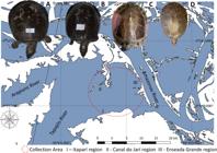
Map of location of the sampling area and species sampled. I - Itaparí Community; II - Igarapé do Jari region; III - Enseada Grande region; A - Podocnemis expansa; B – P. sextuberculata; C – P. unifilis; D – P. erythrocephala.
Turtles of the species P. expansa, P. sextuberculata, P. unifilis and P. erythrocephala (Fig. 1A-D) were captured in crepuscular and night activities using cotton thread gill nets with different mesh sizes, as well as pitfall traps and sets of hooks with natural baits (see Cavalcante et al. 2024 and Table 1). The collection teams were divided into three small boats to ensure timely trap checks more quickly, since the distances among sample points were considerable. This prevented the death of animals caught in the traps, as they could sink and suffocate. Once the animals were captured, they were sent to the advanced field laboratory for procedures of samples. To identify the specimens of chelonians, we used it as a base taxonomic key of Amazonian chelonians (Ferrara et al. 2017). The specimens were physically examined for ectoparasites, and the length (L) and width (W) of the carapace were measured, as well as the sex of each specimen was determined. The ectoparasites found, were removed with forceps and preserved in the alcoholic solution (70%) or in no clorade water for the maintenance of specimens. Additionally, was registered total abundance and sites of infestation by hirudines in the hosts. The slides of leeches were deposited in the collection of the Coleção Parasitológica, Laboratório de Ecologia e Comportamento Animal (LECAN), Santarém, PA, Brazil [UFOPA-P (Hir)0010].
Number of Unoculubranchiobdella expansa per infection site in species of the genus Podocnemis analysed in the present study.
The statistical analysis of the hemoparasites was performed by calculating the parasite indices prevalence (P), intensity (I), mean intensity (MI) and mean abundance (MA) according to the concepts proposed by Bush et al. (1997).
1.1 Preparation of ectoparasites for morphological studies
1.1.1. External morphology
We use an experimental method to keep the ectoparasites fully distended, as we do for dipteran larvae (Nigoghosian et al. 2021, Martín-Vega et al. 2025). Given this, the specimens underwent a rigorous preparation process, including immersion in hot water at 60°C for one minute and fixation in 70% alcohol, followed by staining in a ready-to-use 1% thiazine solution (LB 170117, Laborclin) for 1 minute and diaphanization in 70% alcohol for 5 minutes. The specimens were then examined and photographed using a Zeiss stereomicroscope, Stereo Discovery V8, with an Axiocam Erc5s camera under darkfield diascopic illumination (DDI).
1.1.2. Internal morphology
The hirudines were fixed with alcohol, were rehydrated in distilled water for two hours, and then compressed between slides. Next, they were submerged in a fixation solution consisting of 5% formaldehyde, 2% acetic acid, and 93% saline solution for 12 hours (Pessoa & Martins 1988).
To prepare the specimens, we used the modified hydrochloric carmine method (Eiras et al. 2000), which involves diluting the solution in acetic alcohol in a 1:1 ratio. Specimens were left in the staining solution for five minutes, were dehydrated in acetic alcohol, and washed in 70%, 80%, 90%, and 100% alcohol solutions for three minutes each solution. After the alcohol wash, bathe the specimens in xylene and permanently fix them between slides and coverslips with alkyd resin. The internal structures were thoroughly examined using a Zeiss Axioplan optical microscope at 400 and 1000x magnification. High-quality photographs were expertly captured with an Axiocam ERc 5s camera.
1.1.3. Scanning electron microscopy
External morphology was evaluated by scanning electron microscopy (SEM) previously fixed in ethylic alcohol (70%), transferred to glutar-aldehyde solution (2.5 %) in 0.15 M phosphate buffer (pH 7.3), and then subsequently fixed in osmium tetroxide (1%) in the same buffer, for 2h. The samples were dehydrated in an increasing sequence of ethyl alcohol solutions and washed in a solution of distilled water and filtered water (1:1). Dehydration was carried out with an increasing sequence of ethyl alcohol solutions, and drying was carried out employing a critical point in CPD 020 (Balzer Union), with liquid CO2. The samples were placed on double-sided tape in Stub and covered with a gold-palladium jet.
Results
The analysis of external and internal morphology of leeches showed taxonomic compatibility with Unoculubranchiobdella expansa, described by Peralta et al., (1998) (Fig. 2 and Fig. 3). A prevalence of 66.66% was determined, with MI of 3.39 and MA of 2.61 for specimens of P. expansa; a prevalence of 77.77%, with MI of 0.77 and MA of 5.55 for specimens of P. unifilis; and a prevalence of 100%, with MI of 5.00 and MA of 5.00 for specimens of P. sextuberculata. Only individuals of the species P. erythrocephala were not infested with these ectoparasites.

Detail of the external morphology of Unoculubranchiobdella expansa. A – ventral view; B – dorsal view with detail of annelation; C – lateral view; D – lateral view, detail of the gills; E – apical portion, detail of the anterior sucker; F – detail of the eyespot; G – anal region; H – detail of the posterior sucker.

External and internal anatomical detail of Unoculubranchiobdella expansa. AS – anterior sucker; Pb – proboscis; SG – salivary gland; Es – esophagus; OS – ovarian sac; Cr – Crop; Gi – Gills; Is – Intestine; PC – post-cecum; T – tesstisacs; PS – posterior sucker.
1. Taxonomic summary
Unoculubranchiobdella expansa
Phylum: Annelida Lamarck, 1809
Class: Clitellata Michaelsen, 1919
Subclass: Hirudinea Lamarck, 1816
Order: Hirudinida Siddal et al. 2001
Suborder: Oceanobdelliformes Tessler and de Carle 2018
Family: Ozobranchidae Pinto, 1921
Genus: Unoculubranchiobdella Peralta, Matos & Serra-Freire, 1998
Species: Unoculubranchiobdella expansa Peralta, Matos & Serra-Freire, 1998
Hosts: P. expansa, P. unifilis and P. sextuberculata
Prevalence:P. expansa (66.66%); P. unifilis (77.77%); P. sextuberculata (100%).
Sites of infestation: Base of the limbs for locomotion, base of the neck and base of the tail.
Location: Mouth of the Tapajós River and mouth of the Arapiuns River.
2. External morphology
The hirudines presented are small (Fig. 2a, 2b and 2c), with a ringed body (Fig. 2b), subdivided in the trachelosoma and urosoma. The anterior portion (trachelosoma) features a sucker with an opening (Fig. 2e) and a single eyespot (Fig. 2f). The abdominal region boasts five pairs of digitiform gills with a rough appearance (Fig. 2d), which protruding from the base of the body with slight attenuation at their ends (Fig. 2d). Notably, the reproductive system structures are visible in the central region, with the testisacs standing out (Fig. 2b). At the posterior end, a globose sucker (Fig. 2a, h). Futhermore, the post-cecum and rectal ampulla structures stand out in the body.
3. Internal morphology
Under the light microscope, the eyespot in the apical anterior portion resembles a mathematical plus symbol. The oral sucker is adjacent to the eyespot, followed by the proboscis, which has a tubular structure leading to a wider region where the salivary gland is located. The reproductive system, including four pairs of testisacs, is present below the salivary gland. The pharynx, located in the central area of the body adjacent to the testisacs, connects to the esophagus which leads to the posterior region of the body where the post-cecal region and rectal ampulla are located (Fig. 3).
4. Scanning electron microscopy
In SEM it is possible to observe a body completely covered by circular rings (Fig. 4a), with five pairs of gills formed by small rings, with its distal portion globose (Fig. 4b, c), the oral sucker appears as a triangular invagination in the ventral portion, with a structure similar to a lip marked by striations at different angles (Fig. 4b). At the end of the body there is a globose sucker (Fig. 4d) of concave shape, internally marked by numerous striations that appear to start from the center of the shape towards the edges.

External morphological detail of Unoculubranchiobdella expansa in scanning electron microscopy. a, b. ventral view; c. lateral view with detail of gills; d. detail of the posterior sucker; AS – anterior sucker; PC – post-cecum; Gi – Gills.
5. Sites of infestation
The analysis showed that leeches infested 11 different sites on the hosts, distributed on the locomotor parts at the base of the femur, at the basis of the humerus and at the base of the neck on the ventral and dorsal side, while the base of the tail showed infestation only in the dorsal region.
Considering the genus Podocnemis, the site of infection with the highest number of ectoparasites was the base of the femur in the ventral region with 15.88% of the hirudines, followed by the base of the humerus on the ventral right side with 14.95% and the ventral left side with 14. The base of the neck and the dorsal regions of the femur and humerus showed values between 5.60% and 7.47%. The dorsal base of the tail showed the lowest concentration of bloodsuckers with only 1.89% (Fig. 5a).

Heat points for the distribution of Unoculubranchiobdella expansa by infection site in their hosts. A – General distribution of infection sites for the genus Podocnemis; B – Percentage distribution of infection sites for Podocnemis expansa; C – Percentage distribution of infection sites for Podocnemis unifilis; D – Percentage distribution of infection sites for Podocnemis sextuberculata.
6. Podocnemis expansa
The ventral and dorsal regions of the left humerus had 7.27% and 5.45% of ectoparasites, respectively, the ventral and dorsal regions of the right humerus had 10.90% and 12.72%, the base of the neck on the dorsal side had 10.9% of leeches, the ventral side had 9.09%. The left femur had 7.27% on the dorsal side and 14.54% on the ventral side, while the right femur had 10.9% of ectoparasites on the dorsal side and 7.27% on the ventral side. The base of the tail was only infested on the dorsal side with 3.63% of the hirudines found (Table 1; Fig.5b).
7. Podocnemis unifilis
The percentage distribution of ectoparasites was left humerus ventral and dorsal 4% and 26%, right humerus ventral 14%, neck base ventral 6%, left femur base dorsal 4%, ventral 26%, right femur base dorsal 2% and ventral 18% (Table 1; Fig. 5c).
8. Podocnemis sextuberculata
The examined hosts had ectoparasites in the dorsal region of the left humerus, which represented 6.6% of the parasites found, in the ventral region of the right humerus, which represented 6.6% of the total, in the ventral region of the left femur, which represented 40%, and in the ventral region of the right femur, the area with the highest number of bloodsuckers, which represented 46.6% (Table 1; Fig. 5d).
Discussion
This study presents new information on the distribution of the leech U. expansa by site of infestation in Podocnemis hosts from natural environments in the Brazilian Amazon. The findings contribute to the understanding of host-parasite relationships between these organisms. Additionally, this study present record of new host for neotropical hirudines: P. sextuberculata.
Our study supports the findings presented by Peralta et al. (1998) regarding the parasitic association in the freshwater turtle P. expansa in captivity in the state of Pará and Úngari et al. (2024) for the association in the freshwater turtle P. unifilis in state of Góias, Brazil. It is noteworthy that no other host species of U. expansa are documented apart from the genus Podocnemis. Another species known to parasitize chelonians is B. ringueleti, which was recorded in Colombia parasitizing Podocnemis volgli Müller, 1935, and has not been recorded parasitizing other chelonian species. Additionally, Carvalho & Malvasio (2018) observed this parasitism in free-living P. expansa in the Javaés River in the state of Tocantins, Brazil. The absence of U. expansa in P. erythrocephala may be associated with physical environmental variables. These hosts were caught in an inlet environment where the water temperature characteristics differ from the mouth of the Tapajós River.
Leeches from the Glossiphoniidae, Ozobranchidae, and Piscicolidae families have been identified as vectors of blood parasites for vertebrates (Siddall & Desser 1991, 2001, 2004). In a study by Carvalho & Malvasio (2018), U. expansa was tested for its vector competence in transmitting Haemogregarina sp. and Sauroplasma sp. through experimental infection. Morphological forms compatible with Sauroplasma sp. were observed at the end of the experimental cycle, indicating that U. expansa is a competent vector. However, the studies conducted lack support from molecular biology tools and a well-defined negative control, as well as a precise taxonomic definition of the intraerythrocytic parasite in question.
In captivity, infestation by U. expansa can cause cachexia, severe anemia, prostration, and death, as well as a reduction in oviposition and hatching of host eggs (Peralta et al. 1998). In free-living conditions, large infestations of marine Ozobranchidae can cause erosive diseases in turtles. This degenerative pathology can lead to severe damage to the hosts’ skin and eventual erosion of the plastron (Bunkley-Williams et al. 2008).
The specimens examined in this study did not show any signs of pathologies related to dermal erosion, prostration, or cachexia. The failure to identify debilitated animals in this study can be attributed to natural predation events. Debilitated animals are more susceptible to these events and can be targeted by big cats, crocodilians, and human predators (De La Ossa et al. 2010).
The present study’s identification and quantification of infection sites support Köhnk et al. (2021) findings for Ozobranchus margoi (Apáthy, 1890) parasitizing olive ridley turtles in the Indian Ocean, with infestation found in the locomotor limbs and tail. In our study, we found U. expansa infection in three of the four species of the genus Podocnemis that occur in the Brazilian Amazon.
Studies on the specific parasitism of this leech in freshwater chelonians are unknown, but oceanic species with this level of parasitic specialization have been identified (Sawyer 1986, Yamauchi & Suzuki 2008). Biological information on U. expansa such as life cycle, oviposition rates, survival, physical tolerance to temperature is not available, and the diversity of its hosts and their habitats are unknown, as are the possible detrimental consequences of its parasitic action.
The studies in which this hirudine is mentioned examine infection by blood parasites, treat the leeches observed as secondary information, and generally present records of occurrence without details. Therefore, studies with more objective designs for these leeches are necessary, since this group of annelids has been recognized for its veterinary importance for turtles, and understanding the results of the parasite-host relationship is relevant for future conservation measures for freshwater chelonians.
Acknowledgments
This study was funded by the Fundação de Amparo à Pesquisa do Estado de São Paulo (FAPESP grant # 2018/24980-8), in-part by Coordenação de Aperfeiçoamento de Pessoal de Nível Superior (CAPES Finance Code 001). This work was supported by the Conselho Nacional de Desenvolvimento Científico e Tecnológico (CNPq) under Project number 401968/2025-1 (RB-S), by the Fundação de Amparo à Pesquisa do Estado de São Paulo (FAPESP) under Project no. 2024/14248-9 (R. Bassini-Silva) and E. A. Adriano received a research productivity grant from the CNPq (grant # 304687/2020-0). D. Chagas-de-Souza and T. Alves-Coêlho were supported with scholarships provided by CAPES (Respectively Process # 88887.636892/2021-00 and #88887.598663/2021-00). L. L. Corrêa had guaranteed financial support granted by the CAPES / FAPESPA N. 06/2015 - Process number 88881.160660 / 2017-01.
Data Availability
The data collected and generated during this study are available in the FigShare at https://figshare.com/. The dataset includes specimens’ vouchers used in the analysis and can be accessed at https://doi.org/10.6084/m9.figshare.29344070. The authors confirm that all data necessary for reproducing the study findings are available in the designated dataset.
References
-
BUNKLEY-WILLIAMS, L., WILLIAMS, E.H., HORROCKS, J.A., HORTA, H.C., MIGNUCCI-GIANNONI, A.A., – POPONI, A.C. 2008. New leeches and diseases for the hawksbill sea turtle and the West Indies. Comparative Parasitology, 75(2), 263–270. https://doi. org/10.1654/4335.1
» https://doi.org/10.1654/4335.1 -
BURGIN, S., – BETTS, J. 2012. Epibionts of the Australian Eastern Longnecked Turtle (Chelodina longicollis Shaw) from farm dams. Australian Zoologist, 36(2), 153–158. https://doi.org/10.7882/AZ.2012.015
» https://doi.org/10.7882/AZ.2012.015 -
BUSH, A.O., LAFFERTY, K.D., LOTZ, J.M., – SHOSTAK, A.W. 1997. Parasitology meets ecology on its own terms: Margolis et al. revisited. The Journal of Parasitology, 83(4), 575–583. https://doi. org/10.2307/3284227
» https://doi.org/10.2307/3284227 -
CARVALHO, A.V., – MALVASIO, A. 2018. Transmissão de Sauroplasma sp. (Piroplasmorida: Haemohormidiidae) pela sanguessuga Unoculubranchiobdella expansa (Hirudinea: Ozobranchidae) em Podocnemis expansa (Tartaruga da Amazônia). Revista Ibero-Americana de Ciências Ambientais, 9(2), 41–53. https://doi.org/10.6008/CBPC2179-6858.2018.002.0004
» https://doi.org/10.6008/CBPC2179-6858.2018.002.0004 -
CAVALCANTE, M.C., CHAGAS-DE-SOUZA, D., COÊLHO, T.A., VIANA, L.A., ADRIANO, E., – CORRÊA, L.L. 2024. Hemoparasite analysis and hematological parameters of four freshwater turtle species (Testudines: Podocnemididae) from the Brazilian Amazon. Acta Parasitologica, 69, 1058–1066. https://doi.org/10.1007/s11686-024-00816-z
» https://doi.org/10.1007/s11686-024-00816-z -
CHRISTOFFERSEN, M.L. 2009. A catalogue of Helobdella (Annelida, Clitellata, Hirudinea, Glossiphoniidae), with a summary of leech diversity, from South America. Neotropical Biology and Conservation, 4(2), 89–98. https://doi.org/10.4013/nbc.2009.42.07
» https://doi.org/10.4013/nbc.2009.42.07 -
DE LA OSSA, J., VOGT, R.C., – FERRARA, C.R. (2010). Melanosuchus niger (Crocodylia: Alligatoridae) as opportunistic turtle consumer in its natural environment. Revista Colombiana de Ciencia Animal, 2(2), 244–252. https://doi.org/10.24188/recia.v2.n2.2010.290
» https://doi.org/10.24188/recia.v2.n2.2010.290 - EIRAS, J.D.C., TAKEMOTO, R.M., – PAVANELLI, G.C. (2000). Métodos de estudo e técnicas laboratoriais em parasitologia de peixes. EDUEM.
-
E.L., DA SILVA, R.J. – O´DWYER, L.H. 2024. Unoculubranchiobdella sp. (Hirudinea: Ozobranchidae) as a vector for Haemogregarina spp. in freshwater turtles from Brazil. Parasitology Research 123: 249. https://doi. org/10.1007/s00436-024-08271-5
» https://doi.org/10.1007/s00436-024-08271-5 - FERRARA, C.R., FAGUNDES, C.K., MORCATTY, T.Q. – VOGT, R.C. 2017. Quelônios Amazônicos: Guia de identificação e distribuição. Manaus, Brazil. Wildlife Conservation Society Brasil.
-
GAFFNEY, E.S., MEYLAN, P.A., WOOD, R.C. SIMONS, E. – CAMPOS, D.A. 2011. Evolution of the Side-Necked Turtles: The Family Podocnemididae. Bulletin of the American Museum of Natural History, 2011(350):1-237. https://doi.org/10.1206/350.1
» https://doi.org/10.1206/350.1 -
KÖHNK, S., PETROS, C., LOMAS, C., RIYAD, E.M., SHAMEEL, I., HAWLITSCHEK, O., – STELFOX, M. 2021. Stowaways: Marine Leeches Infecting Olive Ridley Sea Turtles Entangled in Ghost Nets in Maldivian Waters. Comparative Parasitology, 88(2), 169–176. https://doi. org/10.1654/1525-2647-88.2.169
» https://doi.org/10.1654/1525-2647-88.2.169 - MACCALLUM, W.G., – MACCALLUM, G.A. 1918. On the Anatomy of Ozobranchus branchiatus (Menzies) (Vol. 38). American Museum of Natural History.
-
MAGALHÃES, W.F., HUTCHINGS, P., OCEGUERA-FIGUEROA, A., MARTIN, P., SCHEMLZ, R.M., WETZEL, M.J., WIKLUND, H. – WILLIAMS, J.D. 2021. Segmented worms (Phylum Annelida): a celebration of twenty years of progress through Zootaxa and call for action on the taxonomic work that remains. Zootaxa, 4979(1): 190–211. https://doi.org/10.11646/ZOOTAXA.4979.1.18.
» https://doi.org/10.11646/ZOOTAXA.4979.1.18 - MANE-GARZON, F. 1973. Un nuevo tipo de Hirudinea Colombobdella ringueleti n. gen., n. sp. parasito de una tortuga de Colombia. In Trabajos V Congress Latinamericano Zoologia, Montevideo (Vol. 1, pp. 129–137).
-
MARTÍN-VEGA, D., HALL, M. J.R., ANTONY, A., CLARK, B., BARIGYE, R., MOHAMMED, T., HAMDAN, L. – GAJIC, B. 2025. Larval anatomy and intra-puparial development of the camel nasal bot fly, Cephalopina titillator (Diptera: Oestridae). Veterinary Parasitology, 337: 110476. https://doi.org/10.1016/j.vetpar.2025.110476
» https://doi.org/10.1016/j.vetpar.2025.110476 -
NIGOGHOSIAN, G., WEIDNER, L.M. – STAMPER, T.I. 2021. Technical note: A technique to mount Sarcophagidae and Calliphoridae (Diptera) larvae for forensic identification using geometric morphometrics. Forensic Science International: Synergy, 3: 100135. https://doi.org/10.1016/j.fsisyn.2021.100135
» https://doi.org/10.1016/j.fsisyn.2021.100135 - PERALTA, A.S.L., MATOS, E.R., – SERRA-FREIRE, N.M. 1998. Unoculubranchiobdella expansa gen. nov., sp. n. (Hirudinea: Ozobranchidae) parasito de Podocnemis expansa (Chelonia: Pelomedusidae). Entomologia y Vectores, 5(5), 161–177.
- PESSOA, S.B. and MARTINS, A.V. 1988. Parasitologia Médica. (11th ed.). Guanabara Koogan.
- RHODIN, A.G.J., IVERSON, J.B., BOUR, R., FRITZ, U., GEORGES, A., SHAFFER, H.B. – VAN DIJK, P.P. (2017). Turtles of the world: annotated checklist and atlas of taxonomy, synonymy, distribution, and conservation status. (8th ed.) Chelonian Research Monograph, 7(8):1–292.
-
SALERA JÚNIOR, G., PORTELINHA, T.C.L., – MALVASIO, A. 2009. Predação de fêmeas adultas de Podocnemis expansa Schweigger (Testudines, Podocnemididae) por Panthera onca Linnaeus (Carnivora, Felidae), no Estado do Tocantins. Biota Neotropica, 9(3), 387–391. https://doi. org/10.1590/S1676-06032009000300033
» https://doi.org/10.1590/S1676-06032009000300033 - SAWYER, R.T. (1986). Leech Biology and Behavior Clarendon Press.
-
SIDDALL, M.E., – DESSER, S.S. 1991. Merogonic development of Haemogregarina balli (Apicomplexa: Adeleina: Haemogregarinidae) in the leech Placobdella ornata (Glossiphoniidae), its transmission to a chelonian intermediate host and phylogenetic implications. The Journal of Parasitology, 77(3), 426–436. https://doi.org/10.2307/3283130
» https://doi.org/10.2307/3283130 -
SIDDALL, M.E., – DESSER, S.S. 2001. Transmission of Haemogregarina balli from painted turtles to snapping turtles through the leech Placobdella ornata Journal of Parasitology, 87(5), 1217–1218. https://doi. org/10.1645/0022-3395(2001)087[1217:TOHBFPT]2.0.CO;2
» https://doi.org/10.1645/0022-3395(2001)087[1217:TOHBFPT]2.0.CO;2 -
SIDDALL, M.E., – GAFFNEY, E.S. 2004. Observations on the leech Placobdella ornata feeding from bony tissues of turtles. Journal of Parasitology, 90(5), 1186–1188. https://doi.org/10.1645/GE-281R
» https://doi.org/10.1645/GE-281R -
SKET, B., – TRONTELJ, P. 2008. Global diversity of leeches (Hirudinea) in freshwater (pp. 129–137). Springer Netherlands. https://doi.org/10.1007/978-1-4020-8259-7_14
» https://doi.org/10.1007/978-1-4020-8259-7_14 -
TESSLER, M., CARLE, D., VOIKLIS, M.L., GRESHAM, O.A., NEUMANN, J., CIOS, S. – SIDDAL, M.E. 2018. Worms that suck: Phylogenetic analysis of Hirudinea solidifies the position of Acanthobdellida and necessitates the dissolution of Rhynchobdellida, Molecular Phylogenetics and Evolution, 127: 129–134. https://doi.org/10.1016/j.ympev.2018.05.001.
» https://doi.org/10.1016/j.ympev.2018.05.001 - ÚNGARI, L., Oceguera-Figueroa, A., Torres-Carrera, G., Netherlans, E. C., Quagliatto Santos.
- VOGT, R.C. (2008). Tartarugas da Amazônia. Editora INPA.
-
YAMAUCHI, T., – SUZUKI, D. 2008. Geographical distribution of Ozobranchus jantseanus (Annelida: Hirudinida: Ozobranchidae) in Japan. Medical Entomology and Zoology, 59(4), 345–349. https://doi.org/10.7601/mez.59.345
» https://doi.org/10.7601/mez.59.345 -
ZOOTAXA, 4979(1): 190–211. https://doi.org/10.11646/ZOOTAXA.4979.1.18
» https://doi.org/10.11646/ZOOTAXA.4979.1.18


