 ANTERIOR CERVICAL ARTHRODESIS WITH INTERVERTEBRAL SPACER AND CYLINDRIC BONE SUPPORT
ABSTRACT
The anterior cervical discectomy and fusion is considered an established technique in cervical degenerative disease treatment, in which the final goal is the fusion of the segments involved. There are options for spacer grafts, such as bone grafts and synthetic bone substitutes like hydroxyapatite and tricalcium phosphate. This technical note describes the combination of the stand-alone anchored intervertebral spacer and iliac graft, which is used as a pillar between the vertebrae. The iliac crest removal is performed with a 10 mm or 12 mm diameter trephine. The trephine is employed to make a graft in the same format as the inner portion of the intervertebral spacer. The cylindrical bone graft removed must be 1 millimeter higher than the cage that will be implanted so that it is 0.5 mm above and below the upper and lower edges of the cage. The objective is to establish support between the vertebral plateaus and the bone cylinder, as they present similar resistance and biological aspects, which are fundamental characteristics for proper osseointegration and fusion. When the conventional technique is used, the interaction between the cage and the plateaus occurs more intensely, and a plateau fracture may occur, a phenomenon known as subsidence. This technical note describes a feasible way to obtain autograft bone to be used in ACDF stand-alone surgery. This type of graft may be associated with lower rates of pseudoarthrosis. Level of Evidence IV; Case Series.
Keywords:
Diskectomy; Iliac Crest; Transplantation; Autologous; Surgical Procedures; Operative
RESUMO
A discectomia cervical anterior com fusão é considerada uma técnica consagrada no tratamento de doenças degenerativas cervicais, cujo objetivo final é a fusão dos segmentos envolvidos. Existem opções de enxertos para os espaçadores, como enxertos ósseos e substitutos ósseos sintéticos como hidroxiapatita e fosfato tricálcico. Esta nota técnica descreve a combinação entre o espaçador intervertebral autobloqueante e o enxerto de ilíaco, que é utilizado como pilar entre as vértebras. A retirada da crista ilíaca é realizada com uma trefina de 10 mm ou 12 mm de diâmetro. A trefina é empregada para fazer um enxerto no mesmo formato da porção interna do espaçador intervertebral. O enxerto ósseo cilíndrico retirado deve ficar 1 milímetro maior que o cage que será implantado, de modo que fique 0,5 mm acima e abaixo das bordas superior e inferior do cage. O objetivo é estabelecer um suporte entre os platôs vertebrais e o cilindro ósseo, pois apresentam resistência e aspectos biológicos semelhantes, características fundamentais para a adequada osseointegração e fusão. Quando utilizada a técnica convencional, a interação entre o espaçador intervertebral e os platôs ocorre de forma mais intensa, podendo ocorrer fratura do platô, fenômeno conhecido como subsidência. Esta nota técnica descreve uma forma viável de obtenção de autoenxerto ósseo para utilização em cirurgia. Esse tipo de enxerto pode estar associado a menores taxas de pseudoartrose. Nível de Evidência IV; Série de casos.
Descritores:
Discotomia; Crista Ilíaca; Transplante Autólogo; Procedimentos Cirúrgicos Operatórios
RESUMEN
La discectomía cervical anterior con fusión se considera una técnica consolidada en el tratamiento de las enfermedades degenerativas cervicales, cuyo objetivo final es la fusión de los segmentos implicados. Existen opciones de injertos para espaciadores, como injertos óseos y sustitutos óseos sintéticos como hidroxiapatita y fosfato tricálcico. Esta nota técnica describe la combinación entre el espaciador intervertebral autobloqueante y el injerto ilíaco, que se utiliza como pilar entre las vértebras. La extirpación de la cresta ilíaca se realiza con una trefina de 10 mm o 12 mm de diámetro. La trefina se utiliza para realizar un injerto con la misma forma que la porción interna del espaciador intervertebral. El injerto óseo cilíndrico extraído debe ser 1 milímetro más grande que el espaciador que se va a implantar, de modo que quede 0,5 mm por encima y por debajo de los bordes superior e inferior del espaciador. El objetivo es establecer un soporte entre los platillos vertebrales y el cilindro óseo, ya que tienen similares resistencias y aspectos biológicos, características fundamentales para una adecuada osteointegración y fusión. Cuando se utiliza la técnica convencional, la interacción entre el espaciador intervertebral y los platillos vertebrales se produce más intensamente, y los platillos pueden fracturarse, fenómeno conocido como subsidente. Esta nota técnica describe una forma viable de obtener un autoinjerto óseo para su uso em cirugía. Este tipo de injerto puede estar asociado con tasas más bajas de pseudoartrosis. Nivel de Evidencia IV; Serie de Casos.
Descriptores:
Discectomía; Cresta Ilíaca; Trasplante Autólogo; Procedimientos Quirúrgicos Operativos
INTRODUCTION
First described in 1955 by Smith and Robinson, the anterior cervical discectomy and fusion (ACDF) is considered an established technique in cervical degenerative disease treatment1-3.
The more levels operated, the higher the failed fusion risk, so-called pseudoarthrosis. Literature data shows pseudoarthrosis rates from 10 to 12% regarding single-level fusion, 20 to 27% in two-level cases, and 30 to 56% for three levels4.
The objective of this technical note is to describe in detail the combination of the stand-alone anchored intervertebral spacer and iliac graft, which is removed to provide a foothold between the vertebrae, allowing a faster fusion without a cervical plate.
METHODS
Surgical positioning
After general anesthesia, the patient is positioned as usual for anterior cervicotomy, in a supine position on the surgical table with slight head extension, no rotation, and shoulders pulled caudally with adhesive plaster. Based on cadaveric, anatomical, and clinical studies5-7, the anterior superior iliac spine (ASIS) in the selected side must be palpated, and the skin marked 3 cm posterior to it, avoiding lateral femoral cutaneous nerve injury.
Timing of removal of anterior iliac crest
The graft removal may be done simultaneously, before the cervicotomy or after the discectomy, depending on the experience and preferences of the surgical team. However, removing before the discectomy demands precise knowledge of the spacers that shall be implanted.
Anterior iliac crest graft removal (Figure 1)
After asepsis and antisepsis, subcutaneous and periosteum infiltration with local anesthetic with vasoconstrictor (bupivacaine 7.5%) is performed to reduce blood loss and post-operative pain. The skin, subcutaneous tissue, and fascia are incised with the 23rd blade, followed by gentle periosteum detachment with blunt dissection and hemostasis with electrocautery until the bone is seen.

Anterior superior iliac crest marking (A), Incision by planes (B), Iliac crest exposure (C), Bone graft with trephine (D), Trephine diameter equal to the inner portion of the cage (E), Preservation of internal and external bone plates (F).
The iliac crest removal is performed with a 10 mm or 12 mm diameter trephine, depending on the cage that will be implanted. The trephine is employed to make a graft in the same format as the inner portion of the intervertebral spacer and also avoids fracture and violation of the bone tables, as hematoma, infection, and chronic pain are related to bloody and reckless removal7-9.
The cylindrical bone graft removed must be 1 millimeter higher than the cage that will be implanted. For example: a 6 mm cylinder is employed in a 5 mm height cage, and it is important to leave a safety margin ranging from 0.5 to 1 mm so as to adjust the graft when introducing the cylinder in the cage. In this hypothetical example, the cylinder would be 6.5 to 7 mm tall, so it would reach 6 mm after excessive removal with a delicate bone rongeur.
The following bone cylinders are removed side by side, and depending on the patient’s anatomy, a bigger cylinder can be removed for two levels.
Hemostasis is easily obtained after applying bone wax inside the bone cavity and the surgical field is extensively washed with normal saline. After bleeding control, the incision is closed by layers: Vicryl™ 2-0 single interrupted suture in muscle fascia, Vicryl™ 3-0 in the subcutaneous tissue, and Monocryl™ 3-0 subcuticular suture. We do not use surgical drains unless there is active bleeding that is difficult to control.
Cage preparation with bone cylinder and insertion in the disc space (Figure 2)
After the standard procedure of discectomy and preparation of the vertebrae end plates, the implant is tested and chosen separately at each level. The bone cylinder 1 mm higher than the cage is then inserted into the intervertebral spacer so that it is 0.5 mm above and below the upper and lower edges of the cage.

Bone cylinder measurement (A), Prepared bone end plates and measured disc heights (B), Bone graft cylinder 0.5 mm above the cage (C), Bone graft cylinder 0.5 mm below the cage (D).
The objective is to establish support between the vertebral plateaus and the bone cylinder, as they present similar resistance and biological aspects, which are fundamental characteristics for proper osseointegration and fusion. When the conventional technique is used, the interaction between the cage and the plateaus occurs more intensely, and a plateau fracture may occur, a phenomenon known as subsidence.
The cylinder must be positioned in the center of the vertebrae, fitting anatomically to the upper plateau of the caudal vertebra and the concavity proper to the lower vertebral plateau of the cranial vertebra. Distraction with a Caspar retractor is necessary to properly insert the intervertebral spacer at this step.
Anchoring self-locking cage
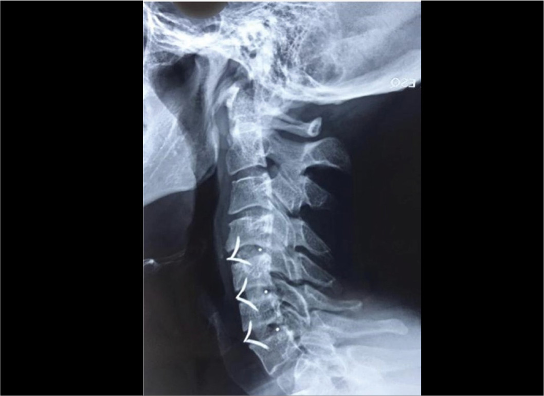
Compression is performed, bringing the vertebral plateaus as close as possible to the bone cylinder and the cage margins and then locking the cage to the plateaus with anchors, flaps, or screws according to the device type.(Figure 3)

The bone graft cylinder must be inserted and then compressed in an anatomical manner in the center of the disc space.
Ethical Approval
Ethical approval was unnecessary for this technical note, equivalent to a case report. The patient provided informed consent for the publication of this case.
DISCUSSION
The pseudoarthrosis etiology is not well defined. Risk factors associated with the patient include smoking, diabetes, chronic use of corticoids, elderly status, poor nutrition, poor bone quality, and metabolic and vascular abnormalities. However, surgery-related factors include multiple-level fusions, bone graft type, and the sort of instrumentation used9-10.
The vertebrae fixation with intervertebral spacers (cage) combined with plate and screw instrumentation reduces cervical spine micromotion and increases the fusion rate, besides helping to recover the physiological lordosis. This system reduces the risk of non-union in multiple levels of surgical procedures1,11,12. It should be pointed out, however, that anterior cervical plates are associated with various complications, such as screws loosening or breakage, nerve injuries, spinal cord or nerve root injuries due to poor implant placement, dysphonia, Horner’s syndrome, adjacent segment disease, in addition to esophageal injury or perforation and dysphagia3,12-15.
The anchored intervertebral spacers are cervical intervertebral fusion devices consisting of polyether ether ketone or titanium cage (intervertebral spacer) and integrated anchored clips (anchors or flaps) designed to provide fixation between the cage and adjacent vertebral bodies. The characteristic design of these spacers restricts the need for additional instrumentation devices4,16.
The main difference between anchored intervertebral spacers and conventional devices (plate and cage) is that, with spacers, no additional plate is attached to the anterior surface of the vertebral body, which reduces complications occurrence. Moreover, using spacers allows for a smaller cervicotomy and reduces surgical time. The studies showed equivalent fusion rates when comparing the use of intervertebral spacers and instrumentation techniques that use anterior plaque, with a lower incidence of dysphagia17-21.
The graft type and the biological agents used in the surgical procedure have also been associated with pseudoarthrosis development2. The grafts are commonly autograft (coming from the patient iliac crest and considered the gold standard in the category because they present the three properties necessary for bone formation: osteogenesis, osteoinduction, and osteoconduction) or allograft type (from cadaveric iliac crest or fibula)22-23.
Studies show that while the first type of graft is associated with complications such as pain, infection, and hematoma23-24, the second can generate an immune response, graft rejection, infection, and low fusion rate25-26. A recent meta-analysis showed that, when using the allograft, the mean pseudoarthrosis rate was 4.8%, while this average drops to only 0.9% using autograft27.
Synthetic bone substitutes such as hydroxyapatite and tricalcium phosphate have become an alternative in spine surgical procedures, with or without anterior plates28-29. However, regarding cervical discectomy, the scientific evidence is limited, and these substitutes present only osteoconduction properties28-30.
CONCLUSION
This technical note describes a feasible way to obtain autograft bone for use in ACDF stand-alone surgery. This type of graft is associated with lower rates of pseudoarthrosis.
-
Study conducted by the Hospital Ortopédico AACD, São Paulo, SP, Brazil.
REFERENCES
- 1 Robinson R, Smith G. Anterolateral disc removal and interbody fusion for cervical disc syndrome. Bull Johns Hopkins Hosp. 1955;96:223-4.
- 2 Leven D, Cho SK. Pseudarthrosis of the cervical spine: risk factors, diagnosis and management. Asian Spine J. 2016;10 (4):776-86.
- 3 Fujibayashi S, Shikata J, Kamiya N, Tanaka C. Missing anterior cervical plate and screws: a case report. Spine (Phila Pa 1976). 2000;25(17):2258-61.
- 4 Cho DY, Lee WY, Sheu PC. Treatment of multilevel cervical fusion with cages. Surg Neurol. 2004;62(5):378-86.
- 5 Mischkowski RA, Selbach I, Neugebauer J, Koebke J, Zöller JE. Lateral femoral cutaneous nerve and iliac crest bone grafts-anatomical and clinical considerations. Int J Oral Maxillofac Surg. 2006;35(4):366-72.
- 6 Ebraheim NA, Yang H, Lu J, Biyani A, Yeasting RA. Anterior iliac crest bone graft. Anatomic considerations. Spine (PhilaPa 1976). 1997;22(8):847-9.
- 7 Silva RB, Cavali PTM, Veiga IG, Risso-Neto MI, Pasqualini W, Santos MAM, et al. Avaliação da dor e morbidade local da retirada do enxerto ósseo da crista ilíaca para artrodese cervical anterior. Coluna/Columna. 2010;9(4):424-9.
- 8 Sasso RC, LeHuec JC, Shaffrey C, Spine Interbody Research Group. Iliac crest bone graft donor site pain after anterior lumbar interbody fusion: a prospective patient satisfaction outcome assessment. J Spinal Disord Tech. 2005;18 Suppl:S77-81.
- 9 David R, Folman Y, Pikarsky I, Leitner Y, Catz A, Gepstein R. Harvesting bone graft from the posterior iliac crest by less traumatic, midline approach. J Spinal Disord Tech. 2003;16(1):27-30.
- 10 Phillips FM, Carlson G, Emery SE, Bohlman HH. Anterior cervical pseudarthrosis. Natural history and treatment. Spine (Phila Pa 1976). 1997;22(14):1585-9.
- 11 Song KJ, Taghavi CE, Lee KB, Song JH, Eun JP. The efficacy of plate construct augmentation versus cage alone in anterior cervical fusion. Spine (Phila Pa 1976). 2009;34(26):2886-92.
- 12 Lowery GL, McDonough RF. The significance of hardware failure in anterior cervical plate fixation. Patients with 2- to 7-year follow-up. Spine (Phila Pa 1976). 1998;23(2):181-6.
- 13 Fountas KN, Kapsalaki EZ, Nikolakakos LG, Smisson HF, Johnston KW, Grigorian AA, et al. Anterior cervical discectomy and fusion associated complications. Spine (Phila Pa 1976). 2007;32(21):2310-7.
- 14 Traynelis VC, Malone HR, Smith ZA, Hsu WK, Kanter AS, Qureshi SA, et al. Rare Complications of Cervical Spine Surgery: Horner’s Syndrome. Global Spine J. 2017;7(1 Suppl):S103-8.
- 15 Zeng J, Duan Y, Yang Y, Wang B, Hong Y, Lou J, et al. Anterior corpectomy and reconstruction using dynamic cervical plate and titanium mesh cage for cervical spondylotic myelopathy: A minimum 5-year follow-up study. Medicine (Baltimore). 2018;97(5):e9724.
- 16 Kaiser MG, Haid RW Jr, Subach BR, Barnes B, Rodts GE Jr. Anterior cervical plating enhances arthrodesis after discectomy and fusion with cortical allograft. Neurosurgery. 2002;50(2):229-36.
- 17 Wang Z, Jiang W, Li X, Wang H, Shi J, Chen J, et al. The application of zero-profile anchored spacer in anterior cervical discectomy and fusion. Eur Spine J. 2015;24(1):148-54.
- 18 Zhang Z, Li Y, Jiang W. A comparison of zero-profile anchored spacer (ROI-C) and plate fixation in 2-level noncontiguous anterior cervical discectomy and fusiona retrospective study. BMC Musculoskelet Disord. 2018;19(1):119.
- 19 Bucci MN, Oh D, Cowan RS, Davis RJ, Jackson RJ, Tyndall DS, et al. The ROI-C zero-profile anchored spacer for anterior cervical discectomy and fusion: biomechanical profile and clinical outcomes. Med Devices (Auckl). 2017;10:61-9.
- 20 Lu Y, Bao W, Wang Z, Zhou F, Zou J, Jiang W, et al. Comparison of the clinical effects of zero-profile anchored spacer (ROI-C) and conventional cage-plate construct for the treatment of noncontiguous bilevel of cervical degenerative disc disease (CDDD): A minimum 2-year follow-up. Medicine (Baltimore). 2018;97(5):e9808.
- 21 Barbagallo GM, Romano D, Certo F, Milone P, Albanese V. Zero-P: a new zero-profile cage-plate device for single and multilevel ACDF. A single institution series with four years maximum follow-up and review of the literature on zero-profile devices. Eur Spine J. 2013;22(Suppl 6):S868-78.
- 22 Kani KK, Chew FS. Anterior cervical discectomy and fusion: review and update for radiologists. Skeletal Radiol. 2018;47(1):7-17.
- 23 Chau AM, Mobbs RJ. Bone graft substitutes in anterior cervical discectomy and fusion. Eur Spine J. 2009;18(4):449-64.
- 24 Emery SE. Cervical spondylotic myelopathy: diagnosis and treatment. J Am AcadOrthop Surg. 2001;9(6):376-88.
- 25 Ryken TC, Heary RF, Matz PG, Anderson PA, Groff MW, Holly LT, et al. Techniques for cervical interbody grafting. J Neurosurg Spine. 2009;11(2):203-20.
- 26 Beaman FD, Bancroft LW, Peterson JJ, Kransdorf MJ, Menke DM, DeOrio JK. Imaging characteristics of bone graft materials. Radiographics. 2006;26(2):373-88.
- 27 Shriver MF, Lewis DJ, Kshettry VR, Rosenbaum BP, Benzel EC, Mroz TE. Pseudoarthrosis rates in anterior cervical discectomy and fusion: a meta-analysis. Spine J. 2015;15(9):2016-27.
- 28 Buser Z, Brodke DS, Youssef JA, Meisel HJ, Myhre SL, Hashimoto R, et al. Synthetic bone graft versus autograft or allograft for spinal fusion: a systematic review. J Neurosurg Spine. 2016;25(4):509-16.
- 29 Dai LY, Jiang LS. Anterior cervical fusion with interbody Cage containing beta-tricalcium phosphate augmented with plate fixation: a prospective randomized study with 2-year follow-up. Eur Spine J. 2018;17(5):698-705.
- 30 Lavrador JP, Livraghi S, Pereira P, Pimentel J. Foreign body reaction to hydroxyapatite after anterior cervical discectomy with fusion. Acta Neurochir (Wien). 2013;155(2):321-2.
Edited by
-
Reviewed by:
Aluízio Augusto Arantes