 ENDOSCOPIC TREATMENT OF ACUTE CAUDA EQUINA SYNDROME: CASE SERIES
ABSTRACT
Cauda Equina Syndrome (CES) is a severe neurological emergency resulting from the compression of the lumbosacral nerve roots, potentially causing motor and sensory deficits, as well as sphincter dysfunctions. Early surgical intervention is essential to prevent permanent neurological sequelae. Traditionally, open laminectomy has been the treatment of choice for decompression. Still, minimally invasive techniques, such as endoscopic discectomy, are gaining popularity due to lower morbidity, smaller incisions, and faster recovery. This study reports three cases of patients with CES treated with endoscopic discectomy. Two cases were caused by a large, extruded disc herniation and one by an intracanal facet cyst. Pain intensity was assessed using the Visual Analog Scale (VAS), and functional disability was measured with the Oswestry Disability Index (ODI) in the preoperative period and at follow-ups of 1, 6, and 12 months. All three patients showed significant improvement in symptoms, with pain reduction and functional improvement. At 12 months, the patients maintained their functional and neurological recovery without reported complications. The endoscopic technique proved to be a safe and effective method for treating CES in these cases, offering early recovery and fewer postoperative complications. Future studies with larger populations are needed to validate these findings. Level of Evidence IV; Case Report.
Keywords:
Cauda Equina Syndrome; Endoscopic Surgical Procedures; Disk Herniation; Decompression; Surgical; Neurosurgery.
RESUMO
A Síndrome da Cauda Equina (SCE) é uma emergência neurológica grave resultante da compressão das raízes nervosas lombossacrais, podendo ocasionar déficits motores, sensoriais e disfunções esfincterianas. A intervenção cirúrgica precoce é fundamental para evitar sequelas neurológicas permanentes. Tradicionalmente, a laminectomia aberta tem sido a abordagem de escolha para descompressão, mas as técnicas minimamente invasivas, como a discectomia endoscópica, estão ganhando espaço por apresentarem menor morbidade, incisões menores e recuperação mais rápida. Este estudo relata três casos de pacientes com SCE tratados com discectomia endoscópica. Dois dos casos foram causados por hérnia discal extrusa volumosa e um por cisto facetário intracanal. A intensidade da dor foi avaliada pela Escala Visual Analógica (EVA) e a incapacidade funcional pelo Oswestry Disability Index (ODI) no período pré-operatório e em seguimentos de 1, 6 e 12 meses. Os três pacientes apresentaram melhora significativa dos sintomas, com redução da dor e melhora funcional. Aos 12 meses, os pacientes mantiveram a recuperação funcional e neurológica, sem complicações relatadas. A técnica endoscópica mostrou-se uma técnica segura e eficaz no tratamento da SCE nos casos relatados, oferecendo recuperação precoce e menos complicações pós-operatórias. Estudos futuros com populações maiores são necessários para validar esses achados. Nível de Evidência IV; Relato de Caso.
Descritores:
Síndrome da Cauda Equina; Procedimentos Cirúrgicos Endoscópicos; Hérnia de Disco; Descompressão Cirúrgica; Neurocirurgia.
RESUMEN
El síndrome de cola de caballo (SCC) es una emergencia neurológica grave resultante de la compresión de las raíces nerviosas lumbosacras, lo que puede ocasionar déficits motores, sensoriales y disfunciones esfinterianas. La intervención quirúrgica precoz es fundamental para evitar secuelas neurológicas permanentes. Tradicionalmente, la laminectomía abierta ha sido el enfoque de elección para la descompresión, pero las técnicas mínimamente invasivas, como la discectomía endoscópica, están ganando popularidad debido a su menor morbilidad, incisiones más pequeñas y recuperación más rápida. Este estudio presenta tres casos de pacientes con SCC tratados con discectomía endoscópica. Dos de los casos fueron causados por una hernia discal extrusa volumétrica y uno por un quiste facetario intracanal. La intensidad del dolor se evaluó mediante la Escala Visual Analógica (EVA) y la discapacidad funcional mediante el Oswestry Disability Index (ODI) en el período preoperatorio y en los seguimientos de 1, 6 y 12 meses. Los tres pacientes mostraron una mejora significativa en los síntomas, con reducción del dolor y mejora funcional. A los 12 meses, los pacientes mantuvieron su recuperación funcional y neurológica sin complicaciones reportadas. La técnica endoscópica demostró ser un método seguro y eficaz para el tratamiento del SCC en estos casos, ofreciendo una recuperación precoz y menos complicaciones postoperatorias. Se necesitan estudios futuros con poblaciones más grandes para validar estos hallazgos. Nivel de Evidencia IV; Reporte de Caso.
Descriptores:
Síndrome de Cauda Equina; Procedimientos Quirúrgicos Endoscópicos; Hernia Discal; Descompresión Quirúrgica; Neurocirugía.
INTRODUCTION
Cauda Equina Syndrome (CES) is a rare but serious condition characterized by the compression of the lumbosacral nerve roots within the spinal canal, resulting in potentially severe neurological deficits1. Early diagnosis and immediate surgical intervention are crucial to avoid permanent sequelae, such as motor, sensory, and sphincter dysfunctions. Traditionally, the surgical approach for CES has been open total laminectomy, with or without arthrodesis, aiming to decompress the affected nerve roots. This technique has been widely used and accepted, given its effectiveness in broad and direct decompression, despite the risks associated with higher surgical morbidity, such as excessive bleeding, greater soft tissue injury, and prolonged recovery time2.
With the advancement of minimally invasive surgical techniques, the need for methods that provide effective decompression with less tissue trauma has become a priority. Minimally invasive techniques, especially the endoscopic technique, have become viable alternatives. These approaches offer potential benefits, including smaller incisions, less damage to paravertebral structures, less postoperative pain, reduced surgical time, and a quicker return to normal activities3. Furthermore, the preservation of spinal stability, associated with lower rates of perioperative complications, makes these techniques attractive in the management of patients with SCE3,4.
In recent years, some authors have explored endoscopic surgery as an alternative option for the treatment of SCE, despite the limited number of cases described in the literature 1,3. The few available studies suggest that endoscopic discectomy may provide results comparable to conventional techniques, both in terms of pain relief and neurological functional recovery 5. Moreover, the minimally invasive nature of this technique can represent a significant advantage in terms of faster postoperative recovery and lower surgical morbidity.6
This study aims to report a series of cases of CES treated with the endoscopic technique.
REPORT OF CASES
This observational, descriptive, and retrospective study presents a series of three cases of CES surgically treated by the endoscopic technique. Two cases were caused by a large lumbar disc extrusion and one by a large intracanalar facet cyst. All patients provided informed consent through the Informed Consent Form (ICF), and the Research Ethics Committee approved the study (CAAE 70416223.8.0000.5487, opinion number 6.339.106).
The intensity of pain was measured using the visual analog scale (VAS) at the preoperative periods (PRE), one month (PO1m), six months (PO6m), and 12 months (PO12m) of follow-up. Functional incapacity was assessed by the Oswestry Disability Index (ODI) at the same time intervals.
Case 1
A 25-year-old male patient presented with intense low back pain radiating to the left lower limb (LLL) for three months, accompanied by hypesthesia in the left leg, without initial motor deficits. After three months of progress, the patient returned with complaints of loss of sensitivity in the perineal region and the inner thighs, as well as sphincter dysfunction with episodes of urinary and fecal incontinence the day before the appointment. The pain from the VAS was rated as 10/10 in both the lumbar region and the LLL. The ODI indicated 74% disability, classifying the patient as “disabled.” The magnetic resonance imaging (MRI) revealed a large extruded disc herniation at L5-S1, occupying about 90% of the spinal canal (Figures 1A, 1B). The simple X-rays did not show significant changes, and the computed tomography (CT) did not show disc or yellow ligament calcification. The patient underwent emergency surgery 20 hours after the onset of severe symptoms, with general anesthesia and intraoperative neurophysiological monitoring (IONM). The endoscopic technique with interlaminar access was chosen. The patient was positioned in a prone position, and the entry point was marked with the help of fluoroscopy. After introducing the endoscope with a working channel of 4.3mm in diameter and 30 degrees of angulation, it was decided to enlarge the interlaminar bony window with a cutting drill to facilitate disc manipulation. The yellow ligament was then opened with scissors (Figure 1C) and a large extruded disc fragment was already identified (Figure 1D). Removing the extruded fragment allowed the visualization of the decompressed neural structures (Figure 1E). During the procedure, retractable bipolar was used for hemostasis, and continuous irrigation of saline was elevated to 1.5m above the patient’s level. The surgical time was 52 minutes, and the patient was discharged 12 hours after the procedure. In the immediate postoperative follow-up, there was complete resolution of sphincter dysfunction, with significant reduction of pain (VAS 2/10 in the lower back and 1/10 in the LLL). The ODI was reduced to 18% in the first month, with a sustained improvement in the 6-month (6%) and 12-month (6%) segments. Total recovery of perineal sensitivity occurred in eight weeks, and at 12 months, the patient reported no pain in the LLL and referred to minimal low back pain (VAS 2/10).

Case 1: sagittal (A) and axial (B) cuts of the pre-operative MRI; the opening of the yellow ligament with scissors (C); identification of a large extruded disc fragment (D); and visualization of the free roots E).
Case 2
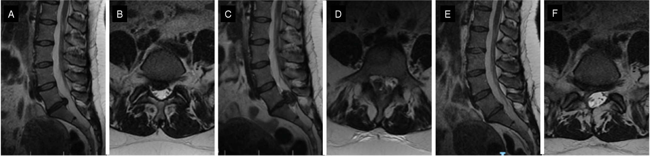
A 43-year-old female patient reported low back pain radiating to the right lower limb (RLL) for 3 months. Positive Lasegue test at 30 degrees on the right, hypesthesia in the L5 dermatome on the right, with no alteration of muscle strength in the lower limbs and reflexes. Initial RM showed right centerlateral extruded disc herniation L5-S1. After two weeks, there was a worsening of pain, loss of sensitivity in the thighs, positive Lasegue test at 20 degrees on the right side and 40 degrees on the LLL, muscle strength grade III of the anterior tibial and long extensor of the right big toe, urinary incontinence, and saddle anesthesia. New MRI showed a significant increase in disc extrusion at L5-S1, thus indicating urgent surgical treatment. The pain intensity on the VAS in the lumbar region was 10/10, and in the MID 10/10. The functional incapacity by ODI was 60% (intense). The endoscopic technique and interlaminar access at L5-S1 on the right side were chosen. The time between the onset of symptoms and the surgery was 18 hours. The procedure was also performed with general anesthesia and IONM. The endoscope was used with a working channel of 4.3mm in diameter and 30 degrees of angulation, retractable bipolar for hemostasis, and continuous saline irrigation elevated to 1.5m above the patient’s level. It was also decided to increase the interlaminar bone window with a cutting drill and create more space for disc manipulation. After removing the extruded disc fragments, it was possible to visualize the decompressed dural sac. The surgical time was 46 minutes, and the patient was discharged 14 hours after the procedure. In the immediate postoperative period, there was partial improvement in sensitivity (80%) in the right lower limb, and pain was reduced to VAS 2/10 in the lower back and the right lower limb. The sphincter function normalized three days after the surgery, and the muscle strength in the MID fully recovered after 14 days. Functional incapacity, measured by the ODI, reduced to 22% in the first month, 14% at 6 months, and 16% at 12 months. Despite the significant improvement, the patient reported some sensitivity reduction in the S1 dermatome on the right side at the 12-month follow-up. The postoperative MRI confirmed the complete removal of the extruded fragment (Figure 2).

Case 2: sagittal (A) and axial (B) cut of the initial MRI; sagittal C) and axial (D) cut of the pre-operative MRI; sagittal (E) and axial (F) cut of the post-operative MRI.
Case 3
Female patient, 53 years old, presented with acute bilateral lumbosciatica (for 3 days), accompanied by saddle anesthesia and sudden onset urinary incontinence eight hours ago. On physical examination, hypesthesia was identified in the L5 and S1 dermatomes on the left, muscle strength grade III of the left anterior tibial and long extensor of the hallux, decreased left Achilles reflex, and positive Lasegue test at 30 degrees in the LLL. The pain intensity was 10/10 in the lower back and the LLL according to the VAS. At that moment, the ODI was 68% (crippled). An MRI showed a large intracanalar facet cyst with gas inside, measuring 3.6x1.2x1.2cm, and causing significant compression of the dural sac. The surgical treatment was performed 20 hours after the onset of neurological symptoms, opting for the endoscopic technique and interlaminar access L4-L5 on the left with central decompression and over-the-top. The procedure was performed under general anesthesia and IONM. Initially, left hemi-hemilaminectomy was performed to access the spinal canal, with identification and complete removal of the synovial cyst. The surgery lasted 79 minutes, and the patient was discharged 24 hours after the procedure. In the immediate postoperative period, a significant improvement in pain was observed by the VAS to 1/10 in the lumbar region and zero in the LLL. There was a complete improvement of the sensory and motor neurological symptoms. Post-operative RM showed satisfactory decompression of the dural sac, and the patient remains asymptomatic after 12 months of follow-up. The functional disability, according to the ODI, was 8%, 4%, and 4% in the PO1m, PO6m, and PO12m periods, respectively. (Figure 3)

Case 3: sagittal (A) and axial (B) cut of the pre-operative MRI; intra-operative image of the synovial cyst (C) and after its removal (D); axial (E) and sagittal (F) cut of the post-operative MRI showing satisfactory central decompression.
DISCUSSION
The CES represents a condition of high severity, requiring immediate surgical interventions to prevent permanent neurological sequelae. Traditionally, the treatment of choice involved open laminectomy, providing effective decompression but accompanied by greater morbidity. With the advancement of minimally invasive techniques, there is a growing search for alternatives that reduce surgical trauma and accelerate postoperative recovery. In this context, endoscopic discectomy has emerged as a viable option, although its applicability in CES is still limited in the literature1,2,5,7.
CES was first described in 1934 as a rare complication of lumbar disc herniation, occurring in 2 to 4% of all types of herniations, and is characterized by the compression of the lumbar, sacral, and distal coccygeal nerve roots to the conus medullaris 1,8. Your main clinical findings include acute pain in the lumbar region, sciatica, saddle paresthesia or anesthesia, motor weakness, sphincter and sexual dysfunction, which may progress to a condition of permanent urinary and fecal incontinence and/or paraplegia9.
The etiology of CES is, in most cases, compressive and can present different causes, such as extruded disc herniation, spinal canal stenosis, ankylosing spondylitis, tumor lesions, cysts, vertebral fractures, or complications after surgical procedures, such as the development of infections or hematomas. It can also occur due to external factors, such as gunshot wounds10-12.
The diagnosis requires a combination of thorough clinical evaluation and imaging tests, particularly magnetic resonance imaging (MRI), which is considered the gold standard 12. Surgical urgency is essential, as the therapeutic window to avoid irreversible sequelae is narrow. It is widely accepted that intervention within 24 hours of the onset of symptoms results in a better prognosis.13
Some authors recommend open total laminectomy and discectomy to reduce the chances of damage to the dural sac and neural structures. However, comparative studies indicate that microdecompression and/or microdiscectomy are safe and effective techniques for treating SCE. They result in complete decompression of the spinal canal without increasing the risks of postoperative complications.14
More recently, some authors have considered endoscopic surgery as an alternative method. Although there are few cases described in the literature of endoscopic treatment for SCE15,16, the surgical treatment performed with endoscopic discectomy has proven to be safe, effective, and minimally invasive17.
The prognosis of CES is related to several factors, such as etiology, degree, and duration of compression, neurological deficit, and stage of disease progression. Different stages of CES manifest with different symptomatologies, which may require various treatment techniques. Some classification systems can be adopted to standardize and stage the degree of disease progression and neurological impairment based on the level of injury, degree of neurological compression, bladder and intestinal symptoms, and sexual dysfunction18.
The diagnosis may be difficult to conclude for patients with early-stage and preclinical CES, and surgical treatment may not be necessary. For patients with advanced-stage CES, conservative treatment may result in severe and permanent neurological deficiencies and wide laminectomy with discectomy should be considered the first treatment indication. For patients with early and intermediate-stage CES, the prognosis is significantly better with surgical treatment, especially when performed within 24 hours of the onset of severe neurological symptoms19. The techniques of percutaneous endoscopic approach and open laminectomy associated with traditional discectomy for CES due to disc extrusion have proven effective, with similar results and prognoses in preliminary studies.7,20
Many surgeons do not recommend percutaneous endoscopic discectomy for the treatment of SCE, considering the risk of complications such as insufficient decompression and dural injury21. The vast majority of authors advocate that open decompression techniques (wide laminectomy, hemilaminectomy, fenestration) and microscopic discectomy are ideal for optimal decompression. However, with the advancement and development of new endoscopes and instruments, percutaneous endoscopic discectomy is becoming an increasingly feasible and safe surgical technique for treating SCE1,2.
Liu Yankang et al. (2021), when comparing the clinical efficacy of percutaneous endoscopic discectomy and laminectomy in the treatment of CES caused by lumbar disc herniation, found no statistically significant difference in postoperative clinical improvement between the two groups in the 6-month and 1-year follow-up. However, they showed a statistically significant advantage in the group treated endoscopically, with less estimated blood loss, reduced surgical time and hospitalization, and in the immediate postoperative period22.
Krishnan et al.21 (reported excellent outcomes in 15 patients undergoing transforaminal percutaneous endoscopic discectomy in the treatment of CES due to lumbar disc herniation 21. Li et al. (2016)17 obtained very similar results (81.2% of good to excellent outcomes) to those of open surgical techniques after using percutaneous endoscopic discectomy in the treatment of CES in 16 patients17. Subash Jha et al. (2015) 16 and Kim et al. (2018)23 presentedcases of CES due to large disc herniation treated by the percutaneous endoscopic technique with good results.
The favorable conclusions regarding the endoscopic technique are complemented by the study of Wang et al. (2020)24, which analyzed the results of percutaneous endoscopic discectomy in the surgical treatment of CES in an emergency setting (time elapsed between diagnosis and surgery averaging 16 hours, with all cases under 24 hours), concluding positive outcomes in shortand long-term postoperative results. This reinforces that the minimally invasive technique, in experienced hands, is capable of providing sufficient decompression, resolving symptoms, and avoiding sequelae24.
The long learning curve is a limitation of the endoscopic technique, which requires extensive surgical training to ensure safety and effectiveness. The longer learning curve, associated with the need for specific instrumentation, can be a limiting factor in centers with lower endoscopic surgery cases25-27. In addition, complications such as dural injury, radiculopathy, and transient intracranial hypertension are possible and should be considered when opting for the endoscopic approach.
CONCLUSION
The endoscopic technique has proven safe and effective in the surgical treatment of CES in the reported cases. New studies should be conducted to confirm its advantages over other techniques in the treatment of SCE.
-
Study conducted by the Clínica Atualli Spine Care, São Paulo, SP, Brasil.
REFERENCES
-
1 Swadling W, Shah S, Harker R. Cauda equina syndrome. Surgery (Oxford). 2024;42(6):424-30. doi: 10.1016/j.mpsur.2024.04.006.
» https://doi.org/10.1016/j.mpsur.2024.04.006. - 2 Estefan M, Munakomi S, Camino Willhuber GO. Laminectomy. Treasure Island (FL): StatPearls Publishing; 2024.
-
3 Gunjotikar S, Pestonji M, Tanaka M, Komatsubara T, Ekade SJ, Heydar AM, et al. Evolution, Current Trends, and Latest Advances of Endoscopic Spine Surgery. J Clin Med. 2024;13(11):3208. doi: 10.3390/jcm13113208.
» https://doi.org/10.3390/jcm13113208. -
4 Park J, Ham DW, Kwon BT, Park SM, Kim HJ, Yeom JS. Minimally Invasive Spine Surgery: Techniques, Technologies, and Indications. Asian Spine J. 2020;14(5):694-701. doi: 10.31616/asj.2020.0384.
» https://doi.org/10.31616/asj.2020.0384. -
5 Krishnan A, Kohli R, Degulmadi D, Mayi S, Ranjan R, Dave B. Cauda equina syndrome: A review of 15 patients who underwent percutaneous transforaminal endoscopic lumbar discectomy (pteld) under local anaesthesia. Malays Orthop J. 2020;14(2):101-10. doi: 10.5704/MOJ.2007.019.
» https://doi.org/10.5704/MOJ.2007.019. -
6 Wirth F, Bergamaschi ECQA, Forti F da S, Bergamaschi JPM. Development of Indications for Endoscopic Spine Surgery: An Overview. Int J Transl Med. 2023;3(3):321-33. doi: 10.3390/ijtm3030023.
» https://doi.org/10.3390/ijtm3030023. -
7 John A, Simjian T, Lamba N, Yuxin Y, Carosella C, Song J, et al. A comparison of the safety and efficacy of minimally invasive surgery versus open surgery in treating cauda equina syndrome: A systematic review and meta-analysis. J Clin Neurosci. 2023;117:98-103. doi: 10.1016/j.jocn.2023.09.016.
» https://doi.org/10.1016/j.jocn.2023.09.016. -
8 Mixter WJ, Barr JS. Rupture of the Intervertebral Disc with Involvement of the Spinal Canal. N Engl J Med. 1934;211(5):210-5. doi: 10.1056/NEJM193408022110506.
» https://doi.org/10.1056/NEJM193408022110506. -
9 Gardner A, Gardner E, Morley T. Cauda equina syndrome: a review of the current clinical and medico-legal position. Eur Spine J. 2011;20(5):690-7. doi: 10.1007/s00586-010-1668-3.
» https://doi.org/10.1007/s00586-010-1668-3. - 10 N Ho DPE. A Case Study of Cauda Equina Syndrome. Perm J. 2003;7(4):13-7.
-
11 Benko MJ, Danison AP, Marvin EA, Saway BF. Distal Cauda equina syndrome: A case report of lumbosacral disc pathology and review of literature. Surg Neurol Int. 2019;10:84. doi: 10.25259/sni-152-2019.
» https://doi.org/10.25259/sni-152-2019. -
12 Scott KW, Trumbull DA, Clifton W, Rahmathulla G. Does Surgical Intervention Help with Neurological Recovery in a Lumbar Spinal Gun Shot Wound? A Case Report and Literature Review. Cureus. 2019;11(6):e4978. doi: 10.7759/cureus.4978.
» https://doi.org/10.7759/cureus.4978. -
13 Wong AP, Shih P, Smith TR, et al. Comparison of symptomatic cerebral spinal fluid leak between patients undergoing minimally invasive versus open lumbar foraminotomy, discectomy, or laminectomy. World Neurosurg. 2014;81(3-4):634-640. doi:10.1016/j.wneu.2013.11.012
» https://doi.org/10.1016/j.wneu.2013.11.012 -
14 Khashan M, Ofir D, Grundshtein A, et al. Minimally invasive discectomy versus open laminectomy and discectomy for the treatment of cauda equina syndrome: A preliminary study and case series. Front Surg. 2022;9. doi:10.3389/fsurg.2022.1031919
» https://doi.org/10.3389/fsurg.2022.1031919 -
15 Shapiro S. Cauda equina syndrome secondary to lumbar disc herniation. Neurosurgery. 1993;32(5):743-7. doi: 10.1227/00006123-199305000-00007.
» https://doi.org/10.1227/00006123-199305000-00007. -
16 Jha SC, Tonogai I, Takata Y, Sakai T, Higashino K, Matsuura T, et al. Percutaneous endoscopic lumbar discectomy for a huge herniated disc causing acute cauda equina syndrome: a case report. J Med Invest. 2015;62(1-2):100-2. doi: 10.2152/jmi.62.100.
» https://doi.org/10.2152/jmi.62.100. -
17 Li X, Dou Q, Hu S, Liu J, Kong Q, Zeng J, et al. Treatment of cauda equina syndrome caused by lumbar disc herniation with percutaneous endoscopic lumbar discectomy. Acta Neurol Belg. 2016;116(2):185-90. doi: 10.1007/s13760-015-0530-0.
» https://doi.org/10.1007/s13760-015-0530-0. -
18 Zeb J, Zaib J, Khan A, Farid M, Ambreen S, Shah SH. Characteristics and clinical features of cauda equina syndrome: insights from a study on 256 patients. Sicot J. 2023;9:22. doi: 10.1051/sicotj/2023019.
» https://doi.org/10.1051/sicotj/2023019. -
19 Chau AMT, Xu LL, Pelzer NR, Gragnaniello C. Timing of surgical intervention in cauda equina syndrome: a systematic critical review. World Neurosurg. 2014;81(3-4):640-50. doi: 10.1016/j.wneu.2013.11.007.
» https://doi.org/10.1016/j.wneu.2013.11.007. -
20 Mahesha K. Percutaneous endoscopic lumbar discectomy: Results of first 100 cases. Indian J Orthop. 2017;51(1):36-42. doi: 10.4103/0019-5413.197520.
» https://doi.org/10.4103/0019-5413.197520. -
21 Krishnan A, Kohli R, Degulmadi D, Mayi S, Ranjan R, Dave B. Cauda equina syndrome: A review of 15 patients who underwent percutaneous transforaminal endoscopic lumbar discectomy (pteld) under local anaesthesia. Malays Orthop J. 2020;14(2):101-10. doi: 10.5704/MOJ.2007.019.
» https://doi.org/10.5704/MOJ.2007.019. -
22 Yankang L, Leiming Z, Lewandrowski KU, Xiangyu T, Zexing Z, Jianbiao X, et al. Full Endoscopic Lumbar Discectomy Versus Laminectomy for Cauda Equina Syndrome. Int J Spine Surg. 2021;15(1):105-12. doi: 10.14444/8014.
» https://doi.org/10.14444/8014. -
23 Kim HS, Adsul N, Ju YS, Ju YS, Kim KJ, Choi SH, et al. Percutaneous Endoscopic Transforaminal Lumbar Discectomy For Massive Disc Herniation Causing Cauda Equina Syndrome: A Case Report. J Minim Invasive Spine Surg Tech. 2018;3(1):27-9. doi: 10.21182/jmisst.2017.00248.
» https://doi.org/10.21182/jmisst.2017.00248. -
24 Wang Y, Chen C, Fan P, Huang L, Zhen H, Liu L. Retrospective Study Percutaneous Endoscopic Lumbar Discectomy as an Emergent Surgery for Cauda Equina Syndrome Caused by Lumbar Disc Herniation. Pain Phys. Available at: https://www.painphysicianjournal.com/
» https://www.painphysicianjournal.com/ -
25 Lee DY, Lee SH. Learning Curve for Percutaneous Endoscopic Lumbar Discectomy. Neurol Med Chir (Tokyo). 2008;48(9):383-9. doi: 10.2176/nmc.48.383.
» https://doi.org/10.2176/nmc.48.383. -
26 Hsu HT, Chang SJ, Yang SS, Chai CL. Learning curve of full-endoscopic lumbar discectomy. Eur Spine J. 2013;22(4):727-33. doi: 10.1007/s00586-012-2540-4.
» https://doi.org/10.1007/s00586-012-2540-4. -
27 Ahn Y, Lee S, Son S, Kim H. Learning Curve for Interlaminar Endoscopic Lumbar Discectomy: A Systematic Review. World Neurosurg. 2021;150:93-100. doi: 10.1016/j.wneu.2021.03.128.
» https://doi.org/10.1016/j.wneu.2021.03.128. -
28 Ju C Il, Lee SM. Complications and Management of Endoscopic Spinal Surgery. Neurospine. 2023;20(1):56-77. doi: 10.14245/ns.2346226.113.
» https://doi.org/10.14245/ns.2346226.113.
Edited by
-
Reviewed by:
Marcelo Risso