 ENDOSCOPIC TREATMENT OF LUMBAR EPIDURAL LIPOMATOSIS: CASE REPORT
ABSTRACT
Spinal epidural lipomatosis (SEL) is a rare cause of lumbar stenosis, characterized by excessive fat accumulation in the epidural space, leading to the dural sac and nerve root compression. This case report describes the endoscopic surgical treatment of a symptomatic lumbar SEL case. A 36-year-old male patient presented with chronic low back pain radiating to the left leg and numbness. MRI revealed nerve root compression at L5-S1 due to increased epidural fat. After conservative treatment failure, the patient underwent endoscopic interlaminar decompression, resulting in immediate symptom relief and functional improvement sustained over 24 months. SEL can be associated with steroid use or occur idiopathically. While laminectomy is the traditional treatment, endoscopic techniques offer a less invasive alternative with effective fat removal and spinal stability preservation. Endoscopic interlaminar decompression effectively treated SEL, providing significant symptom relief and long-term quality of life improvement. Level of Evidence IV; Case Report.
Keywords:
Lipomatosis; Spinal Stenosis; Endoscopic Surgical Procedures; Decompression; Surgical; Spine.
RESUMO
A lipomatose epidural espinhal (LEE) é uma causa rara de estenose lombar, caracterizada pelo acúmulo excessivo de gordura no espaço epidural, levando à compressão do saco dural e raízes nervosas. Este relato descreve o tratamento cirúrgico endoscópico de um caso de LEE lombar sintomática. Paciente masculino de 36 anos, com dor lombar crônica irradiada para o membro inferior esquerdo e dormência nas pernas. A ressonância magnética revelou compressão das raízes nervosas em L5-S1 por aumento de gordura epidural. Após falha do tratamento conservador, o paciente foi submetido a descompressão endoscópica interlaminar, com alívio imediato dos sintomas e melhora funcional mantida por 24 meses. A LEE pode ocorrer associada ao uso de esteroides ou de forma idiopática. O tratamento tradicional é a laminectomia, mas a técnica endoscópica oferece uma alternativa menos invasiva, com resultados eficazes na remoção de gordura epidural e preservação da estabilidade espinhal. A descompressão endoscópica interlaminar foi eficaz no tratamento da LEE, com alívio significativo dos sintomas e melhora da qualidade de vida a longo prazo. Nível de Evidência IV; Relato de Caso.
Descritores:
Lipomatose; Estenose Espinal; Procedimentos Cirúrgicos Endoscópicos; Descompressão Cirúrgica; Coluna Vertebral.
RESUMEN
La lipomatosis epidural espinal (LEE) es una causa rara de estenosis lumbar, caracterizada por la acumulación excesiva de grasa en el espacio epidural, lo que conduce a la compresión del saco dural y las raíces nerviosas. Este informe describe el tratamiento quirúrgico endoscópico de un caso de LEE lumbar sintomática. Paciente masculino de 36 años presentó dolor lumbar crónico irradiado a la pierna izquierda y entumecimiento. La resonancia magnética reveló compresión nerviosa en L5-S1 debido al aumento de grasa epidural. Tras el fracaso del tratamiento conservador, el paciente fue sometido a descompresión interlaminar endoscópica, con alivio inmediato de los síntomas y mejora funcional sostenida durante 24 meses. La LEE puede estar asociada con el uso de esteroides u ocurrir de forma idiopática. Aunque la laminectomía es el tratamiento tradicional, las técnicas endoscópicas ofrecen una alternativa menos invasiva con una eliminación eficaz de la grasa y preservación de la estabilidad espinal. La descompresión interlaminar endoscópica fue eficaz en el tratamiento de la LEE, proporcionando un alivio significativo de los síntomas y una mejora de la calidad de vida a largo plazo. Nivel de Evidencia IV; Reporte de Caso.
Descriptores:
Lipomatosis; Estenosis Espinal; Procedimientos Quirúrgicos Endoscópicos; Descompresión Quirúrgica; Columna Vertebral.
INTRODUCTION
Congenital bony canal stenosis, disc herniations, thickening of the yellow ligament, facet cysts, or a combination of these pathologies generally cause symptoms of lumbar stenosis. However, other rare causes can be observed, such as spinal epidural lipomatosis (SEL)1.
SEL is a rare condition characterized by the excessive growth of epidural adipose tissue within the spinal canal, which can cause compression and present symptoms of spinal and/or radicular stenosis due to nerve root compression.1
Due to the low incidence and prevalence, the surgical options for the treatment of SEL are rarely discussed in the literature.4,5
In this context, the aim of this study is to report a case of symptomatic lumbar SEL treated by the interlaminar endoscopic technique.
CASE REPORT
The patient signed a free and informed consent form (ICF), and the Research Ethics Committee approved the project (CAAE 70416223.8.0000.5487, opinion number 6.339.106).
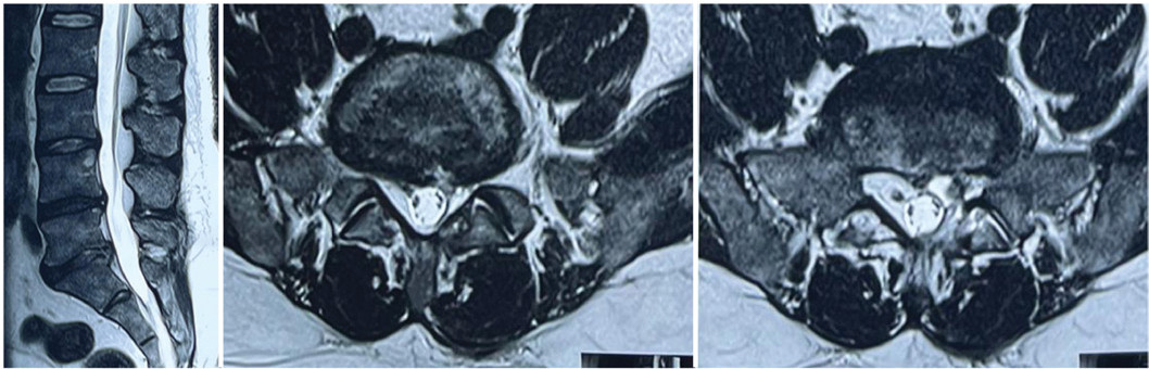
36-year-old male patient with no previous comorbidities, presenting with lower back pain for five years, radiating to the left lower limb, negative Lasegue maneuver, neurogenic claudication, and significant complaints of numbness in the lower limbs, which worsened in the sitting position, with progressive worsening in recent months. The pain intensity was assessed using the Visual Analog Scale (VAS), with 5/10 in the lumbar region and 7/10 in the left lower limb. Functional disability was assessed by the Oswestry Disability Index (ODI), indicating a moderate disability (32%). Simple X-rays without significant changes; dynamic X-rays in flexion and extension without signs of instability; and computed tomography (CT) without evidence of disc or yellow ligament calcifications. Magnetic resonance imaging (MRI) of the lumbar spine revealed increased epidural fat occupying the canal, compressing the dural sac and the cauda equina roots at the L5-S1 level (Figure 1).
Sagittal and axial cuts of the preoperative MRI showed increased fat in the epidural space.
The patient underwent conservative drug treatment and rehabilitation for six weeks without improvement. After the failure of clinical treatment, the persistence of the painful condition, and the patient’s functional incapacity, surgical treatment was chosen. The patient underwent the endoscopic technique of interlaminar decompression of the L5-S1 segment. The surgery was performed under general anesthesia, with the patient in the prone position. The interlaminar window on the left side (more symptomatic side) was identified with radioscopy, and an 8 mm skin incision was made, followed by the positioning of the dilator and the working sleeve. Under endoscopic visualization, the left interlaminar bone window opening at L5-S1 was performed using a cutting drill. After opening the yellow ligament, an increased amount of epidural fat with adherence to the dura mater was visualized (Figure 2a). After removing the fat and this adhesion, the expansion of the dural sac was observed (Figure 2b). Inspection of the posterior edge of the intervertebral disc was performed, with no evidence of extruded fragments.

Intraoperative image showing epidural fat adhered to the dural sac (A) and after its removal (B).
The patient was discharged from the hospital 6 hours after the procedure. In the immediate postoperative period, an improvement in the intensity of lower back pain (VAS 2/10) and pain in the left lower limb (VAS 0/10) was observed, which persisted until the last evaluation with 24 months of follow-up. The patient perceived a 90% improvement in numbness in the lower limbs. After six, 12, and 24 months of the procedure, the ODI was 12% (minimal disability). MRI performed six months after the procedure showed a reduction in the amount of adipose tissue in the epidural space and the dural sac occupying a larger area of the vertebral canal (Figure 3).

Sagittal and axial post-operative MRI cuts show a reduction of epidural fat and expansion of the dural sac.
DISCUSSION
SEL is a condition characterized by the excessive growth of adipose tissue in the epidural space of the spinal canal, causing compression of the spinal cord and/or nerve roots 1 . SEL was first reported in 1975, associated with the use of corticosteroids to prevent rejection of a kidney transplant.2
The most common cause of SEL is long-term exogenous steroid therapy, but it can also be associated with obesity, exposure to endogenous steroids resulting from endocrine abnormalities, surgical interventions, or idiopathic3. According to Fogel et al., its etiology can be classified into four categories: chronic steroid therapy (55.3% of cases), obesity (24.5%), hormonal diseases with endogenous steroid production, such as Cushing’s syndrome (3.2%), and idiopathic (17%).6 The prevalence is higher in middle-aged and elderly men. It is more common in the lumbar spine, followed by the thoracic spine, and rarely in the cervical spine. The vast majority of cases are asymptomatic.7
Possible clinical presentations include back pain, radiculopathy, myelopathy, neurogenic claudication, loss of sensation, difficulty urinating, weakness in the lower limbs, and, rarely, cauda equina syndrome. The exact presentation depends on the location and degree of compression.3
Magnetic resonance imaging is the gold standard for diagnosing and assessing this condition. The diagnosis requires demonstrating an increase in the amount of fat, with hyperintensity of signal on T1-weighted images and intermediate signal intensity on T2-weighted images in the epidural space. Various compressed dural sac formats are also observed in axial MRI scans, ranging from polygonal, star-shaped to the characteristic “Y sign” to almost complete obliteration.8
SEL can be classified through magnetic resonance images, according to Borré et al., based on the proportion of fat occupying the lumbosacral canal: normal (grade zero), mild (grade I: 41-50% of the canal - subtle reduction of the dural sac), these cases being asymptomatic; moderate (grade II: 51-75% of the canal - clear reduction of the dural sac), being symptomatic in 14.5% of the cases; and severe (grade III: greater than 75% of the canal - severe compression of the dural sac), all being symptomatic.9
Clinical treatment is individualized and seeks to address the cause of the disease. Weight loss in obese patients and/or the reduction and suspension of steroids are effective therapeutic strategies, especially when the diagnosis is made in the early stage. Manjila et al. propose, in their study, that bariatric intervention could be a frontline approach to managing obese patients with multifocal and multisystemic lipomatosis (spinal and extraspinal)10. However, when conservative therapies fail or when symptoms are severe, surgical management involving decompressive laminectomy and excision of hypertrophic epidural adipose tissue is indicated.3,6,7,8,9
The patient we present in this case reported lower back pain, pain radiating to the left lower limb, difficulty walking, and especially paresthesias in the legs. He had no history of obesity, steroid use, or endocrine disease, being diagnosed as having idiopathic SEL. Due to progressive clinical worsening, surgical treatment was indicated.
Traditional laminectomy, with the removal of excess epidural fat, is the standard surgical procedure, according to the literature, showing good results. However, it requires a wider incision and more aggressive dissection of the paravertebral musculature, which can cause damage to the posterior structures of the spine and result in instability and deformity in the postoperative segment. This, in addition to increasing the morbidity of the procedure, should be considered since many of these patients have associated comorbidities, which may increase the risk of complications.11
In recent years, endoscopic spine surgery has evolved considerably, with advances in optics, instruments, and endoscopic techniques allowing for excellent-quality images and, through small incisions and minimal tissue invasion, treating various spine pathologies.12
In the surgery of this case, we used an ENDOLINE endoscope with a 30-degree angulation and a 4.3 mm working channel. The high-quality images let us visualize the epidural adipose tissue and several beams encompassing the dural sac, causing compression. As they were removed with forceps, dissectors, and kerrisons, we finally obtained a view of the expanded dural sac and free S1 roots, confirmed with follow-up control exams. In this case of lumbar SEL, the endoscopic technique enabled early discharge and favorable clinical evolution, maintained throughout the 24-month follow-up.
CONCLUSION
The case of lumbar SEL was successfully treated through fully endoscopic interlaminar surgery, with improvement in pain and neurological deficit at 24 months of follow-up. New studies should be conducted to confirm the efficacy and safety of the endoscopic technique in SEL.
-
Study conducted by the Clínica Atualli, São Paulo, SP, Brazil.
REFERENCES
- 1 Ramdani H, Jidal M, Saouab R, Sahri IE, En-Nouali H, El Fenni J. Spinal epidural lipomatosis. Int J Emerg Med. 2022;15(1):14.
- 2 Lee M, Lekias J, Gubbay SS, Hurst PE. Spinal cord compression by extradural fat after renal transplantation. Med J Australia. 1975;1(7):201-3.
- 3 Kim K, Mendelis J, Cho W. Spinal Epidural Lipomatosis: A Review of Pathogenesis, Characteristics, Clinical Presentation, and Management. Global Spine J. 2019;9(6):658-65.
- 4 Hasan S, Härtl R, Hofstetter CP. The benefit zone of full-endoscopic spine surgery. J Spine Surg. 2019;5(Suppl 1):S41-56.
- 5 Gunjotikar S, Pestonji M, Tanaka M, Komatsubara T, Ekade SJ, Heydar AM, et al. Evolution, Current Trends, and Latest Advances of Endoscopic Spine Surgery. J Clin Med. 2024;13(11):3208.
- 6 Fogel GR, Cunningham PY, Esses SI. Spinal epidural lipomatosis: Case reports, literature review and meta-analysis. Spine J. 2005;5(2):202-11.
- 7 Kellett CG, Siva V, Norman ICF, Jung J, Grahovac G, Minhas P. Symptomatic Idiopathic Spinal Epidural Lipomatosis in 9 Patients: Clinical, Radiologic, and Pathogenetic Features. World Neurosurg. 2019;126:e33-40.
- 8 Yasuda T, Suzuki K, Kawaguchi Y, Seki S, Makino H, Watanabe K, et al. Clinical and imaging characteristics in patients undergoing surgery for lumbar epidural lipomatosis. BMC Musculoskelet Disord. 2018;19(1):66.
- 9 Borré DG, Borré GE, Aude F, Palmieri GN. Lumbosacral epidural lipomatosis: MRI grading. Eur Radiol. 2003;13(7):1709-21.
- 10 Manjila S, Fana M, Medani K, Kay MD, Manjila R, Bazil TG, et al. Spinal Epidural Lipomatosis Causing Lumbar Canal Stenosis: A Pictorial Essay on Radiological Grading and the Role of Bariatric Surgery Versus Laminectomy. Cureus. 2022;14(7):e26492.
- 11 Yang K, Ji C, Luo D, Li K, Xu H, Saranathan M. Lumbar laminotomy and replantation for the treatment of lumbar spinal epidural lipomatosis: A case report. Medicine (United States). 2021;100(30):E26795.
- 12 Kim HS, Wu PH, Jang IT. Current and Future of Endoscopic Spine Surgery: What are the Common Procedures We Have Now and What Lies Ahead?. World Neurosurg. 2020;140:642-53.
Edited by
-
Reviewed by:
Robert Meves