 Epidemiological profile of spinal tumor biopsies at a referral center: results over a decade
ABSTRACT
Objective: Define the epidemiological profile of spinal tumors diagnosed by percutaneous biopsy over the past ten years in a Brazilian referral center.
Methods: This was a cross-sectional, descriptive study based on a prospective database of patients who underwent spinal biopsies between 2015 and 2025. Inclusion criteria were histopathological diagnoses of benign, malignant, or pseudotumoral spinal lesions. Cases of infection were excluded. Biopsies were performed percutaneously using Jamshidi needles under fluoroscopic guidance. Variables analyzed included sex, age, histopathology, and lesion location.
Results: Total analyzed: 195 patients (after excluding 26 with infections). Female predominance: 55.8%. Age range: 18 to 65 years (66.1%). Benign lesions: 6.1% (most common: aneurysmal bone cyst and hemangioma). Malignant lesions: 40.5% (44% metastases and 32% multiple myeloma). Pseudotumoral lesions: 7.1% (e.g., Paget’s disease, SAPHO, osteonecrosis). Inconclusive/negative
results: 46.3%. Most affected area: sacrum, followed by the lumbar spine (especially L4). Malignant lesions: mostly lumbosacral; benign lesions: mostly thoracic.
Conclusion: Malignant spinal lesions are more frequent than benign ones, with metastases and myeloma being the most prevalent. The high number of inconclusive biopsies highlights the limitations of percutaneous techniques. Pseudotumoral lesions underscore the importance of biopsy in differential diagnosis. Level of Evidence IV; Case Series.
Keywords:
Spinal Neoplasms; Biopsy; Needle; Neoplasm Metastasis; Multiple Myeloma; Epidemiology; Fluoroscopy.
RESUMO
Objetivo: Definir o perfil epidemiológico dos tumores da coluna diagnosticados por biópsia percutânea em um hospital brasileiro de referência entre 2015 e 2025.
Métodos: Estudo transversal descritivo baseado em um banco de dados prospectivo de biópsias de coluna vertebral realizadas entre 2015 e 2025. Foram incluídos pacientes com diagnóstico histopatológico de tumores benignos, malignos ou pseudotumorais. Excluíram-se os casos com diagnóstico infeccioso. As biópsias foram feitas por via percutânea com agulha de Jamshidi, guiadas por fluoroscopia. Foram avaliados: sexo, idade, diagnóstico histopatológico e localização da lesão.
Resultados: Total analisado: 195 pacientes (após exclusão de 26 com infecção). Predomínio feminino: 55,8%. Faixa etária predominante: 18 a 65 anos (66,1%). Lesões benignas: 6,1% (mais comuns: cisto ósseo aneurismático e hemangioma). Lesões malignas: 40,5% (sendo 44% metástases e 32% mieloma múltiplo). Lesões pseudotumorais: 7,1% (ex: doença de Paget, SAPHO, osteonecrose). Laudos inconclusivos/negativos: 46,3%. Topografia predominante: sacro, seguido da coluna lombar (sobretudo L4). Distribuição das malignas: predomínio lombossacro; benignas, na região torácica.
Conclusões: As lesões malignas são as mais prevalentes entre os tumores da coluna vertebral, destacando-se metástases e mieloma múltiplo. Lesões benignas são menos frequentes, com predominância de cistos ósseos aneurismáticos. O alto número de laudos inconclusivos alerta para limitações das técnicas percutâneas. A presença de lesões pseudotumorais reforça a importância da biópsia para diagnóstico diferencial. Nível de Evidência IV; Série de Casos.
Descritores:
Neoplasias da Coluna Vertebral; Biópsia por Agulha; Metástase Neoplásica; Mieloma Múltiplo; Epidemiologia; Fluoroscopia.
RESUMEN
Objetivo: Definir el perfil epidemiológico de los tumores de la columna vertebral diagnosticados mediante biopsia percutánea en un hospital brasileño de referencia entre 2015 y 2025.
Métodos: Estudio transversal descriptivo basado en una base de datos prospectiva de biopsias de columna vertebral realizadas entre 2015 y 2025. Se incluyeron pacientes con diagnóstico histopatológico de tumores benignos, malignos o pseudotumorales. Se excluyeron los casos con diagnóstico infeccioso. Las biopsias se realizaron por vía percutánea con aguja de Jamshidi, guiadas por fluoroscopía. Se evaluaron: sexo, edad, diagnóstico histopatológico y localización de la lesión.
Resultados: Total analizado: 195 pacientes (tras excluir 26 con infección). Predominio femenino: 55,8%. Grupo etario predominante: entre 18 y 65 años (66,1%). Lesiones benignas: 6,1% (las más comunes: quiste óseo aneurismático y hemangioma). Lesiones malignas: 40,5% (de las cuales 44% fueron metástasis y 32% mieloma múltiple). Lesiones pseudotumorales: 7,1% (ejemplos: enfermedad de Paget, SAPHO, osteonecrosis). Resultados inconclusos/negativos: 46,3%. Topografía predominante: sacro, seguido de columna lumbar (especialmente L4). Distribución de las malignas: predominio lumbosacro; benignas: en región torácica.
Conclusiones: Las lesiones malignas son las más prevalentes entre los tumores de la columna vertebral, destacándose las metástasis y el mieloma múltiple. Las lesiones benignas son menos frecuentes, con predominancia de quistes óseos aneurismáticos. El alto número de resultados inconclusos alerta sobre las limitaciones de las técnicas percutáneas. La presencia de lesiones pseudotumorales refuerza la importancia de la biopsia para el diagnóstico diferencial. Nivel de Evidencia IV; Serie de casos.
Descriptores:
Neoplasias de la Columna Vertebral; Biopsia con Aguja; Metástasis de la Neoplasia; Mieloma Múltiple; Epidemiología; Fluoroscopía.
INTRODUCTION
Spinal tumors can be classified as primary or metastatic, with their location varying according to histological type. Primary tumors are rare, accounting for less than 10% of bone tumors, and their topographical distribution predominates in the cervical spine, followed by the thoracic and lumbar regions. Among the primary tumors, hemangioma stands out as the most common benign type, and plasmacytoma as the most frequent malignant tumor in adults.1
Vertebral metastases are responsible for up to 70% of skeletal involvement in patients with cancer. The most frequent locations are the thoracic spine (approximately 70%), followed by the lumbosacral (20%) and cervical spine (10%).2-4
Diagnosis of these lesions often requires biopsy, which can be performed using percutaneous techniques guided by imaging modalities (CT, fluoroscopy, or MRI), or through open approaches, depending on the clinical scenario and available resources.5,6 CT-guided biopsy is widely used due to its high accuracy and safety, and is especially useful in differentiating between tumoral and infectious etiologies. Magnetic resonance imaging, although free of ionizing radiation, is less commonly used due to its cost and complexity.4
In percutaneous approaches, the choice of access route depends on the anatomical location of the lesion. The transpedicular approach is preferred for vertebral body lesions, while the transforaminal approach is more indicated when there is discal involvement or marginal vertebral erosion.7,8 The instrumentation may vary and includes Jamshidi-type needles, core needles, and semi-rigid grasping forceps, the latter being associated with greater tissue sampling and diagnostic accuracy.9
Recent studies show that even in centers not specialized in oncology, such as quaternary public hospitals in Brazil, patients with spinal tumors tend to present in advanced clinical stages, with a significant proportion already showing neurological deficits upon admission and a hospital mortality rate of up to 13%.10 This reality highlights the importance of understanding the epidemiological profile of these patients to improve diagnosis and guide therapeutic approaches suited to the context of the Brazilian public healthcare system.
This study aims to define the epidemiological profile of spinal tumors diagnosed by percutaneous biopsy over the last 10 years in a Brazilian referral center for the treatment of oncological spinal lesions.
MATERIALS AND METHODS
After approval by the institutional research ethics committee under protocol number CAAE: 56429322.7.0000.5273, a descriptive cross-sectional prevalence study was conducted using a prospective database of patients diagnosed with spinal tumors between 2015 and 2025. Variables analyzed included sex, age, histopathological reports, and tumor location. Biopsies were performed using the percutaneous technique with a Jamshidi needle, via a transpedicular route or through posterior elements, guided by fluoroscopy (Figure 1). Exclusion criteria included patients whose histopathological and culture reports confirmed infection. Inclusion criteria involved histopathological diagnoses of benign, malignant, and pseudotumoral spinal lesions.

Jamshidi bone biopsy needle and cannula system with collection of bone tumor fragment for histopathological examination.
RESULTS
Out of 221 medical records analyzed, 26 were excluded from the study due to a diagnosis of spinal infection. Of the 195 patients included in the study, the demographic profile showed a predominance of females (55.8%) compared to 86 male patients (44.1%). Regarding age, 5% of patients were under 18 years old, 66.1% were between 18 and 65 years old, and 28 patients were over 65 years old.
Benign lesions accounted for 6.1% of the samples, while malignant lesions were found in 40.5% of patients. Additionally, pseudotumoral lesions were diagnosed in 7.1% of patients. Histopathological reports were negative or inconclusive in 46.3% of cases.
Among the benign lesions, aneurysmal bone cysts accounted for 66.7% of the samples, followed by hemangiomas (25%) and giant cell tumors in 8.3% of patients. (Figure 2)

Fourteen patients were diagnosed with pseudotumoral lesions, including Paget’s disease, fibrous dysplasia, histiocytic lesions, osteoporosis, Rosai-Dorfman disease, SAPHO syndrome, and focal osteonecrosis. (Figure 3)

There were 79 diagnoses of malignant lesions, of which 44% were vertebral metastases and 32% were multiple myeloma. (Figure 4)

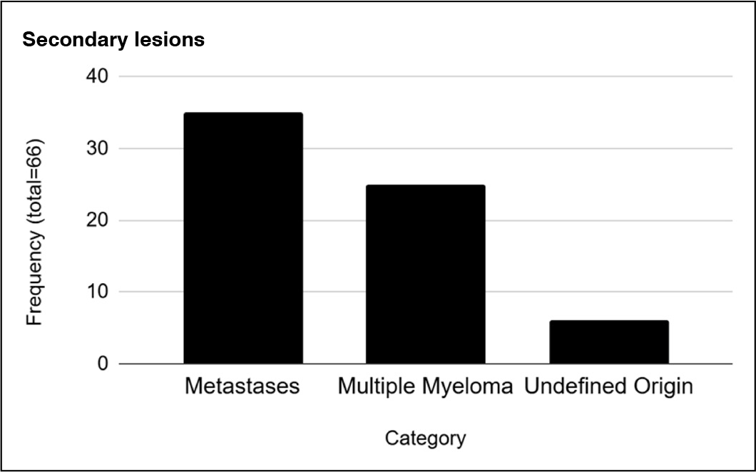
Additionally, 90 samples had negative or inconclusive histopathological reports. Twenty-five patients were diagnosed with primary spinal lesions, of which 12 were benign and 13 malignant. Secondary lesions totaled 66 patients, specifically 35 with metastases, 25 with multiple myeloma, and 6 of undefined origin. (Figure 5)

Distribution of secondary lesions in the spine (Metastases = 53%, Multiple Myelomas = 37.8%, Neoplasm of undefined origin = 9%).
As shown in Figure 6, the overall distribution of lesion incidence, regardless of neoplastic or pseudotumoral nature, showed that multiple myeloma was the most prevalent, followed by metastases, with adenocarcinomas being the most common among them, followed by carcinomas.

Overall distribution of the count of neoplastic lesions in the spine by histopathological type.
Regarding the most affected spinal levels, regardless of histological type, the sacrum had the highest number of lesions, followed by the lumbar vertebrae, especially the fourth lumbar vertebra (L4) (Figure 7). In this study, when the lesion affected more than one vertebral level in the same patient, this involvement was counted and included in the statistics, as one of the study objectives was to assess the predominance of histological type by topography. (Figure 8)


Topographic distribution of the number of neoplastic lesions by affected vertebral segment.
Malignant lesions were more frequent in the lumbosacral spine, followed by the thoracic and cervical spines. Benign lesions were more frequently located in the thoracic spine, followed by the lumbar spine and sacrum. (Figure 8)
DISCUSSION
Spinal tumors can be classified as primary or secondary. Primary lesions are relatively rare, comprising less than 10% of all spinal tumors. The spine is the most common skeletal site for secondary lesions. Tumors can also be classified as benign or malignant.1
In the present study, 12 benign lesions were identified: 8 aneurysmal bone cysts, followed by three hemangiomas and one giant cell tumor. These findings diverge from the literature, which identifies hemangiomas as the most common benign spinal lesion, followed by osteoid osteoma or osteoblastoma, and giant cell tumors in approximately 5% of cases. This discrepancy may be explained by the hospital’s status as a referral center for complex cases, receiving more severe or unresolved cases from other institutions. Hemangiomas are typically diagnosed radiologically, with the “polka-dot sign” on axial CT and hyperintensity on both T1 and T2-weighted MRI images being classic findings. A biopsy is only needed in atypical or uncertain cases.11,12
On the other hand, the data from this study support epidemiological literature by reinforcing the high prevalence of malignant spinal lesions, particularly metastases and multiple myeloma.13 Of the 195 biopsies analyzed, 79 were diagnosed as malignant, with 44% being metastases 32% multiple myeloma, and 24% were distributed among unclassified malignant lesions, chordoma, plasmacytoma, and sarcomas. These figures are similar to those reported by Alexandre et al. (2022), who found 67.3% metastases and 20.7% multiple myeloma in a cohort treated at a quaternary public hospital.
Another relevant finding was the high number of inconclusive or negative pathology reports, totaling 90 samples (46.1%). This may be partially explained by the predominant use of the percutaneous and transpedicular approach with Jamshidi needles, which, while safe and minimally invasive, may have sampling limitations - especially in sclerotic lesions or those with significant extravertebral extension. The use of fluoroscopy instead of CT, which provides greater accuracy, may also contribute to the high number of negative or inconclusive results.3
The presence of 14 cases of pseudotumoral lesions (7.2%) is also noteworthy, as these conditions - such as Paget’s disease, focal osteonecrosis, and SAPHO syndrome - often mimic malignant neoplasms clinically and radiologically. This study underscores the importance of histological biopsy for the differential diagnosis of spinal lesions, particularly when infectious or degenerative diseases are also in the differential, consistent with findings from other studies that included diagnoses beyond metastases and multiple myeloma, as shown by Alexandre et al. (2022).10
In our study, benign tumors occurred more frequently in the thoracic spine, consistent with the epidemiological study conducted by Zhou et al.14 Malignant tumors were more common in the lumbar and sacral regions, differing from literature data, which often indicate the thoracic spine as the most affected region.
CONCLUSION
It is concluded that malignant lesions are more common in the spine, while benign lesions are considerably less frequent. Attention should also be paid to the diagnosis of pseudotumoral lesions, which present a wide range of manifestations and can complicate diagnosis and treatment.
-
Study conducted by the Instituto Nacional de Traumatologia e Ortopedia Jamil Haddad, Av. Brasil, 500, Caju, Rio de Janeiro, RJ, Brazil. 20940-070.
-
Reviewed by:
Robert Meves
REFERENCES
-
1 Chi JH, Bydon A, Hsieh P, Witham T, Wolinsky JP, Gokaslan ZL. Epidemiology and demographics for primary vertebral tumors. Neurosurg Clin N Am. 2008;19(1):1-4. doi: 10.1016/j.nec.2007.10.005.
» https://doi.org/10.1016/j.nec.2007.10.005. -
2 Truong VT, Al-Shakfa F, Phan P, Newman N, Boubez G, Shedid D, et al. Does the Region of the Spine Involved with Metastatic Tumor Affect Outcomes of Surgical Treatments? World Neurosurg. 2021;156:e139-e151. doi: 10.1016/j.wneu.2021.09.014.
» https://doi.org/10.1016/j.wneu.2021.09.014. -
3 Amelot A, Terrier LM, Cristini J, LeNail LR, Buffenoir K, Pascal-Moussellard H, et al. Approaching spinal metastases spread profile. Surg Oncol. 2019;31:61-66. doi: 10.1016/j.suronc.2019.08.007.
» https://doi.org/10.1016/j.suronc.2019.08.007. -
4 Dang L, Liu X, Dang G, Jiang L, Wei F, Yu M, et al. Primary tumors of the spine: a review of clinical features in 438 patients. J Neurooncol. 2015;121(3):513-20. doi: 10.1007/s11060-014-1650-8.
» https://doi.org/10.1007/s11060-014-1650-8. -
5 Saifuddin A, Palloni V, du Preez H, Junaid SE. Review article: the current status of CT-guided needle biopsy of the spine. Skeletal Radiol. 2021;50(2):281-299. doi: 10.1007/s00256-020-03584-9.
» https://doi.org/10.1007/s00256-020-03584-9. -
6 Liu M, Sequeiros RB, Xu Y, He X, Zhu T, Li L, et al. MRI-guided percutaneous transpedicular biopsy of thoracic and lumbar spine using a 0.23t scanner with optical instrument tracking. J Magn Reson Imaging. 2015;42(6):1740-6. doi: 10.1002/jmri.24924.
» https://doi.org/10.1002/jmri.24924. -
7 Basu S, Gala RB, Solanki AM. Percutaneous wide bore needle biopsy for spinal lesions using the kyphoplasty cannula-a technical note. Eur Spine J. 2021 Oct;30(10):2852-2856. doi: 10.1007/s00586-021-06819-y.
» https://doi.org/10.1007/s00586-021-06819-y. -
8 Lee SA, Chiu CK, Chan CYW, Yaakup NA, Wong JHD, Kadir KAA, et al. The clinical utility of fluoroscopic versus CT guided percutaneous transpedicular core needle biopsy for spinal infections and tumours: a randomized trial. Spine J. 2020;20(7):1114-1124. doi: 10.1016/j.spinee.2020.03.015
» https://doi.org/10.1016/j.spinee.2020.03.015 -
9 Marruzzo D, Mancini F, Ricciuti V, Barbieri FR, Preziosi R, Pagano S, et al. Modified percutaneous biopsy of the spine: improvement of the technique. Eur Spine J. 2023;32(1):221-227. doi: 10.1007/s00586-022-07384-8.
» https://doi.org/10.1007/s00586-022-07384-8. -
10 Alexandre M, Santos WZ, Mendonça RGM, Gotfryd AO, Caffaro MFS, Meves R. Profile of patients with spine tumor operated in a South American reference service: epidemiological study. Coluna/Columna. 2022;21(4):262-528. doi:10.1590/S1808-185120222104262528.
» https://doi.org/10.1590/S1808-185120222104262528. -
11 Gaudino S, Martucci M, Colantonio R, Lozupone E, Visconti E, Leone A, et al. A systematic approach to vertebral hemangioma. Skeletal Radiol. 2015;44(1):25-36. doi: 10.1007/s00256-014-2035-y.
» https://doi.org/10.1007/s00256-014-2035-y. -
12 Safaee MM, Carrera DA, Chin CT, Mashhood A, Eisenmenger L, Liang NE, et al. Diagnostic Challenges in Primary Sacral Tumors and the Yield of Computed Tomography-Guided Needle Biopsy in the Modern Era. World Neurosurg. 2020;138:e806-e818. doi: 10.1016/j.wneu.2020.03.094.
» https://doi.org/10.1016/j.wneu.2020.03.094. -
13 Choi D, Crockard A, Bunger C, Harms J, Kawahara N, Mazel C, et al. Review of metastatic spine tumour classification and indications for surgery: the consensus statement of the Global Spine Tumour Study Group. Eur Spine J. 2010;19(2):215-22. doi: 10.1007/s00586-009-1252-x.
» https://doi.org/10.1007/s00586-009-1252-x. -
14 Zhou Z, Wang X, Wu Z, Huang W, Xiao J. Epidemiological characteristics of primary spinal osseous tumors in Eastern China. World J Surg Oncol. 2017;15(1):73. doi: 10.1186/s12957-017-1136-1.
» https://doi.org/10.1186/s12957-017-1136-1.

 Source : Authors.