
 Management of cervical kyphotic deformity after intradural tumor resection in pediatric patients
ABSTRACT
This case series describes pediatric patients with progressive cervical kyphotic deformity after intradural tumor resection, the surgical strategies employed, and their clinical outcomes. A retrospective study was conducted on five patients who underwent surgical correction between March 2002 and November 2024 at a public referral center in Brazil. All patients developed progressive kyphosis (>45°) and underwent corrective surgery. Clinical, radiographic, and follow-up data were collected. The mean age at the time of tumor resection was 5.6 years. All cases involved facetectomy, which was bilateral in 60% of patients. The anterior approach was employed in 80% of cases (used alone or in combination). The mean angular correction achieved was 54.4°. Four patients showed neurological improvement according to the Frankel scale, while one patient had neurological deterioration. Therefore, these findings highlight the high risk of kyphotic deformity progression and neurological deficit after tumor resection in pediatric patients. Preoperative strategies should be adopted to prevent the development of the deformity. Once kyphotic deformity is established, different surgical approaches should be considered and complementary techniques for effective correction. Level of Evidence IV; Case Series.
Keywords:
Kyphosis; Cervical Vertebrae; Spinal Cord Neoplasms.
RESUMO
Essa série de casos descreve os pacientes pediátricos com deformidade cifótica cervical progressiva após ressecção de tumores intradurais, as estratégias cirúrgicas e os seus desfechos clínicos. Estudo retrospectivo foi realizado com cinco pacientes operados entre março de 2002 e novembro de 2024, em instituição pública de referência no Brasil. Todos os pacientes apresentavam cifose progressiva (>45°) e foram submetidos a correção cirúrgica. Foram coletados dados clínicos, radiográficos e evolutivos. A média de idade dos pacientes na ressecção tumoral foi de 5,6 anos. Todos os casos envolveram facetectomia, sendo bilateral em 60%. A abordagem anterior foi empregada em 80%, isolada ou combinada. A média de correção angular foi 54,4°. Quatro pacientes apresentaram melhora neurológica (escala de Frankel) e um apresentou piora. Dessa forma, observa-se que a deformidade cifótica cervical pós-ressecção tumoral em crianças apresenta alto risco de progressão e de déficit neurológico. Durante o planejamento pré-operatório da ressecção inicial, estratégias específicas devem ser adotadas para prevenir o desenvolvimento da deformidade. Uma vez que a cifose está instalada, é fundamental considerar diferentes abordagens cirúrgicas e a associação de técnicas complementares para uma correção eficaz. Nível de Evidência IV; Série de Casos.
Descritores:
Cifose; Vértebras Cervicais; Neoplasias da Medula Espinal.
RESUMEN
Esta serie de casos describe a pacientes pediátricos con deformidad cifótica cervical progresiva tras la resección de tumores intradurales, las estrategias quirúrgicas empleadas y sus desenlaces clínicos. Se realizó un estudio retrospectivo en cinco pacientes que se sometieron a corrección quirúrgica entre marzo de 2002 y noviembre de 2024 en un centro público de referencia en Brasil. Todos los pacientes desarrollaron cifosis progresiva (> 45°) y se sometieron a cirugía correctiva. Se recopilaron datos clínicos, radiográficos y de seguimiento. La edad media en el momento de la resección del tumor fue de 5,6 años. Todos los casos implicaron facetectomía, que fue bilateral en el 60% de los pacientes. El abordaje anterior se empleó en el 80% de los casos (solo o en combinación). La corrección angular media alcanzada fue de 54,4°. Cuatro pacientes mostraron mejoría neurológica según la escala de Frankel, mientras que un paciente tuvo deterioro neurológico. Por lo tanto, estos hallazgos resaltan el alto riesgo de progresión de la deformidad cifótica y déficit neurológico después de la resección del tumor en pacientes pediátricos. Se deben adoptar estrategias preoperatorias para prevenir el desarrollo de la deformidad. Una vez establecida la deformidad cifótica, se deben considerar diferentes abordajes quirúrgicos y técnicas complementarias para una corrección eficaz. Nivel de Evidencia IV; Serie de Casos.
Descriptores:
Cifosis; Vértebras Cervicales; Neoplasias de la Médula Espinal.
INTRODUCTION
Surgical resection is typically the primary treatment option for intradural spinal tumors. The surgical approach performed via the posterior access results in detachment of the paravertebral muscles and affects key structures for cervical spine stability, which is notably frequent with facet joint violation, mainly for tumors treated with laminoplasty and laminectomy.1,2 Additionally, the cervical kyphosis may be accentuated by the effects of radiotherapy, causing anterior or lateral vertebral wedging due to irregular changes in the growth plates.3
Cervical kyphosis following laminectomy occurs gradually with the disruption of the posterior elements, increasing the compressive force on the anterior elements, thus changing the axial plane of the spine.3 This deformity may lead to significant complications, including neurological deficits, dysphagia, and respiratory disorders.1 The incidence of post-laminectomy and post-laminoplasty kyphosis is reported to be higher in children than in adults (22-100% versus 14-30%, respectively).3-5 In addition, other risk factors contribute to post-laminectomy kyphotic deformity, such as tumor size, tumor location in junctional areas, resection involving the facet joints, exposure to radiotherapy, and prior spinal deformity.6,7
Despite the clinical relevance of cervical kyphosis following tumor resection, few studies describe the technical strategies and evolutionary details for the management of pediatric patients treated at public healthcare institutions. Preventive measures during tumor resection are essential to manage this condition and include preoperative identification of spinal deformities, preservation of stabilizing structures such as the semispinalis muscle of C2; postoperative use of spinal orthoses, and evaluation of treatment with posterior, anterior, combined fixation, and osteotomies for the correction of deformities.6-8 In this context, the present study aims to present a case series of five pediatric patients treated at an orthopedic institution within Brazil’s Unified Health System, detailing the surgical strategies and clinical implications based on literature data.
METHODS
Study design and inclusion criteria
This retrospective study of a case series in pediatric patients with cervical kyphotic deformity after intradural tumor resection was treated at a reference public orthopedic institution in Brazil, between March 2002 and November 2024. Patients were identified through the institutional clinical database. Patients included in the study were diagnosed with an intradural tumor, underwent surgical approach via posterior access (laminectomy or laminoplasty), and subsequently developed cervical kyphotic deformity requiring surgical correction. Of the five patients evaluated, three were male. The mean age of participants at the time of tumor resection was 5.6 years (range: 2 to 11 years), and the mean age at the time of the kyphotic deformity correction was 10.4 years (range: 6 to 17 years). This study was approved by the Ethics and Research Committee of the institution (approval number: 6,719,379), and informed consent was obtained from the legal guardians of the patients. Clinical cases with incomplete clinical data or radiographic records were excluded from the study.
Clinical and imaging data
The mean number of spine levels treated by laminoplasty, or laminectomy was 3.4 (range: 2 to 4 levels), while the mean number of levels treated by facetectomy was 1.4 (range: 1 to 2 levels). Clinical, surgical, and radiological information was collected from the medical records and imaging exams. Clinical and imaging data were accessed to collect the following information: tumor type, type of initial surgical approach, number of spine levels addressed, facetectomy (unilateral or bilateral), cervical kyphosis angle (preand postoperative), number of surgical revisions, possible complications, neurological status according to the Frankel scale, and use of adjuvant radiotherapy.
The cervical kyphosis angles were measured based on lateral radiographs in the standing position, using as a reference the angle between the end plates of vertebral bodies at the limits of the deformity. Measurements were performed using digital tools available in the institution. Clinical evolution was assessed based on neurological evolution according to the Frankel scale, the need for reoperations, and the occurrence of postoperative complications.
RESULTS
In this study, patients exhibited a mean preoperative kyphosis angle of 80° (range: 45°-109°). Surgical correction of the kyphotic deformity resulted in an average final angle of 54.4° (range: 25°-69°). Two patients required surgical revision. Regarding neurological evaluations, one patient had a clinical worsening, while the others demonstrated improvement, according to the Frankel scale. (Table 1)
Clinical data of the patients with cervical kyphosis after intradural tumor resection from this study.
Case presentation
Case 1
A 4-year-old male patient presented with a cervicothoracic post-laminectomy kyphotic deformity measuring 109°. He had previously undergone a multilevel laminectomy (C6, C7, T1, T2) and bilateral facetectomy at one level for resection of an intradural tumor at the age of 2. In 2002, posterior spinal fusion from C5 to T8 was performed using Hartshill rods and sublaminar titanium wires, following three weeks of preoperative traction. Two years later, the patient developed progressive kyphosis and rod migration into the laminectomy site, leading to new-onset neurological dysfunction. A posterior revision surgery was then carried out, with replacement of the implants using third-generation instrumentation, including lateral mass screws in the cervical spine and pedicle screws in the thoracic spine. At age 9, the patient experienced a recurrence of the cervicothoracic kyphosis associated with further neurological decline. A third revision procedure was performed using structural autologous fibular strut grafting via a costotransversectomy approach. (Figure 1 e 2)

Preoperative images of case 1. A - Lateral radiograph of the cervicothoracic spine showing kyphosis of 109°. B - Lateral radiograph of the cervicothoracic spine with preoperative traction. C, D - Panoramic AP and lateral radiographs after arthrodesis using Hartshill rod implants. E, F - CT scans in sagittal and axial reconstruction showing rod migration into the medullary canal with spinal cord compression associated with neurological deficit.

Postoperative images of case 1. A - Lateral radiograph of the cervicothoracic spine after first surgical revision with third-generation instrumentation. B - Sagittal CT of the cervicothoracic region after revision with new instrumentation. C - Sagittal CT showing progression of the kyphotic deformity. D - Intraoperative image demonstrating anterior fusion with a fibular structural graft. E - T2-weighted sagittal MRI showing anterior fusion using fibular graft and iliac crest graft. F - Sagittal CT showing fusion with fibular and iliac crest grafts. G - Clinical image of the patient in lateral view. H - Sagittal CT in follow-up reconstruction confirming bone consolidation of the fibular and iliac crest structural grafts.
Case 2
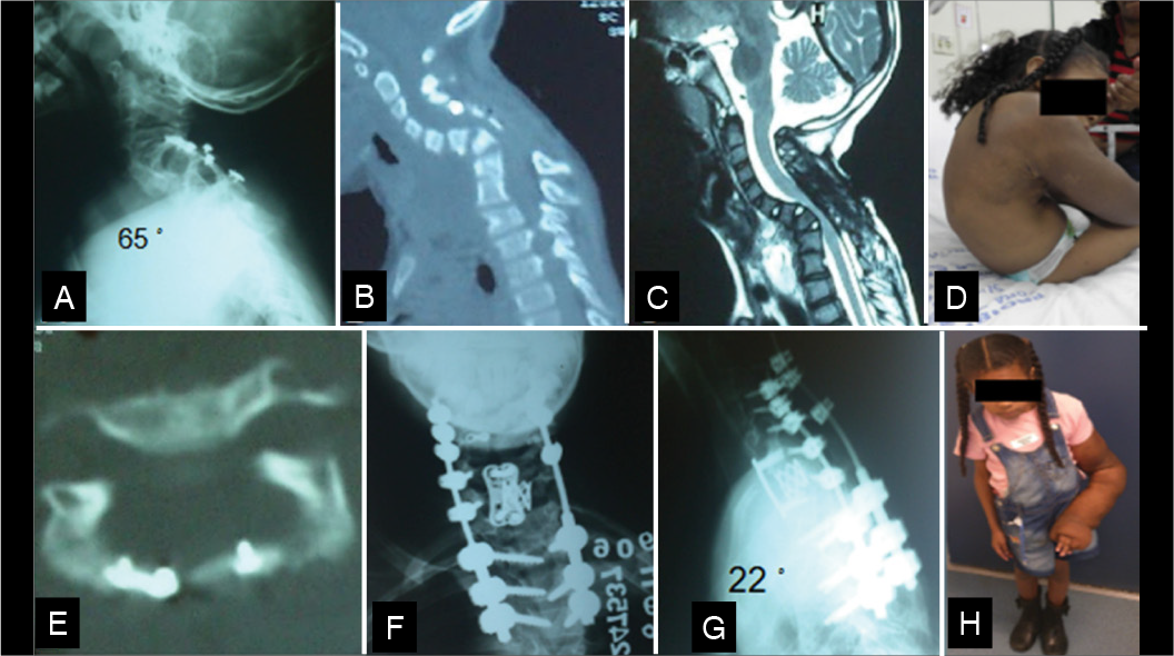
A 7-year-old female patient with neurofibromatosis type 1 underwent neurofibroma resection at the age of 3, performed via a four-level laminoplasty (C4-C7) combined with bilateral facetectomies at two levels. She had vertebral dysplasia. Two years later, the patient developed a swan neck deformity and tetraparesis. Surgical intervention was performed using anterior decompression with C7 corpectomy and anterior fusion, in addition to posterior cervicothoracic fusion, resulting in complete neurological recovery. (Figure 3)

Postoperative and preoperative images of case 2. A - X-ray of the cervicothoracic region in lateral view, showing the deformity after laminoplasty using small reconstruction plates. B - Sagittal CT of the cervicothoracic region showing angular deformity and vertebral dystrophy. C - T2-weighted sagittal MRI of the cervicothoracic region showing compression of the spinal cord. D - Clinical photograph of the patient with inability to ambulate. E - Laminoplasty at C4. F, G - Postoperative AP and lateral radiographs of the cervicothoracic spine, showing the reconstruction cage after C7 corpectomy using an anterior plate and posterior instrumentation with screws in the lateral masses in the cervical spine and pedicle screws in the thoracic region. H - Clinical image of the patient in postoperative follow-up.
Case 3
A 17-year-old female diagnosed with neurofibromatosis presented with intrinsic hand muscle atrophy and gait disturbance on neurological examination. At the age of 6, she underwent a 3-level laminoplasty (C3-C5) with extensive bilateral facetectomy at C4, which subsequently progressed to kyphotic deformity and severe cervical stenosis. Surgical intervention was performed involving anterior C4 corpectomy and C3-C5 fusion, resulting in partial neurological improvement. (Figure 4)

Postoperative and preoperative images of case 3. A - Sagittal CT indicating severe kyphosis and stenosis of the spinal canal. B - T2-weighted sagittal MRI showing spinal cord compression at C4 level. C - Image demonstrating laminoplasty failure with persistent spinal cord compression. D - Intraoperative image of neurofibroma during anterior approach. E - Postoperative AP radiograph showing partial correction of the deformity with a cervical cage and anterior plate, with decompression of the spinal canal. F - Postoperative CT scan confirming partial correction and decompression of the spinal canal.
Case 4
An 11-year-old male patient with neurofibromatosis type 1 presented with left arm monoparesis and swan neck deformity, associated with kyphosis of 102°. Six years earlier, the patient had undergone neurofibroma resection with C3 laminectomy combined with right side facetectomy. Surgical correction was done with an anterior osteotomy approach, including a two-level vertebrectomy with buttress plate and occipitocervical fusion, which resulted in complete neurological recovery. (Figure 5)

Postoperative and preoperative images of case 4. A - Lateral radiograph showing 102° kyphosis. B - Sagittal CT scan showing C3 dysplasia. C - T2-weighted sagittal MRI showing spinal cord compression secondary to the deformity. D - CT scan with posterior 3D reconstruction showing laminectomy of C2 and C3 associated with right facetectomy. E - Axial CT showing laminectomy of C2 and C3 with facetectomy. F - Intraoperative image showing traction positioning. G - Intraoperative fluoroscopy showing anterior structural graft with buttress plate. H - Sagittal CT showing bone consolidation. I, J - Postoperative AP and lateral radiographs showing correction of the deformity and occipitocervical arthrodesis.
Case 5
A 11-year-old male patient presented with severe tetraparesis and impaired hand function due to a swan neck deformity associated with 77° of kyphosis, a condition that developed following resection of an extradural undifferentiated malignant tumor via C4-C6 laminoplasty, complete bilateral facetectomies at all levels, and adjuvant radiotherapy. A three-level anterior osteotomy and posterior cervical fusion performed surgical correction. This patient had two complications: a cerebrospinal fluid (CSF) leak, which was managed with external lumbar drainage; and distal junctional kyphosis, which required extension of the fusion to the thoracic spine. (Figure 6)

Postoperative and preoperative images of case 5. A, B - AP and lateral radiographs of the cervical spine with the patient using a cervical collar. The 77° kyphosis and the presence of reconstruction miniplates are revealed. C - Sagittal CT reconstruction demonstrating severe cervical kyphotic deformity. D - Axial T1-weighted MRI showing spinal cord compression. E - Axial CT showing extensive facetectomy and failure of laminoplasty consolidation at one cervical level. F - Clinical image showing CSF fistula-related wound bulging, successfully managed with conservative treatment. G - Immediate postoperative lateral radiograph of the cervical spine demonstrating partial correction of the deformity.
DISCUSSION
Epidemiology
The incidence of post-laminectomy and post-laminoplasty kyphosis is higher in children than in adults.3,5 This increased susceptibility in pediatric patients is attributed to their growth potential and the higher cartilage content in the vertebral bodies, making them more prone to wedge deformity.1,5 In the present study, this correlation was particularly evident, since all cases treated at our institution occurred in children (mean age of 5.6 years).
Kyphosis Assessment
Simple static radiographs are useful for evaluating the morphology and severity of the deformity, as well as previous instrumentation.9 Flexion and extension radiographs reveal the rigidity and flexibility of the deformity, while panoramic orthostatic radiographs help identify coexisting thoracolumbar deformities.9 It is important to consider the influence of spinopelvic parameters and thoracic deformities on the biomechanics of cervical deformities. Radiographic parameters such as chin-brow vertical angle (CBVA), C2-7 SVA, T1 slope (T1S), neck tilt, and TIA can be measured on lateral radiographs in both neutral and extension positions to aid in classifying the severity of deformity.8,9
Thoracolumbar deformities should be considered when planning treatment for cervical deformities, as they may worsen the overall spinal alignment and lead to future reoperations.10 CT scans are valuable for assessing anatomical details for surgical planning. MRI is essential for evaluating the spinal cord and nerve roots, and is also helpful in assessing spinal cord strain.11 In the present study, CT was used to assess laminoplasty consolidation, the extent of facetectomy, and spinal deformity. In addition, MRI was essential to assess spinal cord strain.
Among the classifications for cervical deformities, the Kim-ISSG classification stands out,11 taking into account both static radiographic findings and the patient’s ability to compensate for misalignment, as assessed by dynamic radiographs. In this classification, patients are divided into three groups. Group 1, referred to as the “Flat Neck” group, is characterized by a large T1S-CL imbalance due to insufficient cervical lordosis. Partial correction is observed with extension, suggesting potential for less invasive treatment. Group 2, defined as the “Focal Deformity” group, is characterized by a significant localized kyphosis between two adjacent vertebrae, with normal cervical SVA and typically small T1S. This represents a focal and rigid deformity limited to the cervical spine. Group 3, referred to as the “Cervicothoracic Deformity” group, is associated with a T1S-CL imbalance not due to inadequate cervical lordosis but rather driven by high T1S. This deformity is usually found in the thoracic or thoracolumbar spine. Group 2 (Focal Deformity) was the pattern observed in our patient cohort.
Also noteworthy is the classification proposed by the Cervical Spine Research Society - Europe (CSRS-Europe),12 which categorizes cervical kyphotic deformity into four groups (A-D). Type A consists of cervical or cervicothoracic kyphosis with preserved regional and global sagittal balance. Type B corresponds to kyphosis associated with increased C2-7 SVA and C2-S1 SVA, indicating global sagittal imbalance. Type C consists of cervicothoracic kyphosis partially compensated by cervical lordosis, but with persistent imbalance. Type D corresponds to patients with adequate global sagittal alignment maintained through compensatory cervical lordosis.
What are the risk factors for kyphosis after a laminectomy?
Risk factors for post-laminectomy kyphosis include young age (particularly under 13 years), extensive laminectomy involving the cervical or upper thoracic spine, facetectomy, malignant tumors, tumor recurrence, need for postoperative radiotherapy, laminectomy involving more than four levels, syringomyelia, and the presence of preexisting cervical deformity.5-7 Certain tumors, beyond the mechanical impact of their resection, may lead to neurological deficits that result in muscular weakness.5,7,13 Radiotherapy contributes to deformity development through its catabolic effects, often causing early-onset kyphosis due to irregular growth of the vertebral endplates, making it an increased risk factor compared to laminectomy or laminoplasty alone.13 In our case series, all patients were under 13 years of age at the time of primary tumor resection. Facetectomy was performed in all cases, with bilateral facetectomies in three patients, in which one of them underwent resection at four levels and the other one at two levels. In addition, one patient received radiotherapy. These findings are consistent with current literature, which associates early-age tumor resection, extensive facetectomy, and radiotherapy as major contributors to the development of postoperative cervical deformity.
Is posterior fixation indicated for the prevention of kyphosis?
The indication for decompression and posterior fixation should consider the presence of neurological deficits since patients with significant neuromuscular dysfunction may benefit from prophylactic instrumentation.13,14 Some studies discourage prophylactic posterior fusion in patients at high risk of tumor recurrence and for those not receiving adjuvant chemotherapy, due to the potential difficulty in assessing recurrence and bone fragility.13 Therefore, when considering posterior fixation, it is essential to evaluate multiple factors, including patient-specific risk factors, radiographic findings, and overall oncological status. In the present study, none of the patients underwent posterior instrumentation during primary tumor resection.
Preventive strategies for kyphosis after tumor resection
The primary goal in surgical treatment should be the complete resection of the intramedullary tumor. Oncological treatment must be prioritized over deformity management, since it directly impacts patient survival. If a preexisting deformity is present, it is important to evaluate whether the patient should be treated in a single or multiple stages.
Reconstruction with a microsurgical plate is recommended during laminoplasty. If a laminectomy is chosen, the suture of the musculature, muscular fascia, and subcutaneous tissue should be performed using continuous non-absorbable sutures to facilitate the identification of the layers up to the dura mater in the case of potential future reoperations.6,8,15
When to treat kyphosis after intradural tumor resection
Two characteristics indicate the need for surgical treatment: progressive kyphosis and clinical symptoms. If the plumb line drawn from the anterior process to the odontoid falls anterior to the manubrium, this suggests upper thoracic spine involvement. Therefore, the vertical and sagittal axes of C2-C7 should be evaluated, in which distances exceeding 4 cm indicate significant cervical disability. In addition, it is essential to determine the reducibility of the deformity through imaging assessments such as CT and extended lateral radiographs.6,8,15
CT also allows the evaluation of facets, disc spaces, fusions, and instrumentation. CT angiography is valuable for the vascular study of vertebral arteries to assist in surgical planning and risk mitigation. In our case series, all patients had progressive kyphotic deformity exceeding 45° at the time of surgical indication. Preoperative neurological status was classified as Frankel C in 60% of cases, Frankel D in 20%, and Frankel E in 20%, indicating that most patients already had neurological impairment before surgical correction.
Rigidity of kyphosis
In rigid kyphosis, the location of ankylosis should be considered. In the case of ankylosed facets, correction may involve compensatory hyperlordosis or disruption of the facet fusion. This can be done via an anterior approach with removal of the anterior disc and body expander, or a posterior approach with facet osteotomy and posterior fixation.
In cases of anterior fusion only, the anterior osteotomies usually correct the deformity. When both anterior and posterior fusions are present, an anterior osteotomy combined with distraction is indicated. Alternatively, a posterior pedicle subtraction osteotomy (PSO) or a three-stage osteotomy (anterior-posterior-anterior or posterior-anterior-posterior) may be required.6,8 In the present study, no patient underwent PSO or three-stage osteotomy.
Flexible kyphosis can be managed with preoperative or intraoperative traction and soft tissue release. Using the anterior approach, discectomies in kyphotic segments with lordotic graft placement can increase the height of the anterior column and restore cervical lordosis. Plating is important to prevent loss of deformity correction and graft migration. Care must be taken to avoid over-distraction during lordosis formation, which can cause neural damage, ligamentum flavum buckling, and thus result in canal stenosis and implant failure in patients with low bone density.8
Posterior instrumentation and fusion depend on the extent of deformity and bone quality. An exception to the use of the posterior approach before anterior reconstruction in patients with rigid cervical kyphosis without ankylosed facet joints is the presence of posterior scar tissue or spinal compression that poses a risk to the patient’s health if an anterior approach is performed.6 Three patients in the present study underwent corpectomy, one underwent anterior uncus osteotomy, and one underwent an exclusively posterior approach.
The use of anterior corpectomy for spinal cord recovery in cases of compression remains debatable. It has been reported that discectomy with reconstruction is sufficient for these cases. Nonetheless, many authors indicate the corpectomy for the treatment of fixed kyphosis or focal angular kyphotic deformity secondary to a wedge-shaped vertebral body.8 It is important to highlight that corpectomies involving more than three levels are associated with high instrumentation failures.8,16,17 In such cases, an intermediate fixation point to reduce the risk of pseudarthrosis should be considered. Reconstruction of the posterior segment with isolated instrumentation may also be indicated, in which the inclusion of all levels is essential. The circumferential approach is indicated in specific situations, such as severe deformity, osteoporosis, risk of pseudarthrosis (e.g., diabetes, smoking, previous radiotherapy), history of multiple surgeries, short life expectancy, fragile bone fixation, severe narcotic dependence, and need for multilevel osteotomy. In our case series, the anterior approach was used in 80% of patients. Among these, a combined approach (anterior and posterior) was performed in three cases. The anterior decompression technique consisted of one-level corpectomy in three patients, two-level vertebrectomy in one patient, and three-level osteotomy in a patient with an undifferentiated malignant tumor. In three cases with the anterior approach, stabilization was achieved using an anterior plate, whereas the case undergoing osteotomy was reconstructed with a cage. The combined approach was performed in three patients, one of whom developed distal junctional kyphosis (DJK) and required a new surgical intervention with extension of the instrumentation to thoracic levels. It is important to note that this patient had a malignant tumor and had undergone radiotherapy, which increases the risk for complications. For one patient, an exclusively posterior approach was performed. Although initially neurologically intact, this patient developed implant migration, requiring a new intervention with reconstruction using a structural graft and new instrumentation.
Use of cervical traction
Cervical traction should be considered for all patients with complex cervical deformities. In flexible cases, traction can reduce the rigidity of deformity and the degree of kyphosis, in addition to promoting some muscular adaptation preoperatively.6 In the present study, only one of the patients underwent preoperative traction for a period of three weeks. Intraoperative traction was performed in two patients (cases 4 and 5).
Osteotomy techniques
Some types of rigid cervical kyphosis can be corrected using exclusively anterior osteotomy through different techniques.18,19 The technique described by Daniel Riew et al. in 201919 involves multiple discectomies associated with uncus osteotomy at the apex of the deformity and adjacent levels, allowing a broad and multilevel correction of a cervical kyphotic deformity. Another technique is corpectomy with replacement by cortical graft or basket of Harms with cancellous graft, associated with anterior fixation with plate and fixed-angle screws.8 Opening wedge osteotomy is reserved for severe deformities and indicated for patients with diffuse cervical ankylosis, allowing an aggressive correction. Posterior approaches include the Smith-Petersen Osteotomy (SPO), which requires a mobile disc with relatively preserved disc height. Pedicle Subtraction Osteotomy (PSO) is a three-column corrective technique used mainly in cases of fixed circumferential deformity.20-23
Ideal extent of arthrodesis
The extent of arthrodesis should be capable of restoring spinal alignment, allowing horizontal gaze, and provide sufficient stability to prevent mechanical complications, such as rod breakage, screw pullout, PJK, or PJD. At the same time, mobility preservation is desirable. Therefore, the use of cervical pedicle screws provides greater resistance than lateral mass screws and may be a strategy to reduce the number of arthrodesis levels.24 There is no consensus in the literature on the ideal extent of arthrodesis in the treatment of kyphotic deformities. The surgical strategy must be achieved with maximal preservation of spinal mobility.
CONCLUSIONS
The management of cervical kyphotic deformities following intradural tumor resection is challenging and requires meticulous preoperative planning. Detailed clinical and imaging evaluation of each case is essential, as is the surgeon’s expertise in the different surgical techniques and available strategies to propose the most appropriate treatment while minimizing the risk of complications. Special attention should be given to preventive measures during the initial tumor resection. It is noted that facetectomy should be avoided during tumor resection, and if necessary, arthrodesis should be performed to prevent deformity. Treatment in cases of kyphosis after tumor resection may involve osteotomies, traction, and combined anterior and posterior approaches.
-
Study conducted by the Instituto Nacional de Traumatologia e Ortopedia Jamil Haddad, Av. Brasil, 500, Caju, Rio de Janeiro, RJ, Brasil. 20940-070.
-
Reviewed by:
Robert Meves
REFERENCES
-
1 Tobin MK, Geraghty JR, Engelhard HH, Linninger AA, Mehta AI. Intramedullary spinal cord tumors: a review of current and future treatment strategies. Neurosurg Focus. 2015;39(2):E14. doi: 10.3171/2015.5.FOCUS15158.
» https://doi.org/10.3171/2015.5.FOCUS15158. -
2 Sciubba DM, Chaichana KL, Woodworth GF, McGirt MJ, Gokaslan ZL, Jallo GI. Factors associated with cervical instability requiring fusion after cervical laminectomy for intradural tumor resection. J Neurosurg Spine. 2008;8(5):413-9. doi: 10.3171/SPI/2008/8/5/413.
» https://doi.org/10.3171/SPI/2008/8/5/413. -
3 de Jonge T, Slullitel H, Dubousset J, Miladi L, Wicart P, Illés T. Late-onset spinal deformities in children treated by laminectomy and radiation therapy for malignant tumours. Eur Spine J. 2005;14(8):765-71. doi: 10.1007/s00586-004-0778-1.
» https://doi.org/10.1007/s00586-004-0778-1. -
4 Ogura Y, Dimar JR, Djurasovic M, Carreon LY. Etiology and treatment of cervical kyphosis: state of the art review-a narrative review. J Spine Surg. 2021;7(3):422-433. doi: 10.21037/jss-21-54.
» https://doi.org/10.21037/jss-21-54. -
5 Furtado SV, Murthy GK, Hegde AS. Cervical spine instability following resection of benign intradural extramedullary tumours in children. Pediatr Neurosurg. 2011;47(1):38-44. doi: 10.1159/000329626.
» https://doi.org/10.1159/000329626. -
6 Joaquim AF, Riew KD. Management of cervical spine deformity after intradural tumor resection. Neurosurg Focus. 2015;39(2):E13. doi: 10.3171/2015.5.FOCUS15134.
» https://doi.org/10.3171/2015.5.FOCUS15134. -
7 Ahmed R, Menezes AH, Awe OO, Mahaney KB, Torner JC, Weinstein SL. Long-term incidence and risk factors for development of spinal deformity following resection of pediatric intramedullary spinal cord tumors. J Neurosurg Pediatr. 2014;13(6):613-21. doi: 10.3171/2014.1.PEDS13317.
» https://doi.org/10.3171/2014.1.PEDS13317. -
8 Dru AB, Lockney DT, Vaziri S, Decker M, Polifka AJ, Fox WC, Hoh DJ. Cervical Spine Deformity Correction Techniques. Neurospine. 2019;16(3):470-482. doi: 10.14245/ns.1938288.144.
» https://doi.org/10.14245/ns.1938288.144. -
9 Sivaganesan A, Smith JS, Kim HJ. Cervical Deformity: Evaluation, Classification, and Surgical Planning. Neurospine. 2020;17(4):833-842. doi: 10.14245/ns.2040524.262.
» https://doi.org/10.14245/ns.2040524.262. -
10 Ames CP, Blondel B, Scheer JK, Schwab FJ, Le Huec JC, Massicotte EM, et al. Cervical radiographical alignment: comprehensive assessment techniques and potential importance in cervical myelopathy. Spine. 2013;38:S149-60. doi: 10.1097/BRS.0b013e3182a7f449.
» https://doi.org/10.1097/BRS.0b013e3182a7f449. -
11 Kim HJ, Virk S, Elysee J, Passias P, Ames C, Shaffrey CI, et al. The morphology of cervical deformities: a two-step cluster analysis to identify cervical deformity patterns. J Neurosurg Spine. 2019;32(3):353-359. doi: 10.3171/2019.9.SPINE19730.
» https://doi.org/10.3171/2019.9.SPINE19730. -
12 Koller H, Ames C, Mehdian H, Bartels R, Ferch R, Deriven V, et al. Characteristics of deformity surgery in patients with severe and rigid cervical kyphosis (CK): results of the CSRS-Europe multi-centre study project. Eur Spine J. 2019;28(2):324-344. doi: 10.1007/s00586-018-5835-2.
» https://doi.org/10.1007/s00586-018-5835-2. -
13 Hersh DS, Iyer RR, Garzon-Muvdi T, Liu A, Jallo GI, Groves ML. Instrumented fusion for spinal deformity after laminectomy or laminoplasty for resection of intramedullary spinal cord tumors in pediatric patients. Neurosurg Focus. 2017;43(4):E12. doi: 10.3171/2017.7.FOCUS17329.
» https://doi.org/10.3171/2017.7.FOCUS17329. -
14 Anakwenze OA, Auerbach JD, Buck DW, Garg S, Simon SL, Sutton LNet al. The role of concurrent fusion to prevent spinal deformity after intramedullary spinal cord tumor excision in children. J Pediatr Orthop. 2011;31(5):475-9. doi: 10.1097/BPO.0b013e318220bb46.
» https://doi.org/10.1097/BPO.0b013e318220bb46. -
15 Steinmetz MP, Stewart TJ, Kager CD, Benzel EC, Vaccaro AR. Cervical deformity correction. Neurosurgery. 2007;60(1 Supp1 1):S90-7. doi: 10.1227/01.NEU.0000215553.49728.B0.
» https://doi.org/10.1227/01.NEU.0000215553.49728.B0. -
16 Boakye M, Patil CG, Ho C, Lad SP. Cervical corpectomy: complications and outcomes. Neurosurgery. 2008;63(4 Suppl 2):295-301; discussion 301-2. doi: 10.1227/01.NEU.0000327028.45886.2E.
» https://doi.org/10.1227/01.NEU.0000327028.45886.2E. -
17 Cheung JP, Luk KD. Complications of Anterior and Posterior Cervical Spine Surgery. Asian Spine J. 2016;10(2):385-400. doi: 10.4184/asj.2016.10.2.385.
» https://doi.org/10.4184/asj.2016.10.2.385. -
18 Kim HJ, Piyaskulkaew C, Riew KD. Anterior cervical osteotomy for fixed cervical deformities. Spine (Phila Pa 1976). 2014;39(21):1751-7. doi: 10.1097/BRS.0000000000000502.
» https://doi.org/10.1097/BRS.0000000000000502. -
19 Safaee MM, Tan LA, Riew KD. Anterior osteotomy for rigid cervical deformity correction. J Spine Surg. 2020;6(1):210-216. doi: 10.21037/jss.2019.12.10.
» https://doi.org/10.21037/jss.2019.12.10. -
20 Dru AB, Lockney DT, Vaziri S, Decker M, Polifka AJ, Fox WC, et al. Cervical Spine Deformity Correction Techniques. Neurospine. 2019;16(3):470-482. doi: 10.14245/ns.1938288.144.
» https://doi.org/10.14245/ns.1938288.144. - 21 Henderson FC, Geddes JF, Vaccaro AR, Woodard E, Berry KJ, Benzel EC. Stretch-associated injury in cervical spondylotic myelopathy: new concept and review. Neurosurgery. 2005;56(5):1101-13; discussion 1101-13.
-
22 Kanter AS, Wang MY, Mummaneni PV. A treatment algorithm for the management of cervical spine fractures and deformity in patients with ankylosing spondylitis. Neurosurg Focus. 2008;24(1):E11. doi: 10.3171/FOC/2008/24/1/E11.
» https://doi.org/10.3171/FOC/2008/24/1/E11. -
23 Mummaneni PV, Dhall SS, Rodts GE, Haid RW. Circumferential fusion for cervical kyphotic deformity. J Neurosurg Spine. 2008;9(6):515-21. doi: 10.3171/SPI.2008.10.08226.
» https://doi.org/10.3171/SPI.2008.10.08226. -
24 Cechin IE, Barros AGC, Khan AA, Silva LECTD. The role of cervical pedicle screw in cervical spine trauma: A single-center retrospective study. J Craniovertebr Junction Spine. 2023;14(3):299-305. doi: 10.4103/jcvjs.jcvjs_19_23.
» https://doi.org/10.4103/jcvjs.jcvjs_19_23.

 Source: Author’s own case.
 Source: Author’s own case.
 Source: Author’s case.
 Source: Author’s case.
 Source: Author’s case.
 Source: Author’s case.