 GIANT CELL TUMOR IN THE SPINE
ABSTRACT
Giant cell tumor (GCT) of the spine represents one of the most complex challenges in spinal oncologic surgery. Despite its classification as a benign neoplasm, its locally aggressive behavior, potential for pulmonary metastasis, and high recurrence rate in cases of incomplete resection make it a delicate condition to manage. The tumor’s close anatomical relationship with critical neurovascular structures limits the possibility of extensive oncological resections and increases the risk of neurological complications. Surgical treatment, especially en bloc resection, continues to be the preferred approach for achieving lasting local control, although its practical application depends on tumor location and technical feasibility. In recent years, denosumab has emerged as a key therapeutic agent, offering the possibility of tumor reduction prior to surgery or control in unresectable cases. However, its use is not without complications, especially regarding its influence on tumor architecture and increased local recurrence rates when used as a neoadjuvant therapy. We present a review of the clinical, radiological, histopathological, and therapeutic aspects of spinal GCT. Current surgical strategies, the emerging role of biological treatment with denosumab, and prognostic factors associated with disease progression are discussed. Multidisciplinary experience and management in specialized centers are key to optimizing oncological and functional outcomes in these patients. Level of Evidence V; Retrospective study of case series and literature review.
Keywords:
Giant Cell Tumors; Tumor Resection; Denosumab.
RESUMO
O tumor de células gigantes (TCG) da coluna vertebral representa um dos desafios mais complexos na cirurgia oncológica espinhal. Apesar de ser classificado como uma neoplasia benigna, seu comportamento localmente agressivo, o potencial de metástase pulmonar e a alta taxa de recorrência em casos de ressecção incompleta tornam seu manejo delicado. A estreita relação anatômica do tumor com estruturas neurovasculares críticas limita a possibilidade de ressecções oncológicas extensas e aumenta o risco de complicações neurológicas. O tratamento cirúrgico, especialmente a ressecção em bloco, continua sendo a abordagem preferencial para alcançar controle local duradouro, embora sua aplicação prática dependa da localização do tumor e da viabilidade técnica. Nos últimos anos, o denosumabe surgiu como um agente terapêutico importante, oferecendo a possibilidade de redução tumoral antes da cirurgia ou controle em casos irressecáveis. No entanto, seu uso não está isento de complicações, principalmente em relação à sua influência na arquitetura tumoral e ao aumento das taxas de recorrência local quando utilizado como terapia neoadjuvante. Apresentamos uma revisão dos aspectos clínicos, radiológicos, histopatológicos e terapêuticos do TCG espinhal. São discutidas as estratégias cirúrgicas atuais, o papel emergente do tratamento biológico com denosumabe e os fatores prognósticos associados à progressão da doença. A experiência multidisciplinar e o manejo em centros especializados são fundamentais para otimizar os resultados oncológicos e funcionais desses pacientes. Nível de Evidencia V; Retrospectivo de série de casos e revisão de literatura.
Descritores:
Tumores de Células Gigantes; Ressecção Tumoral; Denosumabe.
RESUMEN
El tumor de células gigantes (TCG) de la columna vertebral representa uno de los desafíos más complejos dentro de la cirugía oncológica espinal. A pesar de su clasificación como neoplasia benigna, su comportamiento localmente agresivo, el potencial de metástasis pulmonares y la alta tasa de recurrencia en casos de resecciones incompletas lo convierten en una entidad de manejo delicado. La estrecha relación anatómica del tumor con estructuras neurovasculares críticas limita la posibilidad de realizar resecciones oncológicas amplias y aumenta el riesgo de complicaciones neurológicas. El tratamiento quirúrgico, especialmente la resección en bloque, sigue siendo el enfoque preferido para lograr un control local duradero, aunque su aplicación práctica depende de la localización tumoral y la viabilidad técnica. En los últimos años, el denosumab ha surgido como un agente terapéutico clave, con la posibilidad de reducir el volumen tumoral antes de la cirugía o controlar la enfermedad en casos irresecables. Sin embargo, su uso no está exento de complicaciones, particularmente por su efecto sobre la arquitectura tumoral y el aumento en las tasas de recurrencia local cuando se emplea como terapia neoadyuvante. Se presenta una revisión de los aspectos clínicos, radiológicos, histopatológicos y terapéuticos del TCG espinal. Se discuten las estrategias quirúrgicas actuales, el papel emergente del tratamiento biológico con denosumab y los factores pronósticos asociados a la progresión de la enfermedad. La experiencia multidisciplinaria y el manejo en centros especializados son fundamentales para optimizar los resultados oncológicos y funcionales en estos pacientes. Nivel de Evidencia V; Tipo de estudio: Estudio retrospectivo de series de casos y revisión de la literatura.
Descriptores:
Tumores de Células Gigantes; Resección Tumoral; Denosumab.
INTRODUCTION
Giant cell tumor (GCT) of bone is a benign yet locally aggressive neoplasm, histologically characterized by multinucleated osteoclast-like giant cells within a background of mononuclear stromal cells. Spinal involvement is relatively rare, with the sacrum being the most frequently affected region.1
Spinal localization of GCT poses specific challenges due to its proximity to critical neurovascular structures, often limiting the feasibility of complete resection and increasing the risk of local recurrence and surgical complications.
The Enneking classification remains the most widely used system for staging GCT, with stage 3 tumors - characterized by soft tissue extension - carrying a higher risk of recurrence and often requiring en bloc resection. Stage 2 lesions, limited to bone, are usually treated with intralesional curettage. Denosumab, a monoclonal antibody that blocks RANKL, has become a valuable therapeutic tool by inhibiting osteoclast activity and reducing tumor-induced bone destruction.2
This review provides a comprehensive and updated overview of spinal giant cell tumors, including their clinical presentation, histopathology, diagnostic imaging, treatment options, and recent developments in biological therapies.
Epidemiology
Giant cell tumors of the spine are rare neoplasms, with an estimated incidence of approximately 2-15% of all giant cell tumors of bone. They are most commonly found in the metaphyseal region of long bones, with spinal involvement being the least frequent. Among spinal locations, the sacrum is the most commonly affected site, while the mobile segments of the cervical spine (especially C2), thoracic, and lumbar regions represent only a small proportion of bone GCT cases.3
Giant cell tumors typically occur in young adults, usually between the second and fourth decades of life. In the pediatric population, these tumors are even less frequent, with a predominance in patients between 14 and 18 years of age. (Figure 1)

Metastasis
Although GCT is classified as a benign neoplasm, a small percentage of cases (1-9%) may present with pulmonary metastases, suggesting an intermediate biological behavior. Literature reports indicate that the incidence of lung metastases is higher in spinal GCT than in tumors located in the extremities.4
The diagnosis of metastasis may be delayed due to the nonspecific symptoms commonly associated with GCT. While most pulmonary metastases follow an indolent course and can remain stable for long periods, some cases may progress and impair respiratory function.
The presence of metastases is often associated with risk factors such as local recurrence and locally aggressive behavior (Enneking stage 3). Although malignant transformation is exceedingly rare, it should be considered in cases of rapid progression or atypical histological changes.5
Pathophysiology and histology
Giant cell tumor is composed of three main cell types: multinucleated giant cells with osteoclastic features, mononuclear neoplastic stromal cells, and cells of the monocyte/macrophage lineage.3 (Figure 2)

Histological section of a giant cell tumor (hematoxylin-eosin, 200x) showing a mononuclear stromal background with scattered multinucleated giant cells containing uniform, osteoclast-like nuclear features. No atypia or increased mitotic activity is observed. This pattern is characteristic of active-phase bone GCT.
Histologically, the neoplastic component arises from mesenchymal stromal cells, which produce RANKL (receptor activator of nuclear factor-kappa B ligand). This molecule promotes the proliferation of osteoclast precursors and their differentiation into multinucleated giant cells - the primary agents responsible for the bone destruction typical of this tumor.
The stromal cells express RANKL, which binds to the RANK receptor on monocytic precursors, driving their transformation into osteoclast-like cells. In giant cell tumors, this signaling pathway is dysregulated, resulting in exaggerated bone resorption.
Additional factors, such as transforming growth factor-beta (TGF-β) and other cytokines, contribute by modulating RANKL expression and further stimulating osteoclastic activity.
Understanding the molecular basis of the RANK/RANKL interaction has led to the development of targeted therapies. Among them, denosumab (a monoclonal antibody against RANK) offers therapeutic potential as an adjuvant or in cases where surgery is not feasible.3 (Figure 2)
Clinical presentation
Spinal giant cell tumors typically present with pain as the initial symptom. This may result from tumor expansion, bone destruction, or vertebral instability.
Neurological manifestations depend on the location of the lesion and may involve either spinal cord or nerve root compression. In cases of radicular involvement, radicular pain tends to be the dominant feature. When the spinal cord is affected, motor and/or sensory deficits may occur. These neurological symptoms can develop acutely or progressively, particularly when associated with pathological fractures or intratumoral hemorrhage.
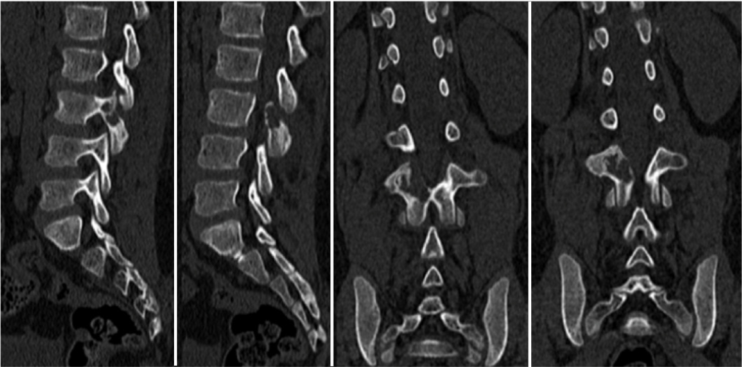
Spinal instability is a notable complication, and may present as mechanical pain, progressive deformity, or vertebral collapse, sometimes leading to acute neurological deterioration. (Figures 3, 4)

Cervical CT scan in a patient with C2 involvement showing instability in the coronal plane and laterolisthesis caused by the tumor lesion.

Lumbosacral CT scan in a patient with L3 facet involvement. Instability in the sagittal plane due to facet involvement is observed.
Unusual clinical presentations have also been reported, such as obstructive hydrocephalus secondary to high spinal cord compression, manifesting with altered consciousness.6
Diagnosis
A multidisciplinary approach is required, combining clinical evaluation, imaging studies, and histological and molecular analysis to accurately characterize the lesion, plan the therapeutic strategy, and determine the stage of the disease.
• Radiography: Typically shows a lytic, expansile lesion with bone destruction and poorly defined margins, without mineralized matrix or periosteal reaction. Depending on the extent of involvement, vertebral collapse may also be evident. (Figure 5)

Left: Cervical profile X-ray. Right: Lumbosacral and pelvic view. Both X-rays show an osteolytic lesion invading C2 and the right sacrum, respectively.
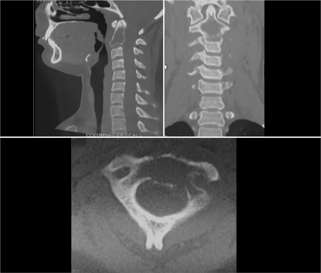
• Computed Tomography (CT) is crucial for assessing bone involvement, pathological fractures, posterior element compromise, and extension into adjacent vertebrae. It also aids in biopsy planning and surgical approach selection. (Figure 6)
CT scan of the cervical spine with sagittal, coronal, and axial sections. A lytic image can be observed, and the bone margins of the lesion can be delimited.
Thoracic CT offers superior specificity over plain radiographs for detecting pulmonary metastases. While its sensitivity in identifying lung involvement in GCT approaches 100%, it may be less specific when distinguishing benign from metastatic nodules.7
• Magnetic resonance imaging typically shows isoor hypointense signals on T1 and heterogeneous intensity on T2, with contrast enhancement after gadolinium. Low T2 signal may indicate fibrosis (Figure 7). MRI is essential for evaluating epidural extension, spinal cord compression, and surgical planning. It also characterizes solid, cystic, and hemorrhagic components of the tumor. In follow-up, it enables early detection of recurrence and monitoring of treatment response. In patients treated with denosumab, increased sclerosis and reduced soft tissue components are common findings.8 (Figure 7)

Sagittal MRI in T2, T1, and STIR sequences with contrast. Note the lesion in the L3L4 region, hypointense in T1 and hyperintense in T2 and STIR.
• PET CT: Useful for evaluating the metabolic activity of the tumor, detecting recurrences or metastases, and assessing postoperative response. It is especially valuable when MRI detail is limited due to metal artifacts.
Giant cell tumors demonstrate high uptake of FDG (fluorodeoxyglucose) in both primary and recurrent lesions, aiding in metabolic characterization. However, their hypermetabolic behavior can resemble malignant bone tumors, which reinforces the need for histopathological confirmation.9
An additional benefit of PET/CT is its ability to assess early therapeutic response, particularly to treatments such as radiotherapy. By relying on metabolic criteria, it can detect treatment efficacy with greater sensitivity and earlier than morphological changes.
• Bone Scintigraphy: While not specific, it can help rule out multifocal disease. However, its sensitivity and specificity for spinal GCT are limited. (Figure 8)

Bone scintigraphy with 99mTc-HDP shows a focal area of intense uptake in L4, involving the vertebral body and ipsilateral pedicle, consistent with increased osteoblastic activity of a giant cell tumor. No additional hypermetabolic lesions are identified. This pattern supports lesion localization and complements MRI in surgical planning.
One finding sometimes observed is the “donut sign”, a pattern of peripheral tracer uptake with a hypoactive center. Though occasionally seen, it is not pathognomonic and limits the diagnostic value and specificity of bone scintigraphy in this context.10
• Biopsy: The definitive diagnosis of giant cell tumor requires histological confirmation.
Percutaneous biopsy has shown diagnostic yields greater than 85% across multiple studies.11 It should be performed only after careful planning of the surgical approach, in order to avoid contaminating future operative fields. Ideally, the biopsy should be carried out at the same institution where surgical treatment will take place.
The primary aim of biopsy is to obtain sufficient tissue to evaluate the lesion’s cellular architecture and differentiate GCT from other giant cell-containing malignancies. Immunohistochemistry is essential for establishing an accurate diagnosis.
In the thoracic and lumbar spine, a transpedicular approach is generally recommended, as it provides safe access and allows for representative sampling using 14-17 gauge needles. In selected cases, a transforaminal route may be used as an alternative.12
For sacral tumors or lesions extending into soft tissue, the approach should be adapted individually to ensure access while minimizing the risk of tumor cell seeding beyond the surgical field.
The use of coaxial systems and semi-rigid cutting tools enhances sample quality and improves diagnostic yield, especially in the typically lytic areas of GCT.
Differential diagnosis
The differential diagnosis of giant cell tumor includes a range of benign and malignant bone lesions that may share overlapping clinical, radiological, or histological features.
Among benign conditions, aneurysmal bone cyst, chondroblastoma, and fibrous dysplasia should be considered. Malignant entities include telangiectatic osteosarcoma, giant cell-rich sarcoma, and metastatic renal cell carcinoma.
Aneurysmal bone cyst may present with clinical and radiological findings similar to GCT, such as expansile lytic lesions with cystic components. However, histologically it is distinguished by the presence of blood-filled vascular spaces.
Telangiectatic osteosarcoma can also contain scattered giant cells and cystic areas, but shows greater cellular atypia, increased mitotic activity, and the presence of osteoid matrix production.
Giant cell-rich sarcoma is a rare tumor that may resemble GCT in cellular morphology but typically exhibits more aggressive behavior and a higher tendency for metastasis. Metastatic renal cell carcinoma may show clear cells and a prominent vascular pattern, features that help distinguish it histologically from GCT.
A comprehensive assessment combining clinical presentation, imaging, histology, and immunohistochemistry is essential for reaching an accurate diagnosis and ruling out other lesions that may mimic a giant cell tumor. (Table 1)
Differential diagnosis of spinal giant cell tumor, highlighting key clinical, radiological, and pathological characteristics.
Surgical treatment
The surgical management of spinal giant cell tumors (GCT) depends on vertebral location, tumor extension, involvement of adjacent structures, neurological preservation, and spinal stability. The primary goal is complete resection with appropriate oncologic margins. To this end, two main strategies are employed: en bloc resection and intralesional curettage.
Several studies support that complete resection with negative margins lowers recurrence rates and improves disease-free survival compared to intralesional approaches. In a retrospective series of 102 patients, en bloc resection was significantly associated with reduced recurrence (p = 0.05), and surgery performed before age 40 correlated with better outcomes (p < 0.01).13 However, this technique carries risks, particularly neurological and vascular complications.
Boriani et al.14 stratified spinal GCTs using the Enneking system. Stage 2 tumors treated with curettage had a 5-year recurrence of just 6%, whereas stage 3 tumors reached 61%. In contrast, en bloc resection achieved local control in 90% of these advanced cases.
Management in high-complexity centers is strongly recommended, as these institutions offer coordinated, multidisciplinary care from initial biopsy to definitive treatment planning. Hart et al. reported a recurrence rate of 18% in patients treated at such centers, compared to 83% in those managed elsewhere.15
Luksanapruksa et al. proposed a treatment algorithm guided by anatomical involvement and clinical presentation. The initial step is to determine whether complete resection is feasible. If so, en bloc resection with wide margins is advised - ideally preceded by selective embolization and followed by postoperative denosumab. When resection is not possible, a more conservative surgical approach combined with local and systemic adjuvant therapies is recommended.16
In patients with unresectable tumors or who are not surgical candidates, the therapeutic focus shifts to medical or palliative management, with the aim of achieving disease control and preserving quality of life.
Choice of surgical technique
When selecting the surgical approach, the WBB (Weinstein-Boriani-Biagini) staging system should be incorporated into preoperative planning to determine the safest strategy for tumor resection. This allows the surgeon to assess whether a posterior approach alone is appropriate, or if a combined anterior-posterior route is required.
The tumor’s location and size are key factors that guide surgical decision-making in terms of both approach and resection strategy. Several reconstructive techniques have been described that combine anterior support with posterior stabilization following tumor removal. Samartzis et al.17 reported favorable outcomes (disease-free survival and preserved neurological function) using anterior expandable cages together with posterior segmental fixation.
The sacrum presents a particularly complex scenario, often requiring a combined approach. While posterior access allows for stabilization, anterior exposure is frequently necessary for adequate tumor resection and bleeding control. Li et al. analyzed 32 cases of sacral GCT treated with various strategies and found that recurrence rates were significantly lower in patients who underwent marginal resection compared to those treated with intralesional curettage alone (18% vs. 71%, p = 0.05).18 In cases of extensive resection requiring soft tissue coverage, preoperative planning should involve plastic surgeons for flap closure and reconstruction.
En bloc resection
En bloc resection involves removing the tumor with oncologically safe margins, avoiding capsule violation to reduce the risk of local spread and recurrence. The surgical approach depends on vertebral location, tumor size and extent, and proximity to vital structures.
This is a technically challenging procedure, associated with higher risks of bleeding, neurological injury, and instrumentation failure, particularly in unstable segments. The main techniques include total en bloc spondylectomy and its variations, adapted to the tumor’s anatomy. This procedure entails complete removal of one or more vertebral bodies with their posterior elements to achieve clear margins and minimize recurrence.19 The need to preserve critical structures (such as major vessels, nerve roots, and the spinal cord) adds to its complexity and risk.
For sacral tumors, en bloc resection may be performed via a posterior approach or a combined anterior-posterior route when necessary. Root sacrifice may or may not be required, depending on the extent of involvement and the need to preserve function in relation to pelvic anatomy. A posterior-only approach has been shown to be safe when there is limited anterior invasion and no or only partial sacroiliac joint involvement. Lateral osteotomies performed through the sacral foramina allow for segmental resection. Preservation of nerve roots above S2 is critical for maintaining motor and sphincter function.
In cases involving large tumor volume or adherence to anterior structures, a combined anterior-posterior approach should be considered. Preoperative anterior dissection with ligation of the internal iliac arteries has been shown to reduce intraoperative bleeding. Lumbopelvic reconstruction is essential for maintaining biomechanical stability, particularly in high sacrectomies, and typically involves the use of spinal-pelvic fixation systems combined with bone grafts. (Figure 9 and 10)

30-year-old male with sacral GTC undergoing intralesional resection and spinopelvic reconstruction. A- Sagittal MRI showing anterior tumor mass in the pelvis, B- Coronal CT scan showing extensive involvement of the sacrum, C- Arteriography and CT embolization, D- Intraoperative image of reconstruction with bone allograft, E and F- Postoperative CT and radiography images.

Case of a 26-year-old woman with prior emergency double-approach surgery for a pathological fracture. (A-B) CT scans (axial and sagittal) showing recurrence of the giant cell tumor. (C) MRI reveals a large tumor mass. (D-E) Posterior en bloc resection of T12. (F) Postoperative CT reconstruction. (G-H) MRI (axial and sagittal) demonstrating effective spinal cord decompression with no signs of recurrence. (I) Intraoperative radiograph showing the T12 recurrence, anterior cage, and adjacent endplates of T11 and L1.
Figure 9 30-year-old male with sacral GTC undergoing intralesional resection and spinopelvic reconstruction. A- Sagittal MRI showing anterior tumor mass in the pelvis, B- Coronal CT scan showing extensive involvement of the sacrum, C- Arteriography and CT embolization, D- Intraoperative image of reconstruction with bone allograft, E and F- Postoperative CT and radiography images.
The most common complications in this surgical procedure include bleeding, wound infection, wound dehiscence, neurological dysfunction, and, to a lesser extent, fistulas or visceral lesions.20 (Figure 10)
In the cervical spine, en bloc resection is a highly demanding procedure due to the proximity to neurovascular structures (spinal cord, vertebral artery, and brachial plexus). Surgical planning should include CT angiography evaluation of the relationship with neurovascular structures and tumor vascularization, which may require preoperative embolization of the affected vertebral artery.
The most commonly used approach in this area is a combined anterior and posterior approach. In cases where the tumor invades critical structures and en bloc resection is not possible, extensive piecemeal resection may be performed, although the latter is associated with a higher rate of local recurrence. In these cases, adjuvant radiotherapy may be used, although its use is still controversial due to the risk of malignant transformation.21
Intralesional curettage associated with adjuvants
Intralesional curettage supplemented with adjuvants (such as phenol, alcohol, liquid nitrogen, or bone cement) is a minimally invasive approach aimed at maximizing local control while minimizing neurosurgical risks. However, this comes at the cost of a higher rate of local recurrence. Studies have shown that when intralesional curettage is performed without adjuvants, recurrence rates can exceed 80% in some cases, particularly in sacral tumors. The addition of local adjuvants has been demonstrated to significantly reduce these rates.22
Phenol, a caustic agent causing protein necrosis, has proven effective in lowering recurrence rates when used with curettage, particularly alongside bone cement.23 Although it is the most studied adjuvant, ethanol offers similar local control with potentially lower toxicity.
Cryotherapy with liquid nitrogen induces tumor cell death through freeze-thaw cycles, and is similarly effective, though it carries higher risks of complications like fractures and soft tissue damage. Its therapeutic effect is linked to the depth of necrosis it produces.
Polymethylmethacrylate (PMMA) cement provides mechanical support and a thermal cytotoxic effect. When used after curettage, it significantly reduces recurrence, especially when combined with phenol or cryotherapy. The sacrum represents a particular challenge. In this location, the use of adjuvants following intralesional curettage has not always correlated with reduced recurrence rates. On the contrary, significant complications (including massive hemorrhage, neurological injury, and wound healing issues) have been observed.
Radiotherapy and preoperative embolization are considered when complete resection is unfeasible or surgical risk is excessively high. Combined, they have shown promising disease-free survival outcomes.24
Radiotherapy offers effective local control in inoperable cases, with 1- and 2-year control rates over 80% and overall survival approaching 98%, based on retrospective series and systematic reviews. It may also be useful as an adjuvant in subtotal resections or when margins are positive. In recurrent disease, it can help slow tumor progression.25
Modern techniques - such as 3D-CRT, IMRT, and particle therapy with protons or carbon ions - allow high-dose delivery (typically 40-70 Gy) while sparing nearby structures and bone marrow. Although uncommon, there remains concern about the risk of post-radiation malignant transformation.
Denosumab
Due to its locally aggressive nature, spinal giant cell tumors (GCTs) represent a significant therapeutic challenge, particularly given the complexity of surgical access and the inherent morbidity and mortality risks associated with intervention.
Although surgery (whether en bloc resection or intralesional curettage) remains the cornerstone of treatment, denosumab has gained increasing relevance in complex or unresectable cases. Phase II multicenter studies have shown that most patients with unresectable GCTs, or those in whom surgery would entail high morbidity, achieve sustained tumor control, with low progression or recurrence rates at 1, 3, and 5 years.26
Denosumab is a monoclonal antibody that targets the receptor activator of nuclear factor kappa-B ligand (RANKL), blocking its interaction with the RANK receptor expressed on osteoclastic giant cells within the tumor. This inhibition leads to reduced osteolysis and, secondarily, progressive ossification of lytic lesions.27 (Figure 11)

Presents the histological progression of a spinal giant cell tumor treated with denosumab. Initial biopsy samples (A-B) show high cellularity with numerous osteoclast-like giant cells in an active stromal background. At 8 and 12 weeks (C-D), there is a notable reduction in giant cells, increased fibrosis, and early peripheral ossification. After six months (E-F), the tissue exhibits minimal cellularity, near-complete absence of giant cells, and replacement by fibrous tissue and new trabecular bone. These findings indicate a favorable histological response to denosumab, with tumor regression and stromal remodeling.
Denosumab may be used as neoadjuvant therapy to shrink the tumor and better define its margins, facilitating less invasive surgery, or as palliative treatment in unresectable cases. However, when planned before en bloc resection, caution is necessary due to its unpredictable effects on tumor consistency.
Yonezawa et al.28 described a case where preoperative denosumab led to extensive peripheral ossification, making posterior dissection of segmental vessels unfeasible and forcing an unplanned intralesional resection. (Figure 12)

Multimodal evaluation of a giant cell tumor lesion in C2 involving the vertebral body and odontoid process.
Several multicenter studies29 have reported stabilization or regression of tumor mass in over 80% of patients treated with neoadjuvant denosumab. Histologically, these cases demonstrate a marked reduction in giant cell populations, increased fibrosis, and new bone formation. (Figure 13)

Radiological evolution of a C2 giant cell tumor after C1-C2 stabilization and denosumab therapy. The lesion, initially osteolytic, shows progressive marginal ossification and loss of the lytic pattern, indicating a favorable therapeutic response following occipito-cervical fixation.
However, neoplastic mononuclear stromal cells may persist, indicating a continued risk of recurrence if complete resection is not achieved. Optimal histologic response (defined as ≥50% reduction in giant cells with fibrosis and ossification) has been associated with improved progression-free survival. (Figure 13)
Local recurrence after denosumab use in GCT remains a subject of debate. While the drug may enable less invasive surgical approaches, several studies suggest it can compromise the completeness of resection. This is attributed to the formation of a fibrous and ossified rim around the lesion, which may hinder full tumor removal. Some reports indicate recurrence rates exceeding 40% in patients treated with neoadjuvant denosumab.30
The adjuvant use of denosumab following surgery, has been proposed for patients with uncertain resection margins or residual disease, with the goal of delaying potential recurrence.
The recommended regimen consists of 120 mg administered subcutaneously every four weeks, with additional 120 mg doses on days 8 and 15 of the initial treatment cycle. Supplementation with calcium and vitamin D are essential to prevent hypocalcemia.31
In 2016, Dubory et al.32 observed that denosumab appeared to achieve its maximum therapeutic effect within the first six months of treatment, after which its efficacy seemed to stabilize or decline. However, to date, the literature lacks conclusive evidence regarding its long-term benefit in terms of progression-free survival, as well as the optimal duration of therapy. In response to this uncertainty, Gasbarrini et al.,2 in a systematic review of denosumab use for spinal GCT, proposed the following therapeutic scheme:
Exclusive treatment with denosumab (“stand-alone”)
GCT with contraindication for en bloc resection due to high morbidity
Recurrence after surgery and/or radiotherapy, with no possibility of further resection
Tumor control is assessed clinically and by persistence of symptomatic remission
Preoperative treatment with denosumab
• If en bloc resection is feasible (based on WBB criteria):
Denosumab for at least 6 months (up to 12 months if the response is delayed)
En bloc surgery
If margins are negative, denosumab may be discontinued
If tumor invasion is observed, treatment should be resumed
• If en bloc resection is not feasible:
Denosumab for 6 to 12 months
Intralesional surgery
Postoperative denosumab should be restarted
Management of recurrences
Although most of the available data on giant cell tumors (GCTs) originate from relatively small case series, several studies suggest that recurrence rates are lower in the mobile spine than in the sacrum. However, interpretation of these data is limited by variations in surgical techniques and adjuvant therapies used across different spinal regions.
Tumors involving both the vertebral body, and the posterior elements of the spine have shown higher recurrence rates than those confined to the vertebral body alone. Furthermore, tumor extension into the spinal canal or paraspinal soft tissues has been associated with an increased risk of recurrence.15
In rare cases, GCTs may be associated with the formation of aneurysmal bone cysts, which tend to exhibit a more aggressive local behavior. In such scenarios, complete surgical resection combined with systemic radiotherapy is recommended. This approach has been linked to favorable outcomes in terms of local tumor control.
CONCLUSION
Giant cell tumors of the spine represent a significant clinical and therapeutic challenge due to their locally aggressive behavior and proximity to critical neurovascular structures - factors that play a key role in planning surgical resection.
Surgical excision with clear oncologic margins remains the standard of care for achieving tumor eradication. The surgical approach should be tailored to each case based on tumor location, extent, and stage.
Denosumab has broadened the therapeutic landscape, offering an alternative in cases where surgery is not feasible or entails high morbidity. Its use must be carefully planned and closely monitored, given its potential for inducing peritumoral ossification and its association with local recurrence.
Finally, we emphasize the importance of multidisciplinary management in specialized centers to optimize surgical and functional outcomes, reduce the risk of recurrence, and ensure rigorous long-term follow-up of this complex spinal pathology.
-
Study conducted by the Hospital Central, Mendoza, Argentina; Hospital Naval Pedro Mallo, Buenos Aires, Argentina; and INTO, Rio de Janeiro, RJ, Brazil.
REFERENCES
- 1 Scotto di Carlo F, Whyte MP, Gianfrancesco F. The two faces of giant cell tumor of bone. Cancer Lett. 2020;489:1-8.
- 2 Bukata SV, Blay JY, Rutkowski P, Skubitz K, Henshaw R, Seeger L, et al. Denosumab treatment for giant cell tumor of the spine. Spine (Phila Pa 1976). 2021;46(5):277-84.
- 3 Tsukamoto S, Mavrogenis AF, Kido A, Errani C. Current treatment concepts in giant cell tumors. Cancers (Basel). 2021;13(15):3647.
- 4 Donthineni R, Boriani L, Ofluoglu O, Bandiera S. Metastatic behavior of giant cell tumor of the spine. Int Orthop. 2009;33(2):497-501.
- 5 Tsukamoto S, Righi A, Mavrogenis AF, Akahane M, Honoki K, Tanaka Y, Donati DM, et al. Late recurrence and risk of malignancy in bone GCT. Cancers (Basel). 2021;13(14):3644.
- 6 Wei CY, Chen ST, Tai HC, Wang WB, Chang CC, Wang YC, et al. Obstructive hydrocephalus from thoracic GCT: case report. Oncol Lett. 2016;11(1):39-44.
- 7 Chang AE, Schaner EG, Conkle DM, Flye MW, Doppman JL, Rosenberg SA. et al. CT in detection of pulmonary metastases: prospective study. Cancer. 1979;43(3):913-6.
- 8 Furuta T, Kubo T, Sakuda T, Saito T, Kurisu K, Muragaki Y, et al. Intraoperative MRI in GCT after denosumab: pilot study. Acta Radiol. 2022;63(2):176-81.
- 9 Muheremu A, Ma Y, Huang Z, Shan H, Li Y, Niu X. et al. Diagnosing giant cell tumor of the bone using positron emission tomography/computed tomography: A retrospective study of 20 patients from a single center. Oncol Lett. 2017;14(2):1985-8.
- 10 Sakurai H, Mitsuhashi N, Hayakawa K, Niibe H. Giant cell tumor of the thoracic spine simulating mediastinal neoplasm. AJNR Am J Neuroradiol. 1999;20(9):1723-6.
- 11 Marruzzo D, Mancini F, Ricciuti V, Barbieri FR, Preziosi R, Pagano S, et al. Modified percutaneous biopsy of the spine: improvement of the technique. Eur Spine J. 2023;32(1):221-7.
- 12 Sucu HK, Bezircioglu H, Ciçek C, Erşahin Y. Computerized tomography-guided percutaneous transforaminodiscal biopsy sampling of vertebral body lesions. J Neurosurg. 2003;99(1 Suppl):51-5.
- 13 Xu W, Li X, Huang W, Wang Y, Han S, Chen S, et al. Factors affecting prognosis of patients with giant cell tumors of the mobile spine: retrospective analysis of 102 patients in a single center. Ann Surg Oncol. 2013;20(3):804-10.
- 14 Boriani S, Cecchinato R, Cuzzocrea F, Bandiera S, Gambarotti M, Gasbarrini A. et al. Denosumab in the treatment of giant cell tumor of the spine. Preliminary report, review of the literature and protocol proposal. Eur Spine J. 2020;29(2):257-71.
- 15 Hart RA, Boriani S, Biagini R, Currier B, Weinstein JN. A system for surgical staging and management of spine tumors. A clinical outcome study of giant cell tumors of the spine. Spine (Phila Pa 1976). 1997;22(15):1773-82.
- 16 Luksanapruksa P, Buchowski JM, Singhatanadgige W, Rose PC, Bumpass DB. Management of spinal giant cell tumors. Spine J. 2016;16(2):259-69.
- 17 Shen FH, Marks I, Shaffrey C, Ouellet J, Arlet V. The use of an expandable cage for corpectomy reconstruction of vertebral body tumors through a posterior extracavitary approach: a multicenter consecutive case series of prospectively followed patients. Spine J. 2008;8(2):329-39.
-
18 Li D, Zhang J, Li Y, Xia J, Yang Y, Ren M, et al. Surgical strategy for the management of sacral giant cel tumors: a 32-case series. Spine J. 2019;19(4):601-8. doi:10.1016/j.spinee.2018.10.005.
» https://doi.org/10.1016/j.spinee.2018.10.005. - 19 Wan W, Zheng W, Wan J, Zhang J, Liu Y, Jia Q, et al. An improved total en bloc spondylectomy for L5 vertebral giant cell tumor through a single-stage posterior approach. Eur Spine J. 2023;32(7):2503-12.
- 20 Weidlich A, Schaser KD, Weitz J, Kirchberg J, Fritzmann J, Reeps C, et al. Surgical and Oncologic Outcome following Sacrectomy for Primary Malignant Bone Tumors and Locally Recurrent Rectal Cancer. Cancers (Basel). 2024;16(13):2334.
- 21 Tu J, Li W, Shu S, Zhang Y, Hua W, Li S, et al. Total spondylectomy of recurrent giant cell tumors in the cervical spine: Two case reports and review of literature. Medicine (Baltimore). 2018;97(20):e10799.
- 22 van der Heijden L, van de Sande MA, van der Geest IC, Schreuder HW, van Royen BJ, Jutte PC, et al. Giant cell tumors of the sacrum--a nationwide study on midterm results in 26 patients after intralesional excision. Eur Spine J. 2014;23(9):1949-62.
- 23 van der Heijden L, van der Geest IC, Schreuder HW, van de Sande MA, Dijkstra PD. et al. Liquid nitrogen or phenolization for giant cell tumor of bone?: a comparative cohort study of various standard treatments at two tertiary referral centers. J Bone Joint Surg Am. 2014;96(5):e35.
- 24 Palmisciano P, Ferini G, Chen AL, Balasubramanian K, Kharbat AF, Sagoo NS, et al. Evaluating the Optimal Management of Inoperable Giant Cell Tumors of the Spine: A Systematic Review and Meta-Analysis. Cancers (Basel). 2022;14(4):937.
- 25 Cao S, Jiang L, Yang S, Liu Z, Wei F, Liu X. Surgical treatment of spinal tenosynovial giant cell tumor: Experience from a single center and literature review. Front Oncol. 2023;12:1063109.
- 26 Chawla S, Blay JY, Rutkowski P, Le Cesne A, Reichardt P, Gelderblom H, et al. enosumab in patients with giant-cell tumour of bone: a multicentre, open-label, phase 2 study. Lancet Oncol. 2019;20(12):1719-29.
- 27 Latorre MR, Albergo JI, Farfalli GL, Roitman PD, Plantalech L, Ayerza MA, et al. [Denosumab as a treatment for giant cell tumor of bone. Indications, results and side effects]. Medicina (B Aires). 2021;81(5):767-73.
- 28 Miao Z, Xu M, Zheng K, Gong H, Yan N, Chen Q, et al. Denosumab combined with precision radiotherapy for recurrent giant cell tumor of the thoracic spine: a case report and literature review. Front Neurol. 2023;14:1308600.
- 29 Tang Q, Lu J, Zhu X, Song G, Wu H, Xu H, et al. The efficacy and safety of short-course neoadjuvant denosumab for en bloc spondylectomy in spinal giant cell tumor of bone: a preliminary report. Eur Spine J. 2023;32(12):4297-305.
- 30 Tsukamoto S, Tanaka Y, Mavrogenis AF, Kido A, Kawaguchi M, Errani C. Is Treatment with Denosumab Associated with Local Recurrence in Patients with Giant Cell Tumor of Bone Treated with Curettage? A Systematic Review. Clin Orthop Relat Res. 2020;478(5):1076-85.
- 31 Puri A, Gulia A, Hegde P, Verma V, Rekhi B. Neoadjuvant denosumab: its role and results in operable cases of giant cell tumour of bone. Bone Joint J. 2019;101-B(2):170-7.
- 32 Dubory A, Missenard G, Domont J, Court C. Interest of Denosumab for the Treatment of Giant-cells Tumors and Aneurysmal Bone Cysts of the Spine. About Nine Cases. Spine (Phila Pa 1976). 2016;41(11):E654-60.
Edited by
-
Reviewed by:
Robert Meves