 Saving enamel: safe removal of residual residues after detaching orthodontic brackets
ABSTRACT
In conventional orthodontics, the adhesive fixation of brackets plays an essential role in the success of the treatment. However, the removal of these devices at the end of the treatment can pose risks of excessive wear to the external surface of the enamel, especially when rotary instruments are indiscriminately used to remove the resin composite employed in cementation. Given that enamel is an inorganic tissue with no regenerative capacity, it becomes imperative to adopt conservative techniques to minimize unnecessary wear. This text aimed to discuss more conservative approaches, exploring the use of multilayered burs instead of diamond tips, and associating them with polishing rubbers, magnification, and consideration of the fluorescence characteristics of adhesive materials. These practices are intended to minimize impact and preserve the integrity of the enamel. Reflecting on the indiscriminate use of diamond tips for enamel polishing emerges as essential in modern dental practice.
Indexing terms
Dental Enamel; Composite resins; Orthodontics
RESUMO
Na ortodontia convencional, a fixação adesiva de braquetes desempenha um papel essencial no sucesso do tratamento. Entretanto, a remoção desses dispositivos ao final do tratamento pode acarretar riscos de desgaste excessivo da superficie externa do esmalte, especialmente quando instrumentos rotatórios são usados indiscriminadamente para remover o compósito resinoso empregado na cimentação. Dado que o esmalte um tecido inorgânico e sem capacidade de regeneração, torna-se imperativo adotar técnicas conservadoras para minimizar desgaste desnecessário. Este texto teve como propósito discutir abordagens mais conservadoras, explorando o uso de brocas multilaminadas em detrimento das pontas diamantadas, e associando-as a borrachas de polimento, magnificação, e consideração das características de fluorescência dos materiais adesivos. Essas práticas têm o intuito de minimizar o impacto e preservar a integridade do esmalte. A reflexão sobre a utilização indiscriminada de pontas diamantadas para polimento do esmalte emerge como essencial na prática odontológica moderna.
Termos de indexação
Esmalte dentário; Resina composta; Ortodontia
INTRODUCTION
Orthodontic treatment typically requires the use of brackets that are adhered to the dental surface to move and align the teeth. For successful treatment, these brackets must remain fixed to the dental structures through adhesive procedures until the final stage of treatment [1]. The first step in the adhesive protocol for bonding the bracket to the dental surface involves the application of a phosphoric acid gel at a concentration of 35% to 37% [2] on the bonding area. This creates surface roughness, which enhances the adhesion of the adhesive and resin composite applied to the conditioned surface [3].
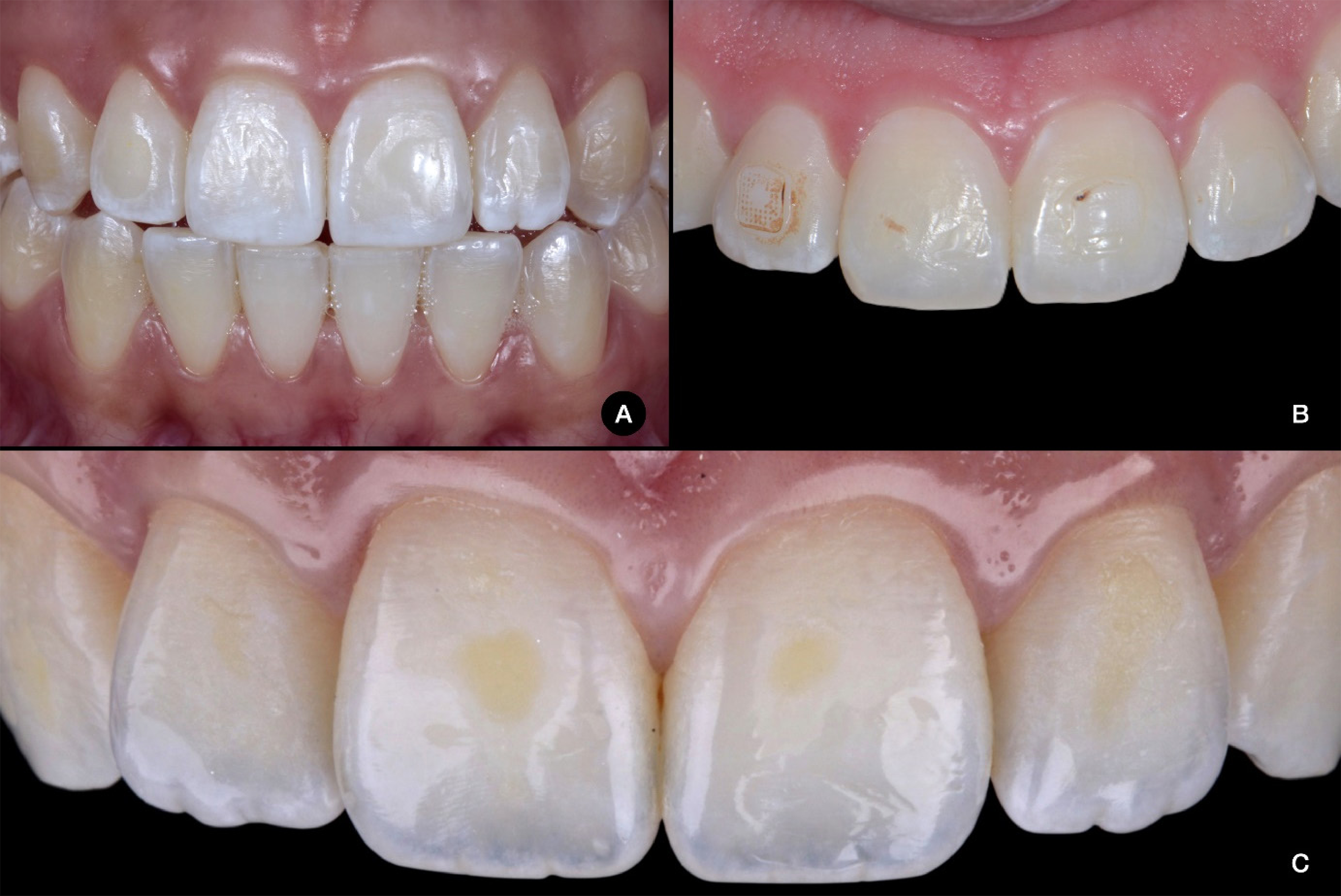
When the treatment is completed, the removal of brackets is performed mechanically using specific orthodontic pliers [4], followed by the removal of resinous material through abrasive mechanical methods. The presence of possible residues of bonding agents over the dental surface favors biofilm formation, which can result in periodontal damage, the development of carious lesions [5], as well as color alteration over time (figure 1) and interference with the aesthetics of the smile.

Examples of composite resin remnants on canines and incisors after bracket removal in young adults often show color alteration and increased surface roughness. In Figure A, composite resin remnants, surface wear, and scratches on the enamel – likely caused by a rotary instrument such as a diamond bur – are evident. Figures B and C show color alteration due to residual composite resin.
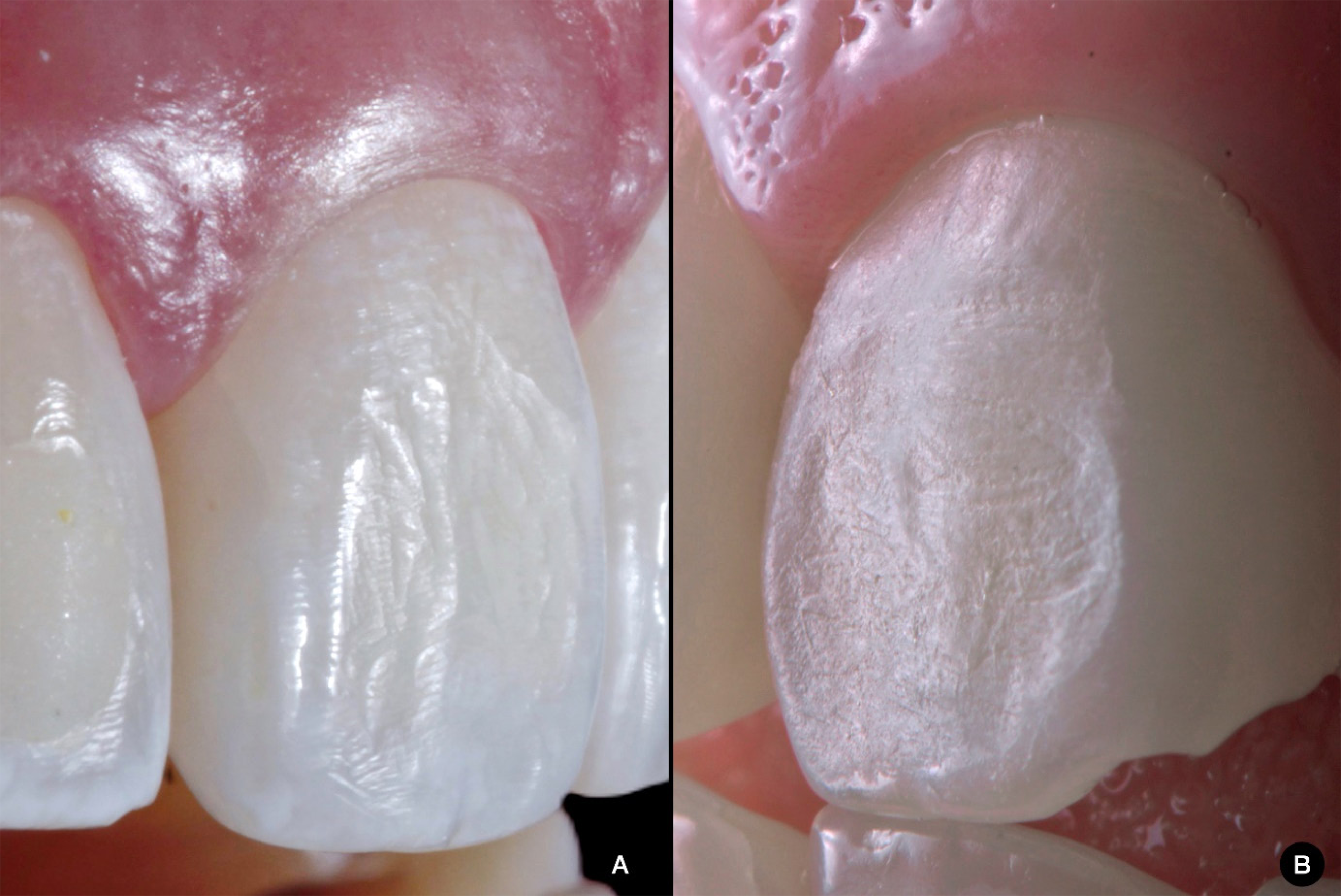
Rotary instruments are frequently used for this purpose, and their abrasive power depends on the size and composition of the particles, the rotational speed, and the pressure applied to the enamel, making it an operator-dependent technique [6,7]. Although no removal method is entirely safe and capable of producing no enamel wear [8,9], these steps must be planned and executed to minimize damage to the dental structure. Among the most common iatrogenic aspects are cracks and fractures [10-12], and wear of the dental surface [5,8], particularly due to the excessive use of high and medium-grit diamond burs and abrasive discs (figure 2). Additionally, using these instruments can increase enamel roughness, creating grooves and fissures that favor extrinsic pigmentation and biofilm accumulation [8,13].

Examples of wear on incisors likely caused by using a diamond bur during orthodontic bracket removal in young adult patients.
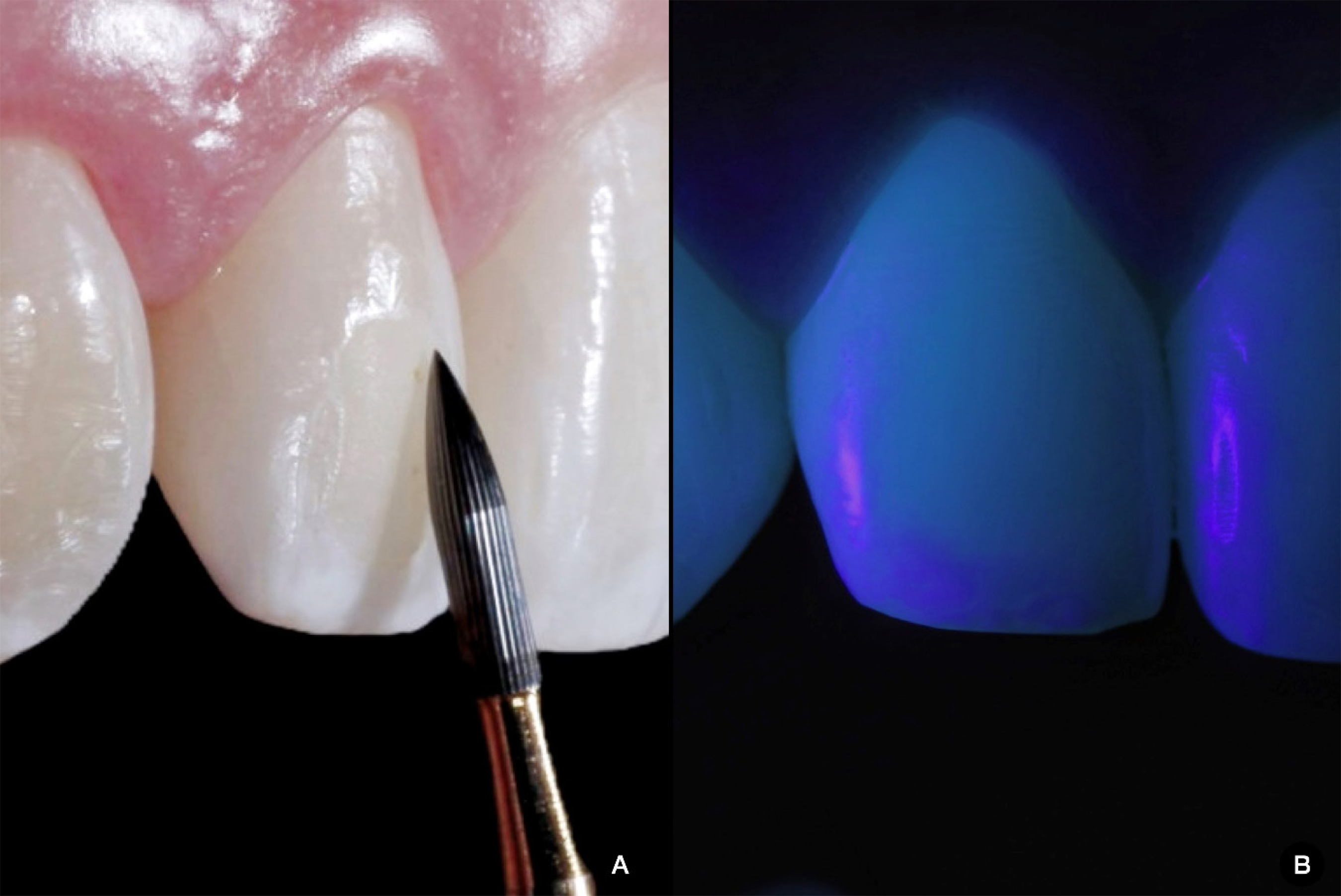
To minimize these enamel imperfections, it is recommended to polish the dental enamel regardless of the choice of polishing agents [8]. A recent systematic literature review found that Arkansas stones, diamond burs, steel burs, and lasers caused the most enamel wear, while multilaminated tungsten carbide burs (figure 3A) were faster and more effective compared to polishing discs and rubber cups, ultrasonic instruments, and polymer burs [14]. Other studies also report that the cutting efficacy of multilaminated burs is related to their morphological characteristics, such as the number and smoothness of blades [15,16]. Additionally, the pressure applied by the operator using the high-speed handpiece can affect the bur’s cutting ability [16]. Currently, the multiplying contra-angle is also available as an option, offering a constant rotational speed and higher torque than conventional high-speed handpieces, facilitating control during abrasion and reducing the risk of excessive wear [17].

A) Removal of resin composite using a high-speed 24-blade multilaminated tungsten carbide bur (Orthometric, São Paulo, Brazil). B) Ultraviolet light illumination shows a slight color difference between the resin and the tooth. In the photo, the Black Light tip (Valo Lens, Ultradent Products) is attached to the photopolymerization unit (Valo Cordless, Ultradent Products).
The use of ultraviolet light, combined with rotating instruments, can be highly effective [18]. It is a non-destructive technique often used to help identify cracks, fractures, and carious lesions [19,20], optimizing clinical care. When analyzing caries, the decalcification of dental structures and loss of organic matter reduces light transmission, resulting in a radiolucent image [21]. Ultraviolet illumination relies on the fluorescence of dental materials and tissues, an optical property where the material or tissue absorbs UV light and reflects it at a visible wavelength [22]. Some composite resins and resinous materials do not exhibit the same fluorescence as tooth structure [23]. Consequently, specific resin composites with modified fluorescence are available for bonding orthodontic devices, facilitating material visualization during removal.
The fluorescence-aided identification technique helps visualize the difference in fluorescence between restorative materials and dental structures using a blue light curing unit [24]. Currently, accessory lenses that attach to a polywave photopolymerizing unit are available, facilitating this process. However, ultraviolet lamps (black lights) can be a more affordable option with similar results and can be used in the office. Whether using a lens or a lamp, the main advantage of this technique is its non-invasive nature, minimizing excess wear on dental structures. Professionals seek conservative protocols for removing resinous materials that do not damage dental structures [24,25].
In addition to ultraviolet light illumination, magnification is an excellent ally in reducing the risks of excessive tooth structure wear. The main advantage of magnification is that it allows better visualization during procedures, enabling satisfactory ergonomics. Magnifying glasses and dental microscopes are commonly used for magnification in dentistry [26-28]. These tools have been widely advocated for maintaining professional ergonomics and facilitating care and visualization by allowing closer inspection of the operative field [28]. Despite the recognized advantages, the use of magnification in Brazil remains limited but has growing market potential. Since working with magnification requires a learning curve, introducing it during dental education would help spread its use, benefiting patients with faster and more precise procedures and improving the ergonomics of dental surgeons. Therefore, democratizing access to magnifying glasses and microscopes is essential to enhance the competence of Brazilian dentistry and improve the quality of life for professionals.
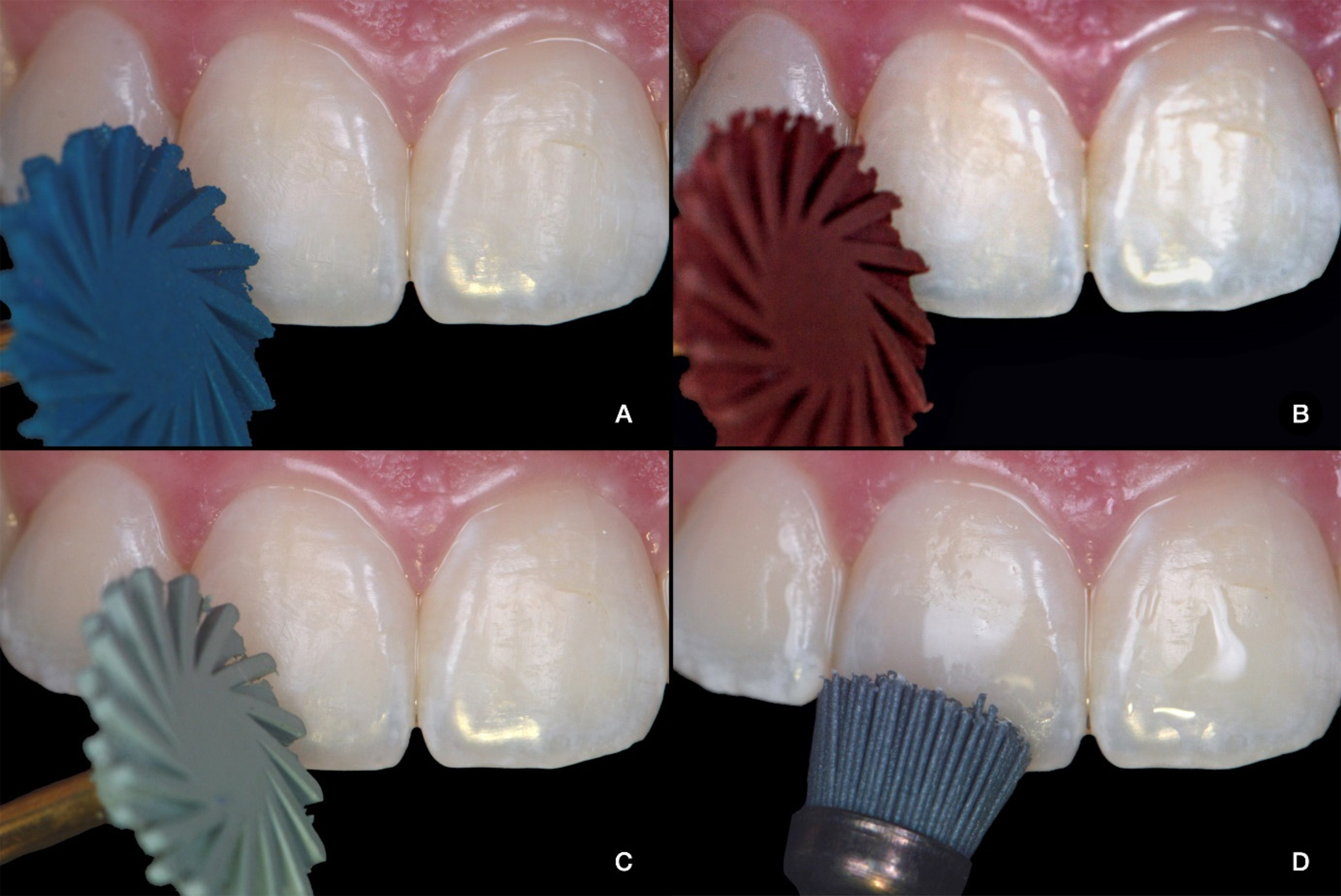
After removing resin residues during bracket debonding, it is necessary to polish the tooth surface to mitigate the risks caused by rotary instruments. Achieving greater enamel smoothness is crucial for improving hygiene, controlling biofilm, and providing patient comfort, as a rough surface can cause discomfort when in contact with the tongue or soft tissues. Additionally, a smooth surface enhances shine, improving the aesthetics of the smile. Evidence shows that spiral systems are more efficient for polishing composite resins compared to polishing discs [7] (figure 4) and that flame-shaped or cup-shaped rubbers are more indicated for polishing enamel [29]. The superior polishing performance of rubbers, mainly spirals, over discs, is attributed to the material’s geometry and flexibility, which provide better surface contact and reduce the necessary pressure [7,30,31]. Some studies have also demonstrated the benefits of polishing composite resins with spirals under irrigation (wet polishing), which reduces surface roughness and temperature during the process [32]. However, studies on the effects of wet polishing on enamel are still limited [33].

Suggested polishing sequence using spiral polishers with decreasing grain size for removing residue from the upper central incisor. A) American Burrs spiral polisher with large particle size. B) American Burrs spiral polisher with medium grain size. C) Fine-grained American Burrs spiral polisher. D) Ultra-fine silicon carbide brush – Dhpro.
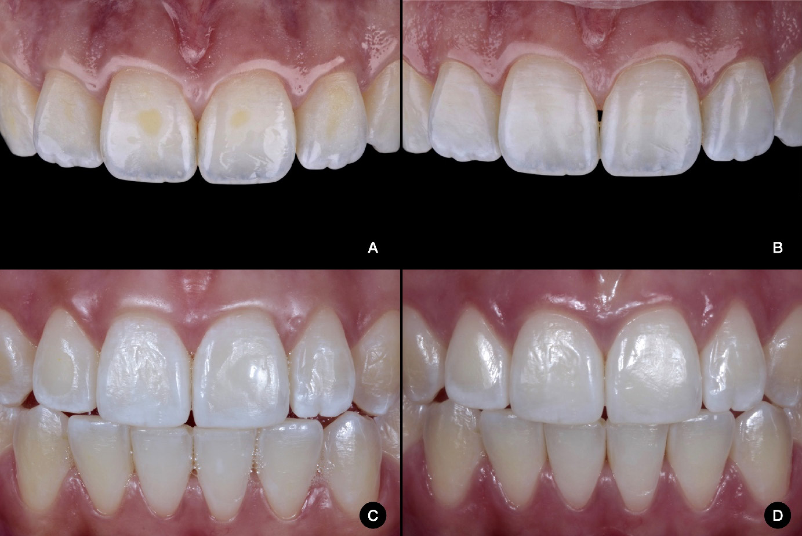
Finally, after the complete removal of orthodontic materials, there must be follow-up for examinations regarding the lack of postoperative sensitivity (figure 5).

Initial and final aspects of two situations in which the removal of excess followed the recommendations described in this text.
FINAL CONSIDERATIONS
Training and knowledge in bracket debonding procedures are crucial to minimize irreversible damage to dental structures, particularly in preserving healthy enamel, especially among young patients. It is imperative for dental surgeons to invest in modern materials and instruments to facilitate minimal intervention and avoid unnecessary invasion of healthy tissues. In today’s society, which values aesthetics and efficiency, the application of the “daughter test,” proposed by Kelleher in 2010 [34] and recently revisited by Sellars in 2022 [35], is increasingly urgent and necessary in patient treatment, regardless of familial relationship.
How to cite this article
-
Almeida LS, Jing JZ, Rodrigues MS, Hilgert LA, Zanatta RF. Saving enamel: safe removal of residual residues after detaching orthodontic brackets. RGO, Rev Gaúch Odontol. 2024;72:e20240020. http://dx.doi.org/10.1590/1981-86372024002020240006
REFERENCES
-
1 Tsichlaki A, Chin SY, Pandis N, Fleming PS. How long does treatment with fixed orthodontic appliances last? A systematic review. Am J Orthod Dentofac Orthop. 2016;149(3):308-18. http://dx.doi.org/10.1016/j.ajodo.2015.09.020
» https://doi.org/10.1016/j.ajodo.2015.09.020 -
2 Brauchli L, Muscillo T, Steineck M, Wichelhaus A. Influence of enamel conditioning on the shear bond strength of different adhesives. J Orofac Orthop / Fortschritte der Kieferorthopädie. 2010;71(6):411-20. http://dx.doi.org/10.1007/s00056-010-1036-2
» https://doi.org/10.1007/s00056-010-1036-2 -
3 Bishara SE, VonWald L, Laffoon JF, Jakobsen JR. Effect of altering the type of enamel conditioner on the shear bond strength of a resin-reinforced glass ionomer adhesive. Am J Orthod Dentofac Orthop. 2000;118(3):288-94. http://dx.doi.org/10.1067/mod.2000.104903
» https://doi.org/10.1067/mod.2000.104903 -
4 Arici S. The force levels required to mechanically debond ceramic brackets: an in vitro comparative study. Eur J Orthod. 2000;22(3):327-34. doi: 10.1093/ejo/22.3.327.
» https://doi.org/10.1093/ejo/22.3.327 -
5 Eliades T. Enamel surface roughness following debonding using two resin grinding methods. Eur J Orthod. 2004;26(3):333-8. http://dx.doi.org/10.1093/ejo/26.3.333
» https://doi.org/10.1093/ejo/26.3.333 -
6 Alessandri Bonetti G, Zanarini M, Incerti Parenti S, Lattuca M, Marchionni S, Gatto MR. Evaluation of enamel surfaces after bracket debonding: An in-vivo study with scanning electron microscopy. Am J Orthod Dentofac Orthop. 2011;140(5):696-702. http://dx.doi.org/10.1016/j.ajodo.2011.02.027
» https://doi.org/10.1016/j.ajodo.2011.02.027 - 7 Batista GR, Zanatta RF, Borges AB, Torres CRG. The effects of polishing techniques on surface roughness and gloss of different composites. Gen Dent. 2021;69(5):46-51.
-
8 Macieski K, Rocha R, Locks A, Ribeiro GU. Avaliação dos efeitos de três métodos de remoção da resina remanescente do braquete na superfície do esmalte. Dental Press J Orthod. 2011;16(5):146-54. http://dx.doi.org/10.1590/S2176-94512011000500021
» https://doi.org/10.1590/S2176-94512011000500021 -
9 Zachrisson BU, Årthun J. Enamel surface appearance after various debonding techniques. Am J Orthod. 1979;75(2):121-37. http://dx.doi.org/10.1016/0002-9416(79)90181-7
» https://doi.org/10.1016/0002-9416(79)90181-7 -
10 Gwinnett AJ, Gorelick L. Microscopic evaluation of enamel after debonding: Clinical application. Am J Orthod. 1977;71(6):651-65. http://dx.doi.org/10.1016/0002-9416(77)90281-0
» https://doi.org/10.1016/0002-9416(77)90281-0 -
11 Habibi M, Nik TH, Hooshmand T. Comparison of debonding characteristics of metal and ceramic orthodontic brackets to enamel: An in-vitro study. Am J Orthod Dentofac Orthop. 2007;132(5):675-9. http://dx.doi.org/10.1016/j.ajodo.2005.11.040
» https://doi.org/10.1016/j.ajodo.2005.11.040 -
12 Zarrinnia K, Eid NM, Kehoe MJ. The effect of different debonding techniques on the enamel surface: An in vitro qualitative study. Am J Orthod Dentofac Orthop. 1995;108(3):284-93. http://dx.doi.org/10.1016/S0889-5406(95)70023-4
» https://doi.org/10.1016/S0889-5406(95)70023-4 -
13 Joo H-J, Lee Y-K, Lee D-Y, Kim Y-J, Lim Y-K. Influence of orthodontic adhesives and clean-up procedures on the stain susceptibility of enamel after debonding. Angle Orthod. 2011;81(2):334-40. http://dx.doi.org/ 10.2319/062610-350.1
» https://doi.org/10.2319/062610-350.1 -
14 Grocholewicz K. Effect of Orthodontic Debonding and Adhesive Removal on the Enamel – Current Knowledge and Future Perspectives – a Systematic Review. Med Sci Monit. 2014;20:1991-2001. http://dx.doi.org/10.12659/MSM.890912
» https://doi.org/10.12659/MSM.890912 -
15 Di Cristofaro RGR, Giner L, Mayoral JR. Comparative Study of the Cutting Efficiency and Working Life of Carbide Burs. J Prosthodont. 2013;22(5):391-6. http://dx.doi.org/10.1111/jopr.12017
» https://doi.org/10.1111/jopr.12017 -
16 Siegel SC, Fraunhofer JA. Dental Cutting with Diamond Burs: Heavy‐Handed or Light‐Touch? J Prosthodont. 1999;8(1):3-9. http://dx.doi.org/10.1111/j.1532-849X.1999.tb00002.x
» https://doi.org/10.1111/j.1532-849X.1999.tb00002.x -
17 Campbell SC. Are friends electric?: a review of the electric handpiece in clinical dental practice. Dent Update. 2013;40(3):194-200. http://dx.doi.org/10.12968/denu.2013.40.3.194
» https://doi.org/10.12968/denu.2013.40.3.194 -
18 Mäkinen E, Lassila L, Varrela J, Vallittu P. Light-curing of orthodontic bracket adhesive by transillumination through dentine and enamel. Biomater Investig Dent. 2019;6(1):6-12. http://dx.doi.org/10.1080/26415275.2019.1632709
» https://doi.org/10.1080/26415275.2019.1632709 - 19 Pacquet W, Delebarre C, Browet S, Gerdolle D. Therapeutic strategy for cracked teeth. Int J Esthet Dent. 2022;17(3):340-55.
-
20 Wang F, Su C, Yang C, von den Hoff J, Bian Z, Meng L. Validity of near‐infrared light transillumination for the assessment of proximal caries in permanent teeth. Aust Dent J. 2022;67(1):46-54. http://dx.doi.org/10.1111/adj.12880
» https://doi.org/10.1111/adj.12880 - 21 Horibe H, Katsura S, Fujimori K, Yamada M. Multiple distribution of the fluorescence in human teeth. Acta Histochem Cytochem. 1974;7(4):334-41
-
22 Vaarkamp J, ten Bosch JJ, Verdonschot EH. Propagation of light through human dental enamel and dentine. Caries Res. 1995;29(1):8-13. http://dx.doi.org/10.1159/000262033
» https://doi.org/10.1159/000262033 -
23 Volpato CAM, Pereira MRC, Silva FS. Fluorescence of natural teeth and restorative materials, methods for analysis and quantification: A literature review. J Esthet Restor Dent. 2018;30(5):397-407. http://dx.doi.org/10.1111/jerd.12421
» https://doi.org/10.1111/jerd.12421 -
24 Albertini P, Tauro R, Barbara L, Albertini E, Lombardo L. Fluorescence-aided removal of orthodontic composites: an in vivo comparative study. Prog Orthod. 2022;23(1):16. http://dx.doi.org/10.1186/s40510-022-00411-w
» https://doi.org/10.1186/s40510-022-00411-w -
25 Stadler O, Dettwiler C, Meller C, Dalstra M, Verna C, Connert T. Evaluation of a Fluorescence-aided Identification Technique (FIT) to assist clean-up after orthodontic bracket debonding. Angle Orthod. 2019;89(6):876-82. http://dx.doi.org/10.2319/100318714.1
» https://doi.org/10.2319/100318714.1 -
26 Christensen GJ. Magnification in dentistry. J Am Dent Assoc. 2003;134(12):1647-50. http://dx.doi.org/10.14219/jada.archive.2003.0111
» https://doi.org/10.14219/jada.archive.2003.0111 -
27 Doppalapudi N, Kumar Burugapalli R. Benefits of Utilization of Magnification in Dentistry: A Review. Dent Res Oral Heal. 2020;03(03). http://dx.doi.org/10.26502/droh.0026
» https://doi.org/10.26502/droh.0026 -
28 Wajngarten D, Botta AC, Garcia PPNS. Magnification loupes in dentistry: A qualitative study of dental students’ perspectives. Eur J Dent Educ. 2021;25(2):305-9. http://dx.doi.org/10.1111/eje.12605
» https://doi.org/10.1111/eje.12605 -
29 Vidor MM, Felix RP, Marchioro EM, Hahn L. Enamel surface evaluation after bracket debonding and different resin removal methods. Dental Press J Orthod. 2015;20(2):61-7. http://dx.doi.org/10.1590/2176-9451.20.2.061-067.oar
» https://doi.org/10.1590/2176-9451.20.2.061-067.oar -
30 da Costa J, Goncalves F, Ferracane J. Comparison of Two-Step Versus Four-Step Composite Finishing/Polishing Disc Systems: Evaluation of a New Two-Step Composite Polishing Disc System. Oper Dent. 2011;36(2):205-12. http://dx.doi.org/10.2341/10-162-L
» https://doi.org/10.2341/10-162-L -
31 Kemaloglu H, Karacolak G, Turkun LS. Can Reduced-Step Polishers Be as Effective as Multiple-Step Polishers in Enhancing Surface Smoothness? J Esthet Restor Dent. 2017;29(1):31-40. http://dx.doi.org/10.1111/jerd.12233
» https://doi.org/10.1111/jerd.12233 -
32 De Freitas MVNM dos RP, De Freitas DTNP, De Almeida LN, Magalhães APR, Cardoso P de C, Decurcio R de A. Influência do uso da irrigação durante o acabamento e polimento de resinas compostas: rugosidade superficial, estabilidade de cor e morfologia de superfície. Rev Odontológica do Bras Cent. 2019;28(85):45-52. http://dx.doi.org/10.36065/robrac.v28i85.1342
» https://doi.org/10.36065/robrac.v28i85.1342 -
33 Silva JP, Coelho A, Paula A, Amaro I, Saraiva J, Ferreira MM, et al. The Influence of Irrigation during the Finishing and Polishing of Composite Resin Restorations–A Systematic Review of In Vitro Studies. Materials (Basel). 2021;14(7):1675. http://dx.doi.org/10.3390/ma14071675
» https://doi.org/10.3390/ma14071675 - 34 Kelleher MGD. The “Daughter Test” in aesthetic (‘esthetic’) or cosmetic dentistry. Dent Update. 2010;37(1):5-11.
-
35 Sellars S. Revisiting “The Daughter Test.” Br Dent J. 2022;233(8):591-591. http://dx.doi.org/10.1038/s41415-022-5146-1
» https://doi.org/10.1038/s41415-022-5146-1
Edited by
-
Assistant editor: Luciana Butini Oliveira