
 Benign maxillary osteochondroma: a clinical case report
ABSTRACT
Osteochondroma is a benign tumor that arises from bone tissue with the presence of cartilage, slow growing, commonly asymptomatic, and can impact structures as the lesion grows. The objective of this study was to report a clinical case of benign maxillary osteochondroma that occurred in a 55-year-old female patient. In the initial clinical examination, no signs and symptoms were found. In imaging studies, a radiopaque lesion was found associated with the buccal bone cortex of the maxilla, above dental element 21, with pedicled insertion and lobulated surface. Due to the particularities of the lesion, surgical excision was proposed. The histopathological examination indicated mature cartilaginous and bone tissue, confirming the suspicion of osteochondroma.
Indexing terms
Benign tumor; Maxillary osteochondroma; Surgical excision; Cartilaginous tissue; Mature bone tissue
RESUMO
Osteocondroma é um tumor benigno que surge do tecido ósseo com presença de cartilagem, de crescimento lento, comumente assintomático, podendo impactar as estruturas conforme o crescimento da lesão. O objetivo deste estudo foi relatar um caso clínico de osteocondroma maxilar benigno ocorrido em um paciente do sexo feminino, de 55 anos. No exame clínico inicial, não foram constatados sinais e sintomas. Nos exames de imagem, foi encontrada uma lesão radiopaca associada à cortical óssea vestibular da maxila, acima do elemento dental 21, com inserção pediculada e superfície lobulada. Em virtude das particularidades da lesão, foi proposta a excisão cirúrgica. O exame histopatológico indicou tecido cartilaginoso e ósseo maduro, confirmando a suspeita de osteocondroma.
Termos de indexação
Tumor benigno; Osteocondroma maxilar; Excisão cirúrgica; Tecido cartilaginoso; Tecido ósseo maduro
INTRODUCTION
The osteochondroma, also known as osteocartilaginous exostosis, is a common tumor of the axial skeleton that typically appears in endochondral bones, representing 35.8% of all benign bone tumors [1]. In the craniofacial region, it is a rare finding, with an incidence of approximately 0.6% [2]. The osteochondroma is a lesion that originates from the bone cortex and is covered by cartilage [1].
Generally, it occurs early in life, affecting young adults between 10-30 years old, but in the craniofacial complex, osteochondromas are often observed in older adult women [3].
Osteochondromas can be found in the skull base, maxillary sinus, zygomatic arch, and mandible [1]. In the mandible, it occurs most frequently in the condyle or coronoid processes, presumably because the embryonic development of the temporomandibular joint takes place through endochondral ossification [2]. Although often asymptomatic, the clinical signs and symptoms depend on the size of the lesion and its relationship with adjacent structures [4].
Imaging studies play a crucial role in the diagnosis and monitoring of osteochondroma [3]. Panoramic radiography can reveal a diffuse radiopaque mass, and cone-beam computed tomography (CBCT) demonstrates a large expansile bony proliferation of varying sizes [5]. Cone-beam computed tomography is essential for proper planning of the therapy to be employed, and depending on the symptoms and extent of the osteochondroma, the treatment involves the excision of the tumor [3].
Among the pathologies that resemble osteochondroma are osteoma, ossifying fibroma, osteosarcoma, and exostosis [5,6].
Therefore, the aim of this study is to describe a rare clinical case of benign maxillary osteochondroma in the anterior region of the maxilla, emphasizing the importance of up-to-date studies on uncommon lesions. It is crucial for dental professionals to have access to information, facilitating early diagnosis and resulting in therapeutic efficiency.
CASE REPORT
This is a qualitative, descriptive, documentary, retrospective study of a case report type. The research was conducted through the analysis of the clinical records of a patient treated at the Dentistry Department of an Integrated Health Clinic at the University, located in the southern region of Santa Catarina. Patient selection was based on inclusion criteria: having been treated at the Dentistry Department of an Integrated Health Clinic at the University, located in the southern region of Santa Catarina, in the years 2022 and 2023; being of legal age; and having benign maxillary osteochondroma. Exclusion criteria included: not having the Institution’s approval for access to the clinical records; the patient not signing the informed consent form; and not having undergone treatment.
Data related to the medical history, extraoral and intraoral clinical evaluation, imaging analysis, and anatomopathological examination were extracted.
The discussion of the results took place based on the diagnostic and surgical procedure and the references found, considering the variables: Etiology, location, radiographic and histopathological diagnosis, differential diagnosis, and recommended therapies.
The project was submitted for evaluation to the Ethics and Research Committee on Human Subjects at the University of Southern Santa Catarina and received approval with the authorization through the Informed Consent Form, under approval number 6.153.838.
RESULTS
Patient S.W., biologically female, 55 years old, attended the Dentistry Department of an Integrated Health Clinic at the University, located in the southern region of Santa Catarina, for an evaluation of the existing dental elements and upper and lower prosthetic rehabilitation.
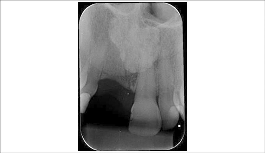
Interproximal radiographs of premolars and molars, and periapical radiograph of upper central and lateral incisors were performed. In the clinical examination, no facial asymmetry, painful symptoms, or history of trauma were observed. In the radiographic examination, using the periapical technique, a radiopaque area with defined borders was noted in the midline region, superimposed on the middle third of the root of tooth 21. Initially, the presence of a bone graft was considered; however, the patient denied undergoing graft surgery (figure 1)

Initial periapical radiograph of the upper central incisors region, showing a radiopaque area in the midline region.
A panoramic radiograph and CBCT of the maxilla were requested. In the panoramic radiograph, a radiopaque area with well-defined borders, slightly rounded, was found in the mesial region to the middle third of the root of tooth 21, without a radiolucent halo, with non-homogeneous content (figure 2). Diagnostic hypotheses included exostosis, peripheral osteoma, advanced ossifying fibroma, or benign maxillary osteochondroma.

Initial panoramic radiograph showing a radiopaque image in the anterior region of the maxilla.
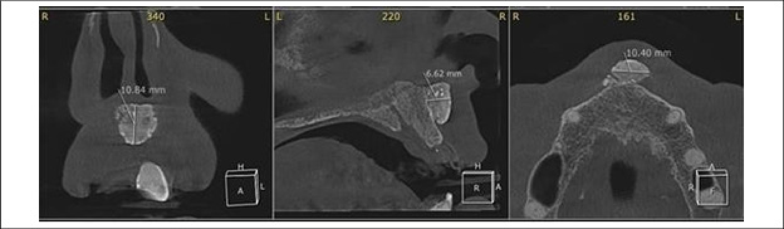
In the CBCT (cone-beam computed tomography) examination, a hyperdense area with well-defined borders, lobulated margin, partially heterogeneous density, exophytic, and pedunculated was observed, associated with the vestibular cortex of the midline (figure 3). The diagnostic hypotheses of exostosis, peripheral osteoma, and benign maxillary osteochondroma were maintained based on the CBCT images.

Initial cone-beam computed tomography. Coronal, sagittal, and axial sections of the anterior region of the maxilla, revealing a hyperdense exophytic lesion.
Due to its benign characteristics, surgical excision of the pathological lesion was proposed, and the removed tissues were referred for histopathological analysis.
The patient was referred to the postgraduate clinic in Minor Oral Surgery at the institution for the excision of the lesion.
The patient underwent the surgical procedure under infraorbital nerve anesthesia using articaine 4%, 1:100,000 (DFL - RJ Brazil). The Wasmund incision was performed, which consists of a festooned line on the attached gingiva with one or two vertical components. This incision was executed using a no. 15c scalpel blade (MEDIX-PR Brazil), gaining access to the lesion. Macroscopically, a bony mass located in the left hemimaxilla was observed, in the mesial region to the middle third of the root of tooth 21, with a hardened consistency and surrounded by fibrous tissue. The lesion showed confirmed sessile attachment and a lobulated surface. The lesion had an approximate diameter of 0.8. The lesion projection was performed with a Molt #9 periosteal elevator (QUINELATO, SP, Brazil) and a Molt 2-4 periosteal elevator (QUINELATO, SP, Brazil), and the lesion was removed in fragments using the elevator. A Molt #9 periosteal elevator (QUINELATO, SP, Brazil) and Kelly Straight Forceps (QUINELATO, SP-Brazil) were used. At the end of the surgery, continuous suturing was performed with 4-0 nylon suture (Shalon, GO-Brazil) to close the tissue edges (figure 4).

Intraoral image (a) Initial clinical examination (b) Bone mass located in the midline of the maxilla exposed during the surgical procedure (c) Removal of the lesion(d) Suturing of the surgical site (e, f) Fragments of the lesion.
The prescribed postoperative medication included Amoxicillin 500mg for seven days, Dipyrone 500mg, and Ibuprofen 600mg for five days.
The pathological lesion was placed in a container with 10% liquid formalin and sent for histopathological examination. It exhibited mucosa of the mucossecretory and respiratory type, associated with cartilaginous tissue showing areas of endochondral ossification, as well as areas of bone tissue, indicating the presence of benign maxillary osteochondroma (figure 5).

The suture removal was performed after seven days postoperatively. The patient reported the absence of painful symptoms, showing satisfactory signs of healing.
A follow-up cone-beam computed tomography (CBCT) was conducted after 4 months, where a slight contour change was observed in the maxillary vestibular bone cortex, presumably due to ongoing repair. The patient continues to be under observation to confirm the bone repair (figure 6).

Cone-beam computed tomography 4 months postoperatively. Axial and sagittal sections of the anterior region of the maxilla.
DISCUSSION
The investigation of rare conditions, such as benign maxillary osteochondroma, plays a crucial role as it provides dental professionals with access to information, contributing to the enhancement of diagnostic skills and enabling more effective therapeutic interventions, thereby minimizing harm and losses.
The etiology of benign maxillary osteochondroma is not fully understood, and the Lichtenstein hypothesis is a predominantly accepted theory. It suggests that the periosteum has a pluripotent capacity to originate chondroblasts or osteoblasts, and benign osteochondroma results from a metaplastic alteration in the periosteum [7]. For some authors, trauma is considered a possible trigger for this alteration [1-4]. Although the location of the lesion in this study may suggest a possible previous trauma, the patient denied any history, reporting no significant trauma in the anterior region of the maxilla. Osteochondromas are benign tumors commonly believed to originate from the proliferation of epiphyseal cartilage. However, this hypothesis cannot explain their occurrence in endochondral bones [4].
The average age of manifestation of osteochondromas in the mandible is higher than that observed in long bones [6], With an average age of 36.4 years and a slower growth, extending well beyond the post-pubertal period [7]. In this study, the patient presents in adulthood, which is consistent with the literature. Although the evolution time cannot be precisely determined, as there is no report of its onset.
Osteochondroma is a bony projection that exhibits potential for cartilaginous growth and is commonly observed near the growth plate, located at the ends of long bones, such as the knee, hip, shoulder, and other joints [5]. Osteochondromas are common tumors in long bones but are rare in the craniofacial region, with an incidence of around 0.6% [2].
The pathology in question is more documented in the skull base, maxillary sinus, zygomatic arch, and mandible, with its occurrence in the mandible being more frequent in the condyle or coronoid process [1-5].It is important to note that osteochondroma in craniofacial bones is extremely rare since facial bones more commonly undergo intramembranous ossification. Therefore, the occurrence of such a condition in the maxilla is rare, with few cases described in the literature [2-8].
In the scope of the analysis conducted for this study, a predominant trend of higher incidence of benign osteochondroma in the mandibular region was identified, with the mandibular condyle being the most frequently affected site, totaling six documented cases [1,2,3-5,9].
Furthermore, it is worth noting that occurrences of osteochondromas affecting the mandible were also recorded, including two cases in the coronoid process [4-7], two cases in the ramus [8-10], one case in the angle [11], and one case in the mandibular symphysis [12].
Benign osteochondromas affecting the maxilla are a rare occurrence in the literature, with records of only one case in the maxillary sinus [6], It is a case in the posterior region of the maxilla [13]. Therefore, in this context, this report represents a rarer case, in which a benign maxillary osteochondroma was diagnosed in the anterior region of the maxilla.
Even though, in the majority of cases, they manifest asymptomatically, depending on size and location, they can result in painful symptoms and an increase in volume in the affected area.
Suspicions of coronoid osteochondroma should arise when patients experience a progressive worsening of the ability to open the mouth and facial deformity. Clinical cases related to condylar osteochondroma have been reported by the authors, involving patients with an average age of 20 to 50 years. These cases exhibited symptoms such as swelling, clicking sounds in the temporomandibular joint, trismus, pain during chewing, hypoacusis [5], and deviation of the jaw with progressive facial asymmetry [1,2,7].
Due to the limitation in the ability to open the mouth, it is important to differentiate this condition from temporomandibular joint disorders [7].
As previously reported in the clinical case of benign maxillary osteochondroma affecting the maxillofacial region, the 55-year-old female patient did not present any clinical symptoms. According to the data from the analyzed clinical cases of maxillary osteochondroma, only one of the authors mentioned the presence of painful symptoms [6].
Early diagnosis is essential to provide appropriate treatment, which can impact the patient’s quality of life [3]. The definition of an accurate diagnosis is based on clinical, radiological, and histological criteria [2].
In panoramic radiography, radiopaque images with well-defined borders can be detected [7,8-13]. In the reported case, panoramic examination and periapical radiography revealed the presence of a well-defined, slightly circular radiopaque lesion in the anterior region of the maxilla. However, as these are two-dimensional images, they did not provide decisive information to define the diagnosis, as initially, there was a possibility that the lesion could be centrally located in relation to the alveolar ridge.
The use of various imaging modalities is of paramount importance, with CBCT being the most relevant for the evaluation of bone lesions. However, it is crucial to emphasize that histopathological examination remains the cornerstone for obtaining a definitive diagnosis in such conditions [13].
CBCT is considered essential for the diagnosis of osteochondroma as it provides precise information about the tumor’s location, density, and its relationship with adjacent structures. Such details are of utmost importance for the treatment planning [1,2,4,7,11,13]. In the reported case, CBCT revealed an exophytic, pedunculated, hyperdense lesion with slight heterogeneity and well-defined borders, providing more information and allowing the formulation of more accurate diagnostic hypotheses.
Considering osteochondroma is considered rare and radiographically similar to other conditions, formulating diagnostic hypotheses is challenging and may encompass various pathologies. Among the possibilities, osteoma would be the primary hypothesis, as it exhibits slow development, radiopaque radiographic density, well-defined margins, and no radiolucent halo [14] reminiscent of the image observed in the initial examinations of this study. The absence of signs and symptoms [15], common in osteoma cases, was also noted in the reported case. Multiple osteomas associated with intestinal polyposis may be related to Gardner syndrome; however, although the studied patient reported a case of intestinal cancer in family members, there was no association with the syndrome.
Ossifying Fibroma is a fibro-osseous pathology with high incidence in the oral and maxillofacial region, which can manifest as central or peripheral [16]. Peripheral ossifying fibroma was considered as a differential diagnosis due to the observed similarity in the reported case, characterized by an exophytic lesion with radiopaque mass. These lesions appear as unilocular with well-defined borders, a radiolucent halo, and mixed radiographic density, encompassing both radiolucent and radiopaque areas [16]. However, upon analyzing the case in question, the presence of a hypodense halo was not observed, and there was no observable swelling on clinical examination. Therefore, the hypothesis of peripheral ossifying fibroma was ruled out.
Exostoses are benign bony protrusions originating from the cortical bone, being self-limiting bony structures, palpably firm, and with normal coloration [3]. Their growth pattern is not continuous, ceasing after puberty [15]. Exostoses are common in the oral cavity, affecting the vestibular side of the alveolar process in the maxilla and/or mandible, where they are classified as palatal torus and mandibular torus [17], this differentiation distinguishes them from the pathology observed in the reported case, thus ruling out the possibility of it being an exostosis.
Another pathology with similar characteristics is osteosarcoma, although even rarer in the gnathic bones, it has a more accelerated development, causing volume increase, pain, paresthesia, and ulcerations [18], which were not observed in the reported case. The periosteal reaction pattern of osteosarcoma can vary, with the “sunburst” pattern being the most common [18]. Although osteosarcoma is listed in the literature as a differential diagnosis, it was not considered in this case due to the absence of signs and symptoms, the presumed slow growth, sessile attachment, and well-defined lobulated borders.
Histopathologically, osteochondromas are characterized by the presence of bony tissue covered by hyaline and fibrous cartilage. In the deeper layer, where the cartilage interfaces with the bone, the process of endochondral ossification is observed [4].
On histopathological examination, microscopic analysis revealed the presence of a mucosasecretory and respiratory mucosa, as well as the coexistence of cartilaginous tissue, along with areas of bony tissue, and the presence of endochondral and intramembranous ossification characterizing benign maxillary osteochondroma.
Unlike osteochondromas in long bones, which generally do not cause symptoms and do not require treatment, craniofacial osteochondromas can result in functional and aesthetic problems that necessitate surgical removal [7]. In this case, the patient did not present aesthetic or functional dysfunctions but opted for surgical removal to prevent potential future damage.
Surgical approaches mainly include intraoral and extraoral, or a combination of both techniques. Intraoral access is more favorable as it allows direct access, eliminating the possibility of facial nerve injury and visible scar formation [7]. In this case, an intraoral approach was advocated, as it provided indirect access, minimizing potential injuries to anatomical structures and scars. Although some authors suggest that extraoral access may be required in situations involving condylar lesions.
Surgical resection is a definitive solution, with recurrence or malignant transformation being extremely rare. Surgery is often indicated in symptomatic cases [6].
It is relevant to emphasize that the surgical resection of an osteochondroma may be associated with potential complications, such as neuropraxia, fracture, and arterial laceration [3]. Regarding the follow-up of the case in question, it is important to note that the patient did not experience any postoperative complications surgical, the patient is currently under follow-up with no evidence of recurrence.
CONCLUSION
Benign maxillary osteochondroma is a tumor that originates cortically to the endochondral bones, with the potential for cartilaginous growth, rarely occurring in the maxilla. The etiology of osteochondroma remains uncertain in the literature.
This study reported a case of benign maxillary osteochondroma associated with the vestibular bone cortex of the maxilla, in the midline region, with a pedunculated and asymptomatic insertion, discovered during a routine examination. However, it may cause asymmetry and discomfort, depending on the size and affected area.
Imaging studies are of utmost importance, with cone-beam computed tomography being more indicated for the evaluation of these lesions. However, histopathological examination remains the cornerstone for the definitive diagnosis.
It is evident that, although benign maxillary osteochondroma is a rare occurrence, it should be studied, as understanding bone conditions is fundamental for early diagnosis and effective treatment.
How to cite this article
-
Klein LB, Cimolin PX, Santos FDB, Muryel RB, Juliana MV, Maragno ÂC. Benign maxillary osteochondroma: a clinical case report. RGO, Rev Gaúch Odontol. 2024;72:e20240024. http://dx.doi.org/10.1590/1981-86372024002320240008
REFERENCES
-
1 Verma N, Kaur J, Warval GS. A simplified approach in the management of osteochondroma of the mandibular condyle. Natl J Maxillofac Surg. 2020;11(1):132-5. https://doi.org/10.4103/njms.NJMS_1_19
» https://doi.org/10.4103/njms.NJMS_1_19 -
2 Mohapatra M, Banushree CS. Osteochondroma condyle: a journey of 20 years in a 52-year-old male patient causing severe facial asymmetry and occlusal derangement. J Oral Maxillofac Pathol. 2019;23(1):162. https://doi.org/10.4103/jomfp.JOMFP_136_17
» https://doi.org/10.4103/jomfp.JOMFP_136_17 -
3 Souza DP, Junqueira JLC, Raitz R. Mandible condyle osteochondroma: clinical case report. RGO, Rev Gaúch Odontol. 2017;65(4):371-5. https://doi.org/10.1590/1981-863720170002000133542
» https://doi.org/10.1590/1981-863720170002000133542 -
4 Mohanty S, Gupta H, Dabas J, Kumar P. Osteochondroma of maxillofacial region: Tumor arising from two different developmental bones. J Oral Maxillofac Pathol. 2016 May-Aug;20(2):329. https://doi.org/10.4103/0973-029X.185904
» https://doi.org/10.4103/0973-029X.185904 -
5 Mahajan A, Patil DJ, Shah V, Mulay M. Giant Osteochondroma of the mandibular condyle and temporomandibular joint: a case report. J Oral Maxillofac Pathol. 2022;26(2):290. https://doi.org/10.4103/jomfp.jomfp_112_22
» https://doi.org/10.4103/jomfp.jomfp_112_22 -
6 Gil Guerra AB, Gómez San Martín E, Jiménez Cuenca MI. Osteochondroma of maxillary sinus, an unusual location. Acta Otorrinolaringol Esp (Engl). 2018;69(3):183-4. https://doi.org/10.1016/j.otorri.2017.04.003
» https://doi.org/10.1016/j.otorri.2017.04.003 -
7 Lan T, Liu X, Liang PS, Tao Q. Osteochondroma of the coronoid process: A case report and review of the literature. Oncol Lett. 2019;18(3):2270-7. https://doi.org/10.3892/ol.2019.10537
» https://doi.org/10.3892/ol.2019.10537 -
8 Kumar PS, Rao DS, Manepalli S, Damera A, Killada JK. Osteochondroma involving the ramus of the mandible: an unusual location. Case Rep Dent. 2020. https://doi.org/10.1155/2020/8603027
» https://doi.org/10.1155/2020/8603027 - 9 Lim W, Weng LK, Tin GB. Osteochondroma of the mandibular condyle: report of two surgical approaches. Ann Maxillofac Surg. 2014;4:215-9.
-
10 Gopinath D, Beena VT, Padmakumar SK, Sugirtharaj G. Osteochondroma of the mandibular ramus: report of a rare case. J Oral Maxillofac Surg Med Pathol. 2014;26(3):432-5. https://doi.org/10.1016/j.ajoms.2013.06.008
» https://doi.org/10.1016/j.ajoms.2013.06.008 -
11 Abe R, Miyamoto I, Sato H, Saitou D, Yamaya G, Yamada H. An unusually large osteochondroma of the mandibular angle: a case report. World. J Surg Oncol. 2017;15(1):201. https://doi.org/10.1186/s12957-017-1270-9
» https://doi.org/10.1186/s12957-017-1270-9 -
12 Navaneetham A, Rao KA, Kumaran S, Baweja HH. A unique case of multiple osteochondroma: mandibular symphysis and femur. Ann Maxillofac Surg. 2012; 2(2):182-4. https://doi.org/10.4103/2231-0746.101356
» https://doi.org/10.4103/2231-0746.101356 -
13 Jain M, Singhal S, Goyal M, Sharma B. Osteochondroma of maxilla posterior region: a unique case. J Clin Diagn Res. 2015;9(4):14-5. https://doi.org/ 10.7860/JCDR/2015/12478.5804
» https://doi.org/10.7860/JCDR/2015/12478.5804 -
14 Blackwell MC, Thakkar B, Flores A, Zhang W. Extracolonic manifestations of Gardner syndrome: a case report. Imaging Sci Dent. 2023;53(2):169-74. https://doi.org/10.5624/isd.20230006
» https://doi.org/10.5624/isd.20230006 -
15 Hasan S. Giant osteoma of the mandible: report of a rare case with review of literature. Indian J Otolaryngol Head Neck Surg. 2022;74(Suppl 3):4535-42. https://doi.org/10.1007/s12070-021-02565-1
» https://doi.org/10.1007/s12070-021-02565-1 -
16 Collins LHC, Zegalie NFT, Sassoon I, Speight PM. A Clinical, radiological and histopathological review of 74 ossifying fibromas. Head Neck Pathol. 2023; 17(2):433-46. https://doi.org/10.1007/s12105-022-01522-w
» https://doi.org/10.1007/s12105-022-01522-w -
17 Costa ALL, Batista ALM, Costa SFS, Bastos JV, Milagres RMC, Amaral TMP. Uncommon bilateral maxillary exostosis: case report. RGO, Rev Gaúch Odontol. 2020;68. https://doi.org/10.1590/1981-863720200002420180026
» https://doi.org/10.1590/1981-863720200002420180026 -
18 Loubna A, Bouchra T. Osteosarcoma of the jaws: a case report. Int J Surg Case Rep. 2022;93. https://doi.org/10.1016/j.ijscr.2022.106909
» https://doi.org/10.1016/j.ijscr.2022.106909
Edited by
-
Assistant editor: Luciana Butini Oliveira

 Source: Patient’s medical record (2023).
 Source: Patient’s medical record (2023).
 Source: Patient’s medical record (2023).
 Source: Patient’s medical record (2023).
 Source: Patient’s medical record (2023).
 Source: Patient’s medical record (2023).