 Intraoral injury by canine bite: a case report
ABSTRACT
Animal bites account for 15% of emergency hospital cases and are more frequent in males and children. These injuries may compromise the esthetics and functionality of the victims. Correct management of these cases, including careful attention to tetanus and rabies immunization, contributed to complete resolution. To report a case of a single deep laceration in the region of the jugal mucosa with a red color and irregular contour caused by a canine bite and discuss the therapeutic approach for its treatment. A 45-year-old white male patient with an intraoral lesion caused by a canine bite was treated at a public family health unit 1 d after trauma. Treatments included antisepsis, immunization, antibiotic prophylaxis, general recommendations, and follow-up. There are controversies regarding the closure of the lesion and antibiotic prophylaxis. The present case report contributes to clarifying a proper treatment approach for lesions that involve the intraoral mucosa, a region highly permeable to viruses and rich in microorganisms.
Indexing terms
Dentistry; Facial Injuries; Wound Infection
RESUMO
As mordidas de animais representam 15% das emergências hospitalares e são mais frequentes em homens e crianças. Este tipo de lesão pode comprometer a estética e funcionalidade dos tecidos envolvidos. O correto manejo desses casos, incluindo a atenção cuidadosa à imunização contra o tétano e a raiva, contribuem para a resolução completa da lesão. O objetivo foi relatar um caso clínico de laceração única, profunda, na região de mucosa jugal, de coloração eritematosa e contorno irregular, causada por mordida canina, e discutir a conduta terapêutica para seu tratamento. Paciente do sexo masculino, leucoderma, 45 anos, compareceu a uma Unidade Básica de Saúde da Família 24 horas após episódio de mordida canina. O tratamento da lesão intraoral compreendeu antissepsia, antibioticoterapia profilática, imunização, recomendações pós-atendimento e proservação do caso. Existem controvérsias na literatura quanto à sutura dos bordos da lesão e profilaxia com antibióticos, e a discussão do presente relato de caso contribui para o esclarecimento da melhor conduta clínica e o tratamento mais adequado de lesões que envolvem a mucosa oral, que representa ser um tecido altamente susceptível à invasão de mircrorganismos.
Termos de indexação
Odontologia; Traumatismos faciais; Infecção dos ferimentos
INTRODUCTION
Raising domestic animals brings several psychological, social, and physiological benefits to caregivers. However, these animals have an instinct to bite, and there are frequent accidents involving animal bites in humans, particularly in the facial region [1-3] Most animal bites are of canine origin (80–90%), affecting more children than adults, with a higher occurrence between 0-9 years of age, when they are more susceptible to critical injuries [3-4]. In cases involving the facial region, which represent approximately 15% of emergency cases, intervention by oral and maxillofacial surgeons is necessary for both adult and pediatric patients [3].
Canine bites are deep, elongated, V-shaped blunt wounds without any traces of suction, and may involve crushing, tearing, laceration, avulsion, or a combination of these types of injuries [2-4], as the force of a dog’s jaw can reach up to 1,800 psi (126.55 kgf/cm2) in one attack [1]. Owing to the greater depth of lesions caused by canine teeth, such a bite has a greater potential for tissue penetration, and considering the polymicrobial nature of this condition, there is a greater risk of developing diseases such as rabies and tetanus [2,3]. Additionally, there may be systemic, functional, psychological, and esthetic involvement of the patient, requiring a multiprofessional approach to the cass [5].
In the case of animal bites, several factors must be evaluated such as the severity and location of the lesion, time between the event and care, patient immunization, and animal condition [6-8] However, there is no consensus in the literature regarding the standard treatment for these cases, particularly concerning primary closure and antibiotic prophylaxix [2-6,9].
This study aimed to report a case of a canine bite that resulted in an intraoral lesion and discuss a therapeutic approach for its treatment.
CASE REPORT
This study was approved by the Ethics Committee with the protocol number: 55389521.0.0000.5544.
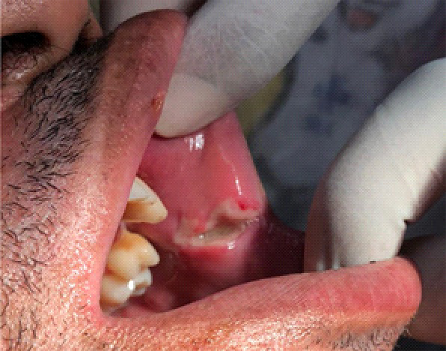
A 45-year-old white male patient sought care at a public family Health Unit for an intraoral wound caused by a canine bite 1 d after trauma. The patient gave a history of pain. He mentioned self-cleaning the wound using vinegar and potassium alum, popularly known as Hume Stone. Additionally, he prepared mouthwashes using the tea leaves of Schinus terebinthifolius and Alternanthera brasiliana, popularly known as aroeira and benzetacil, respectively. An extraoral physical examination revealed no signs of injury, inflammation, or infection of the head or neck. Intraoral examination revealed a blunt lacerated wound. It was a single deep lesion, reddish in color, smooth, with a regular contour in the left cheek jugal mucosa, with signs of edema, erythema, and pain (figure 1).

Immediately after the intraoral examination, irrigation was performed under pressure using a syringe and needle containing approximately 50 mL of 0.9% saline.
The multidisciplinary team prescribed ibuprofen 600 mg every 8 h for 3 days, amoxicillin + potassium clavulanate 500 + 125 mg every 8 h for 7 days, and metronidazole 250 mg 2 tablets every 12 h for 5 days. Additionally, observation of the dog on subsequent days and anti-rabies prophylaxis are recommended. As for vaccination, the prophylaxis schedule was not completed, as the dog displayed usual behavior during the observation period, and the owners presented the updated vaccination card of the animal.
According to oral care guidelines, mouthwashes with 0.12% chlorhexidine were prescribed every 12 hours for 10 days for wound antisepsis. Additionally, he was advised on oral hygiene, sun exposure, and the correct use of medications. After four days, the patient returned for reassessment. Fibrin covered the entire area of the lesion, with evidence of reduced wound extension and the beginning of healing by second intention (figure 2).

Clinical appearance with evident fibrin network formation on physical examination four days after the initial examination.
Follow-up 2 months after the first consultation demonstrated satisfactory functional results, with complete closure of the initial ulcerative lesion (figure 3).

Healing of the buccal mucosal lesion was observed on intraoral clinical examination 2 months after the initial examination during follow-up consultation.
DISCUSSION
Injuries caused by bites represent a large proportion of physical aggression observed in hospitals and emergency services, with domestic animals being the main causative agents. Considering the place of occurrence, a few studies have reported that a greater number of patients are victims of bites in their homes [10-12], which corroborates the aspects described in the present case report.
Dog bites are considered complex injuries owing to the possibility of esthetic and functional sequelae [2,13] the polymicrobial nature of the animal’s saliva, and the possibility of transmitting a variety of bacteria and viruses [6,8]. Wound contamination is characterized by tissue devitalization and the presence of foreign bodies, which can lead to severe infections and even necrosis. Such characteristics also contribute to the development of tetanus bacillus, thus necessitating anti-tetanus prophylaxix [4], which was not required in this case because the patient was already immunized.
All mammals are potential sources of rhabdovirus infection and can transmit it to humans, leading to central nervous system (CNS) infection with a mortality rate of approximately 100%, thus generating a high cost in preventive care [8,14]. Owing to the possible exposure to the rabies virus, the wound must be cleaned using a large amount of running water, soap, or other detergents immediately after the event to reduce the risk of infection. Additionally, as recommended by the Brazilian Ministry of Health (MH), cleaning should be repeated in the health unit regardless of the time elapsed, and antiseptics, such as polyvinylpyrrolidone-iodine, povidone, or chlorhexidine digluconate, should be used [14].
Several authors have found that saline solution is the most suitable for cleaning wounds because it has characteristics similar to those of physiological water and does not harm the tissue or cause deleterious effects on wound cells. Irrigation must be performed using a syringe and needle in deep penetrating wounds [2-13]. In the present case, although there was no advantage of antiseptics over saline solution4, a prior mouthwash with 1% chlorhexidine digluconate was used, as it is a protocol for dental care during the coronavirus disease 2019 pandemic, which can also be considered beneficial in this situation according to the MH protocol [14]. Immediately after the intraoral examination, irrigation was performed under pressure using a syringe and needle containing approximately 50 mL of 0.9% saline.
Complete anamnesis should be performed, with an assessment of exposures according to the wound characteristics and the animal involved, for the correct indication of rhabdovirus prophylaxis. One of the characteristics that requires evaluation is the location of the lesion, as regions close to the CNS (head, face, or neck) or highly innervated regions (hands, digital pulps, and soles of the feet) are considered serious because they facilitate the exposure of the nervous system to viruses. Licking of mucous membranes is a critical occurrence because the mucous membranes, even intact, are permeable to the virus, and the licks generally cover more extensive areas. The depth of the wound must also be analyzed and can be classified as superficial (no bleeding) or deep (presence of bleeding), given that punctate wounds are considered deep even if they do not bleed. Another aspect to be examined is the extent and number of lesions as each perforation is a gateway for the virus. The present case could be classified as a serious lesion because it was an extensive wound in the head and neck region (close to the CNS) and the oral mucosa since the mucosa was permeable to rhabdovirus [14].
As for the animal involved, a few aspects such as the animal’s state of health at the time of the aggression and spontaneous aggression (with no apparent cause), must be analyzed, as it may suggest that the animal was affected by rabies. Additionally, the origin of the animal must be analyzed to determine whether there is access to rabies control measures in its habitat. The animal’s lifestyle habits should also be considered, as dogs that spend long periods outdoors must be considered at-risk animals, even if they have an owner and are vaccinated. In the present case, the patient reported that the aggression occurred after provocation. Although the animal was known and vaccinated, the dog was observed for 10 days, a period encompassing the time of viral excretion into the animal’s saliva. Owing to the absence of changes in the animal’s behavior and signs suggestive of disease, the prophylactic regimen that had been initiated was discontinued according to the MH protocol [14].
According to a few authors, the possibility of wound infection is influenced by several factors: (1) the characteristics of the aggressor animal, with human and cat wounds presenting a higher probability of infection; (2) wound location, as lesions on the face have a lower chance of infection owing to abundant vascularization and postural drainage but are located close to the CNS; (3) factors inherent to the individual, such as the presence of pre-existing diseases (diabetes, immunodeficiencies, malnutrition); (4) characteristics of the lesion, such as the presence of coarse contaminants and the fact that puncture wounds have a greater chance of infection due to the large inoculation of bacteria and the difficulty of performing deep cleaning; and (5) time between the event and treatment, with variation among a few authors from 8 h to 4 days as a permissive period for primary closure; however, the longer the time, the greater is the risk of infection [6,15]. Given these factors, the wound in the present case demonstrated a considerable risk of infection because it was a punctate lesion in the intraoral mucosa, with a period of approximately 24 h between the event and treatment.
Different approaches have been advocated by the authors when dealing with the ideal time for wound closure. Primary closure is especially indicated for head and neck wounds, as most injuries involve structures such as the lips, ears, and other regions of the face where esthetic results are more relevant. Primary suturing is recommended for uninfected and recent bite wounds, whereas delayed suturing should be performed for those already infected or untreated within the first 24 h [4,13]. In the present case, the patient arrived at the health unit 1 d after the trauma, which contraindicated primary suturing. In addition, there was one case of a serious wound in the mucosa, with a high possibility of infection and without esthetic involvement.
Several aerobic and anaerobic organisms are cultivated in dogs and cats, and current literature recommends the use of amoxicillin and potassium clavulanate as the antimicrobial prophylactic agents of choice. This is due to the broad spectrum of action against the bacteria that infect these wounds, which are producers of β-lactamases, such as Streptococcus aureus and Prevotella. In patients with a history of allergic reactions to penicillin, azithromycin is indicated, and has good efficacy [2]. In the present case, ibuprofen was prescribed owing to signs of inflammation, and, as in the case of an intraoral bite, a combination of amoxicillin + potassium clavulanate and metronidazole was used for greater coverage against non-surgical infections of anaerobic microbial etiology; hence, broad-spectrum beta-lactams should be combined with metronidazole [13].
CONCLUSION
Injuries caused by canine bites require essential care, ranging from clinical examination and initial management to wound closure and follow-up, for successful treatment and complete resolution. Attention should be paid to the correct performance of wound antisepsis, indications for tetanus and rabies immunization, and antibiotic prophylaxis. Although controversial, primary surgical closure is the first choice for uninfected esthetic wounds. However, in this case, ibuprofen was prescribed due to signs of inflammation and, as in the case of an intraoral bite, a combination of amoxicillin + potassium clavulanate and metronidazole was used. The discussions in this context and documentation through more studies must be expanded regarding cases that affect unesthetic regions with abundant microbiota and high permeability, such as the oral mucosa.
How to cite this article
-
Assis MFA, Marino AC, Moreno BCL, Medrado ARAP. Intraoral injury by canine bite: a case report. RGO, Rev Gaúch Odontol. 2024;72:e20240036. http://dx.doi.org/10.1590/1981-86372024003620230096
REFERENCES
- 1 Silva LMA, Rocha AFL, Ferreira CS, Cunha G, Pereira Filho VA, Gabrielli MAC, et al. Tratamento de ferimento por mordedura canina: relato de caso [resumo] Rev Odontol UNESP. 2019;48(especial):63.
- 2 Porto GG, Souza BLM, Sampaio DO. Management of animal bite injuries: Case reports. Rev Cir Traumatol Buco-Maxilo-Fac. 2013;13(4):39-44.
- 3 Dantas RMX, Silva RRA, Cunha FP Filho, Dantas AMX, Tormes AKM. Lip injury by animal bite. Rev Cir Traumatol Buco-Maxilo-Fac. 2018;18(1):39-43.
- 4 Alencar MGM, Bortoli MMD, Almeida HCRD, Moraes PKMD, Lima NRD, Vasconcelos BCDE. Reconstruction for injury in upper lip biting in animal in child. Rev Cir Traumatol Buco-Maxilo-Fac. 2015;15(4):53-8.
- 5 Franzin LCDS, Gomes GDS, Casola HD, Marchese LT, Masson V, Camargo WR. Acidente por mordedura canina: relato de caso clínico. Rev Uningá. 2016;9(1):111-7.
-
6 Cavalcanti AL, Porto E, Dos Santos BF, Cavalcanti CL, Cavalcanti AFC. Facial dog bite injuries in children: A case report. Int J Surg Case Rep. 2017;41:57-60. https://doi.org/10.1016/j.ijscr.2017.10.008
» https://doi.org/10.1016/j.ijscr.2017.10.008 - 7 Oliveira VMR, Pereira PLL, Silva JÁ, Miranda CFJ, Rodrigues KO, Rodrigues TO, et al. Dog bite and human anti-rabies healthcare, Minas Gerais. Arq Bras Med Vet Zootec. 2012;64(4):891-8.
- 8 Nascimento AO, Matos RAC, Mendes SC, Corrêa VF. Freire M. Perfil epidemiológico do atendimento antirrábico humano no Brasil: uma revisão de literatura. REME Rev. Min. Enferm. 2019;23:e-1216.
- 9 Benson LS, Edwards SL, Schiff AP, Williams CS, Visotsky JL. Dog and cat bites to the hand: treatment and cost assessment. J Hand Surg Am. 2006;31(3):468-73.
- 10 Carvalho WO, Soares DFPP, Franceschi VCS. Characteristics of attendance rendered by public health services in Maringá-Paraná in 1997 to prevent human rabies. Inf Epidemiol SUS. 2002;11(1):25-35.
- 11 Paranhos NT, Silva EA, Bernardi F, Mendes MCNC, Junqueira DMAG, Souza IOM, et al. Study of aggression by dogs, according to the kind of interaction between dog and victim and motivating circumstances in the accidents, Municipality of São Paulo, 2008 to 2009. Arq Bras Med Vet Zootec. 2013;65(4):1033-40.
- 12 Carvalho CC, Silva BTF. Epidemiological characteristics of dog bite accidents attended at a basic health unit in northeast Brazil. Rev Bras Promoção Saúde 2007;20(1):17-21.
-
13 Zangari A, Cerigioni E, Nino F, Guidi R, Gulia C, Piergentili R, et al. Dog bite injuries in a tertiary care children’s hospital: A seven-year review. Pediatr Int. 2021;63(5):575-80. https://doi.org/10.1111/ped.14484
» https://doi.org/10.1111/ped.14484 - 14 Ministério da Saúde (Brasil). Normas técnicas de profilaxia da raiva humana. Brasília: Secretaria de Vigilância em Saúde; 2014.
- 15 Palmer J, Rees M. Dog bites of the face: A 15 year review. Br J Plast Surg. 1983;36(3):315-8.
Edited by
-
Assistant editor: Luciana Butini Oliveira