 Oral manifestations of Crouzon Syndrome and their implications for dental care: case report
ABSTRACT
Crouzon Syndrome is a rare genetic disease that affects craniofacial development resulting in various manifestations in the head and neck region. This article aimed to report a clinical case of a patient with Crouzon Syndrome, highlighting the relevant implications for dental care. The clinical, radiological aspects and dental treatment performed were evaluated. The dentist plays a fundamental role in the oral rehabilitation of these patients, promoting improvements in aesthetics and quality of life. This study provided insights into the management of the condition in the dental context, considering clinical, radiographic aspects and recommended treatments. Understanding these oral manifestations and their implications seems to be essential to offering adequate care and improving the quality of life of patients with CS.
Indexing terms
Disostose craniofacial; Assistência odontológica; Manifestações bucais
RESUMO
A Síndrome de Crouzon é uma doença genética rara que afeta o desenvolvimento craniofacial resultando em diversas manifestações na região de cabeça e pescoço. Esse artigo teve como objetivo relatar um caso clínico de um paciente com a Síndrome de Crouzon, destacando as implicações relevantes para o atendimento odontológico. Foram avaliados os aspectos clínicos, radiológicos e tratamento odontológicos realizados. O cirurgião-dentista desempenha um papel fundamental na reabilitação bucal desses pacientes, promovendo melhorias na estética e qualidade de vida. Esse estudo forneceu insights sobre o manejo da condição no contexto odontológico, considerando aspectos clínicos, radiográficos e tratamentos indicados. A compreensão dessas manifestações bucais e suas implicações parece ser essencial para oferecer um atendimento adequado e melhorar a qualidade de vida dos pacientes com SC.
Termos de indexação
Craniofacial dysostosis; Dental care; Oral manifestations
INTRODUCTION
Crouzon Syndrome (CS) is characterized by a set of conditions that affect the development of an individual’s skull, leading to facial deformities and exophthalmos [1]. It is a rare genetic syndrome, and although uncommon, it has a 50% chance of being transmitted when one parent carries the disease, regardless of the sex of the offspring [2].
CS was first reported in 1912 by the physician Louis Édouard Octave Crouzon, who identified a mother and son with craniosynostosis, resulting in cranial and facial deformities. There are various syndromes that lead to craniofacial deformities, but CS is considered the mildest among those involving craniosynostoses [3,4].
CS requires multidisciplinary treatment, including dental care, and the dentist must be aware of the causes and limitations of the condition to recognize and adequately treat the patient. In addition to promoting oral health education, dentists should work and collaborate with a multidisciplinary team, including physicians and psychologists [4-6].
This syndrome can be identified prenatally, and surgical intervention should be performed by the first year of life to alleviate increased intracranial pressure and prevent mental retardation, as well as to correct cranial morphological abnormalities [5]. The characteristic feature of this syndrome is the closure of the coronal and sagittal cranial sutures in utero, which can cause various anomalies such as atresic maxilla, exophthalmos, brachycephaly, and hypertelorism [6].
The literature postulates that fibroblast growth factor 2 (FGFR2) is a type of protein with widespread tissue expression, and various mutations in FGFR2 lead to CS [7]. Previous studies have shown that in this syndrome, severe malformations occur, including morphological anomalies of the maxilla and mandible, which have been associated with respiratory, neurological, and psychosocial disorders. When premature fusion of sutures occurs, maxillofacial deformities follow, requiring special attention [8,9].
In this context, the dentist presents an important role in preventing further complications and treating sequelae in patients with this condition. Therefore, this study aimed to report dental treatment in a patient with the mentioned syndrome and highlight its oral manifestations and implications for dental care.
CASE REPORT
This is a clinical case report of a patient treated at the integrated clinic of the Universidade Professor Edson Antônio Vellano, Divinópolis Campus. This research project was submitted to and approved by the Research Ethics Committee of UNIFENAS (CAAE: 68903523.6.0000.5143). The patient was informed about all procedures involved in the study, and all questions were answered prior to treatment. Additionally, the patient signed the Informed Consent Form (ICF) and granted authorization for the use of data, exams, and images.
A 17-year-old male patient, with light brown skin, attended the dental clinic at the Universidade Professor Edson Antônio Vellano, Divinópolis Campus, accompanied by his father for an evaluation. During the anamnesis, the patient reported having a broken tooth. At the beginning of the clinical examination, it was noted that the patient was shy and embarrassed but cooperative.
It was identified that the patient had Crouzon Syndrome (CS), which affects the craniofacial structure and impacts the teeth and uvula. He also reported difficulties in breathing and speaking and had undergone several craniofacial surgeries during childhood to correct the premature fusion of cranial sutures.
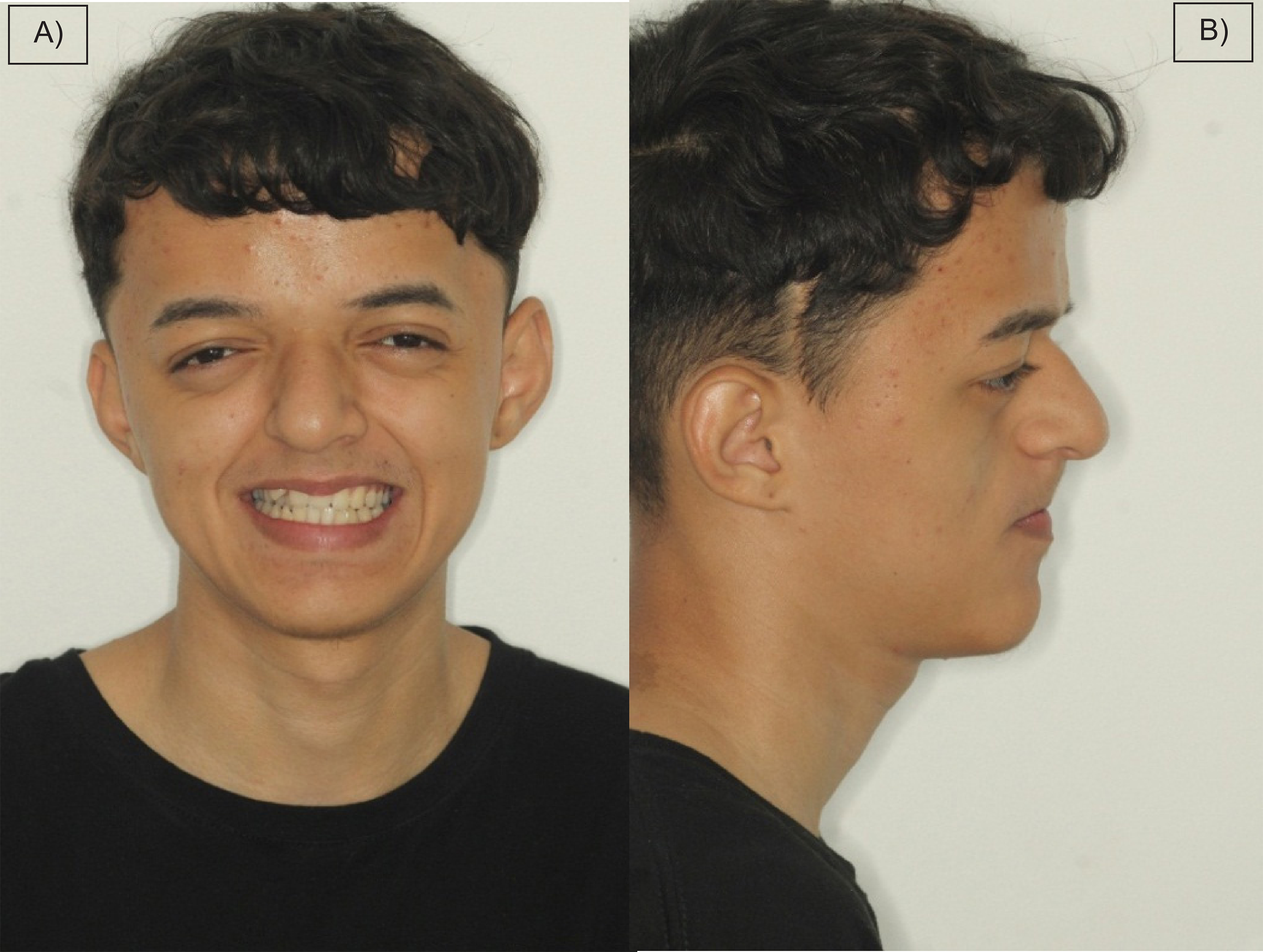
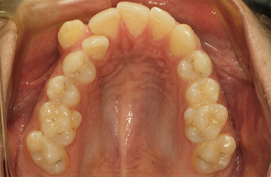
Intraoral examination revealed poor hygiene, dental crowding resulting in bilateral crossbite (figure 1), a narrow and underdeveloped maxilla compared to the face (figure 2), and teeth with a mulberry shape possessing deep and dark pits and fissures (figure 3), in addition to caries and a furcation lesion with pain in tooth 36.


A) Frontal photographic image, exposing the smile. B) Profile photographic image of the patient showing the underdeveloped maxilla.
Photographic image of the atretic palate, crowding and mulberry-shaped molars with deep pigmented grooves.
Soft tissue examination identified an atrophied uvula and a high-arched palate (figure 4). Additional imaging exams were requested (figure 5).


For the oral environment’s adaptation, the initial treatment consisted of dental prophylaxis and supragingival scaling using Gracey and McCall curettes to remove calculus, with the aid of an ultrasonic scaler and a bicarbonate jet. Following this, a Robson brush with prophylactic paste was used on all teeth at low speed, followed by rubber cup polishing. A hygiene kit containing a toothbrush, dental floss, and toothpaste was given to the patient, and proper oral hygiene techniques were taught.
The next stage of treatment required the extraction of tooth 36 due to a furcation lesion causing pain. The anesthesia with 2% Lidocaine with Epinephrine (DFL, Taquara, Rio de Janeiro, Brazil) was performed. After removal of the tooth, the socket was curetted with a Lucas curette, followed by saline irrigation and closure with 3-0 suture and needle holder assistance. Postoperative instructions and medication were provided to the patient.
After a 14-day healing period, the patient underwent molding for the fabrication of a band-type space maintainer, which was then sent to the laboratory. The purpose of the space maintainer installation is to preserve the space for future prosthetic treatment. It was placed after prophylaxis of tooth 37 and cemented with glass ionomer cement (FGM Dental Group, Joinville, SC, Brazil) using cotton roll isolation.
Subsequently, restorations of teeth 16, 17, 26, 27, and 46, which presented with caries, were performed. These procedures required anesthesia and the use of a 1014 spherical bur at high speed (Angelus®, Londrina, PR) and a dentin spoon excavator. After absolute isolation, the complete removal of carious dentin was performed, followed by etching with 37% phosphoric acid (FGM Dental Group, Joinville, SC, Brazil) for 30 seconds, then rinsing and drying. Self-etch adhesive (FGM Dental Group, Joinville, SC, Brazil) was applied to the enamel and dentin, dried, and light cured. 1mm increments of composite resin were added using a resin spatula kit and light-cured until satisfactory anatomical and functional restoration was achieved.
Finishing and polishing were carried out with discs and a rubber cup kit. Occlusion was checked with carbon paper, and necessary adjustments were made. In another session, fissure sealing of the molars with resin sealant (FGM Dental Group, Joinville, SC, Brazil) was performed to prevent future caries lesions, using cotton roll isolation.
DISCUSSION
Dental treatment for patients with Crouzon Syndrome (CS) presents significant challenges, requiring a multidisciplinary and personalized approach [10]. The presented case highlights the complexity of these challenges and illustrates the classic and innovative strategies necessary to provide effective and compassionate care for these patients.
One of the main issues in treating CS is the need to address not only aesthetic concerns but also functional ones. Dental crowding and a narrow maxilla are common problems in these patients, affecting not only appearance but also proper masticatory function. This case demonstrated the importance of identifying and knowing how to treat not only the aesthetics of the smile but also masticatory function, which is essential for the patient’s quality of life [1, 11].
Another distinctive feature of CS is the presence of mulberry-shaped teeth, which have deep pits and fissures. These characteristics make these teeth susceptible to caries and infiltrations. The application of resin sealant, as performed in this case, is an essential preventive strategy. Furthermore, innovative restorative techniques, such as the restoration of teeth 16, 17, 26, 27, and 46, are necessary to ensure the integrity of the teeth and prevent future complications [12].
It is crucial to recognize both the clinical and emotional needs of patients with CS. An empathetic approach and psychosocial support are fundamental components of care for these patients. As discussed, trust between the patient and the dental professional plays a crucial role. Creating a welcoming and supportive treatment environment is essential to ensure that the patient feels safe during procedures, promoting not only cooperation but also the patient’s self-esteem [13].
Interdisciplinary collaboration is undoubtedly the backbone of successful CS treatment. Coordination among dentists, plastic surgeons, orthodontists, and psychologists is vital to provide a comprehensive approach to the patient. Continuous information sharing among team members is essential to plan and execute coordinated interventions, ensuring the best possible outcome for the patient [14].
In summary, dental treatment for CS goes beyond aesthetic restoration; it is a matter of restoring quality of life. The integration of advanced techniques, compassion, and interdisciplinary collaboration is crucial to overcoming the unique challenges presented by this condition. This case highlights not only the clinical complexities but also the resilience and determination of the patient, as well as the dedication of healthcare professionals in providing holistic and high-quality care.
CONCLUSION
Crouzon Syndrome (CS) transcends the realm of physical manifestations; it has a substantial impact on the self-image and psychosocial prognosis of affected individuals. This study underscores the imperative of an integrative and empathetic therapeutic strategy that values the importance of compassionate care and sensitive understanding at every clinical encounter. This holistic approach should not be confined to the physical sphere but must also address the emotional and psychological ramifications inherent in the patient’s condition. The complexity of CS requires that medical interventions be informed by a deep recognition of fundamental human needs, encompassing the overall well-being of the individual.
How to cite this article
-
Sena APO, Silva TC, Monteiro LN, Pardini DS, Silveira FF, Tonelli SQ. Oral manifestations of Crouzon Syndrome and their implications for dental care: case report. RGO, Rev Gaúch Odontol. 2024;72:e20240037. http://dx.doi.org/10.1590/1981-86372024003720240035
REFERENCES
-
1 Pary A, Pedra J, Neto C. Tratamento ortodôntico-cirúrgico da Síndrome de Crouzon em paciente adulto: relato de caso clínico. Rev Clin Ortodon Dental Press. 2018;17(2). https://doi.org/10.14436/1676-6849.17.2.043-054.art
» https://doi.org/10.14436/1676-6849.17.2.043-054.art -
2 Al-Namnam NM, Hariri F, Thong MK, Rahman ZA. Crouzon syndrome: Genetic and intervention review. J Oral Biol Craniofac Res. 2019 Jan-Mar;9(1):37-39. https://doi.org/10.1016/j.jobcr.2018.08.007
» https://doi.org/10.1016/j.jobcr.2018.08.007 -
3 Li XJ, Su JM, Ye XW. Crouzon syndrome in a fraternal twin: A case report and review of the literature. World J Clin Cases. 2022;10(16):5317. https://doi.org/10.12998/wjcc.v10.i16.5317
» https://doi.org/10.12998/wjcc.v10.i16.5317 -
4 Vilan Xavier AC, Pinto Silva LC, Oliveira P, Villamarim Soares R, de Almeida Cruz R. A review and dental management of persons with craniosynostosis anomalies. Spec Care Dentist. 2008 May-Jun;28(3):96-100. https://doi.org/10.1111/j.1754-4505.2008.00019.x
» https://doi.org/10.1111/j.1754-4505.2008.00019.x -
5 Kumar GR, Jyothsna M, Ahmed SB, Lakshmi KS. Crouzon’s Syndrome: A Case Report. Int J Clin Pediatr Dent. 2013 Jan;6(1):33-7. https://doi.org/10.5005/jp-journals-10005-1183
» https://doi.org/10.5005/jp-journals-10005-1183 -
6 Khonsari RH, Way B, Nysjö J, Odri GA, Olszewski R, Evans RD, et al. Fronto-facial advancement and bipartition in Crouzon-Pfeiffer and Apert syndromes: Impact of fronto-facial surgery upon orbital and airway parameters in FGFR2 syndromes. J Craniomaxillofac Surg. 2016 Oct;44(10):1567-1575. https://doi.org/10.1016/j.jcms.2016.08.015
» https://doi.org/10.1016/j.jcms.2016.08.015 -
7 Sicard L, Hennocq Q, Paternoster G, Arnaud E, Dure-Molla M, Khonsari RH. Dental phenotype in Crouzon syndrome: a controlled radiographic study in 22 patients. Arch Oral Biol. 2021 Nov;131:105253. https://doi.org/10.1016/j.archoralbio.2021.105253
» https://doi.org/10.1016/j.archoralbio.2021.105253 -
8 Khominsky A, Yong R, Ranjitkar S, Townsend G, Anderson PJ. Extensive phenotyping of the orofacial and dental complex in Crouzon syndrome. Arch Oral Biol. 2018 Feb;86:123-130. https://doi.org/10.1016/j.archoralbio.2017.10.022
» https://doi.org/10.1016/j.archoralbio.2017.10.022 -
9 Kobayashi Y, Ogura K, Hikita R, Tsuji M, Moriyama K. Craniofacial, oral, and cervical morphological characteristics in Japanese patients with Apert syndrome or Crouzon syndrome. Eur J Orthod. 2021 Jan 29;43(1):36-44. https://doi.org/10.1093/ejo/cjaa015
» https://doi.org/10.1093/ejo/cjaa015 -
10 Bhattacharjee K, Rehman O, Venkatraman V, Kikkawa D, Bhattacharjee H, Gogoi R, et al. Crouzon syndrome and the eye: An overview. Indian J Ophthalmol. 2022 Jul;70(7):2346-2354. https://doi.org/10.4103/ijo.IJO_3207_21
» https://doi.org/10.4103/ijo.IJO_3207_21 -
11 Holongwa P. Tratamento ortodôntico precoce da Síndrome de Crouzon: relato de caso. J Maxillofac Oral Surg. 2009;8(1):74-76. https://doi.org/10.1007/s12663-009-0018-7
» https://doi.org/10.1007/s12663-009-0018-7 -
12 Kurt H, Gençel B, Kader AC. Prosthetic rehabilitation of a Crouzon patient: A case report. Contemp Clin Dent. 2010 Jul;1(3):196-200. https://doi.org/10.4103/0976-237X.72794
» https://doi.org/10.4103/0976-237X.72794 -
13 Alves E dos S, Francisco AL. Ação psicológica em saúde mental: uma abordagem psicossocial. Psicol Ciênc Prof. 2009;29(4):768-779. https://doi.org/10.1590/S1414-98932009000400009
» https://doi.org/10.1590/S1414-98932009000400009 -
14 Tripathi T, Srivastava D, Bhutiani N, Rai P. Comprehensive management of Crouzon syndrome: A case report with three-year follow-up. J Orthod. 2022 Mar;49(1):71-78. https://doi.org/10.1177/14653125211019412
» https://doi.org/10.1177/14653125211019412
Edited by
-
Assistant editor: Luciana Butini Oliveira