
 Dental trauma in permanent teeth: pediatric dentistry rehabilitation strategy
ABSTRACT
This case report aims to describe the clinical management of a dental trauma in an 8-year-old male who suffered a fall while playing sports. Twelve months after the fall, he sought care at the Pediatric Dentistry Emergency Service of the Departamento de Saúde Coletiva, Odontopediatria e Ortodontia of the Piracicaba School of Dentistry, Unicamp, reporting mobility in the upper left central incisor (#21). The patient was immediately seen by a dentist who performed a periapical radiograph and performed immediate aesthetic reconstruction with composite resin on the upper left and right permanent central incisors. During the consultation at the Emergency Service, a clinical examination and periapical radiograph were performed, and a horizontal fracture in the middle third of the root, alveolysis, and exposure of root fragments were found, making endodontic treatment of the tooth related to the complaint unfeasible (#21). The patient’s mother received guidance on the situation, and, after approval, the tooth was extracted. For rehabilitation, a space maintainer with a stock tooth was planned, for aesthetic reasons and the need to preserve the space, with immediate installation. Four months later, at the follow-up appointment, the space was maintained, and the patient approved the aesthetics. Continuous periodic appointments were held to monitor the patient and adjust or replace the space maintainer for 15 months. Hereupon, early diagnosis, immediate intervention, and monitoring of dental trauma are essential to maintain oral health, preserving aesthetics and function until the end of the patient’s growth and development.
Indexing terms
Dentition, permanent; Pediatric dentistry; Tooth injuries
RESUMO
Este relato de caso tem como objetivo descrever o manejo clínico de um traumatismo dentário em uma criança de 8 anos de idade, sexo masculino que sofreu queda durante prática esportiva. Após 12 meses da queda, procurou atendimento no Serviço de Urgência em Odontopediatria do Departamento de Saúde Coletiva, Odontopediatria e Ortodontia da Faculdade de Odontologia de Piracicaba/Unicamp, relatando mobilidade no incisivo central esquerdo superior (21). Na anamnese verificou-se que o paciente foi atendido de imediato por um cirurgião-dentista que realizou uma radiografia periapical e fez a reconstrução estética imediata com resina composta nos incisivos centrais permanentes superiores esquerdo e direito. Na consulta no Serviço de Urgência, foi realizado exame clínico e radiografia periapical e verificou-se fratura horizontal no terço médio da raiz, alveólise e exposição de fragmentos radiculares, inviabilizando o tratamento endodôntico do dente relativo à queixa (21). A mãe do paciente recebeu orientações sobre a situação e, após a aprovação, o dente foi extraído. Para a reabilitação foi planejado um mantenedor de espaço com dente de estoque, por questões estéticas e necessidade de preservação do espaço, com instalação imediata. Quatro meses depois, na consulta de acompanhamento observou-se a manutenção do espaço e a aprovação estética pelo paciente. Foram realizadas consultas periódicas contínuas para acompanhamento do paciente e ajustes ou trocas do mantenedor de espaço durante 15 meses. Com isto, o diagnóstico precoce, intervenção imediata e monitoramento do traumatismo dentário são essenciais para manter a saúde bucal, preservando estética e função até o término do crescimento dos pacientes.
Termos de indexação
Dentição permanente; Odontopediatria; Traumatismos dentários
INTRODUCTION
Root fractures are a rare but clinically significant form of Traumatic Dental Injury (TDI), accounting for approximately 0.5% to 7% of all dental traumas, predominantly affecting permanent teeth [1,2]. These injuries are most associated with falls, sports-related incidents, and recreational activities, particularly in children and adolescents [3,4]. Depending on their location – cervical, middle, or apical third – root fractures present distinct challenges in diagnosis, treatment, and prognosis, depending on root site location and dislodgment of the fragments [5,6].
Prompt and appropriate management of root fractures is critical to avoid severe complications such as pulp necrosis, periapical lesions, pathological root resorption, and tooth loss. Treatment strategies vary widely, ranging from conservative approaches like splinting and endodontic therapy to surgical interventions [7]. Delayed or inadequate treatment may lead to irreversible damage, impacting not only oral health but also the patient’s overall quality of life, nutrition, and psychosocial well-being [8].
Given the rarity and complexity of horizontal root fractures, particularly in the permanent dentition of young patients, the present case report aims to describe the clinical management of a horizontal root fracture, highlighting the treatment protocol, clinical outcomes, and considerations for long-term follow-up.
CASE REPORT
The case report was approved by the local Research Ethics Committee (protocol # CAAE 79343724.0.0000.5418), and informed consent was obtained and duly signed by both the parents and the child, following established ethical guidelines.
An 8-year-old male patient, in good general health, with no history of systemic or infectious diseases or allergies, presented to the Pediatric Dentistry Emergency Service of the Departamento de Saúde Coletiva, Odontopediatria e Ortodontia of the Universidade Estadual de Campinas of Faculdade de Odontologia de Piracicaba (Unicamp-FOP), complaining on mobility and pain of the anterior teeth. The main concern expressed by the patient was the possibility of losing the affected tooth, alongside a fear of being bullied at school. During the anamnesis, the patient’s mother reported that a dental trauma occurred one year prior when the patient fell while playing football with friends, resulting in injury to teeth #11 and #21 (right and left, central superior incisors). Following the accident, the family sought care at a private clinic, where panoramic and periapical radiographs of the affected region were obtained (figure 1). The initial treatment provided by the attending clinician included Class IV composite resin restorations on teeth #11 and #21. However, after one year the patient subsequently reported mobility in tooth #21 during mastication. Faced with this complaint, the family returned to private clinic. A new periapical radiography was taken (figure 1) and the patient was then referred to the Pediatric Dentistry Service of Departamento de Saúde Coletiva, Odontopediatria e Ortodontia at Faculdade de Odontologia de Piracicaba, at Unicamp for specialized evaluation and management.

After obtaining signed informed consent from the patient’s mother, a thorough clinical and radiographic examination was performed to determine the appropriate therapeutic approach. An extensive Class IV composite resin restoration was observed on tooth #21, accompanied by significant mobility and the presence of inflammatory tissue in the buccal region. Additionally, a root fragment was exposed within inflammatory tissue, due to an alveolysis (figure 2). The radiographic evaluation included a periapical radiograph (figure 1), which revealed a horizontal fracture in the middle third of the root of tooth #21, associated with bone loss at the fracture site.

Frontal photograph of the clinical examination of teeth #11 and #21 at the Pediatric Dentistry Emergency Service.
An ongoing process of rizogenesis was also observed (figure 2), as well as the presence of a periapical lesion.
Due to the extent of bone loss in the fractured region and loss of a root fragment as a result of the trauma, endodontic treatment of tooth #21 was contraindicated. Consequently, the treatment plan involved the extraction of the tooth followed by prosthetic rehabilitation (figures 3 and 4), considering the patient’s aesthetic and functional needs as well as his initial complaint.


Therapeutic planning included an initial impression of the dental arch to fabricate a removable upper appliance with stock teeth (figures 4 and 5), aiming to provide immediate rehabilitation. Following the manufacturing of the prosthetic device was accomplished and the tooth #21 was extracted. Postoperative surgical management of the pain, ibuprofen (250 mg/5 ml), was prescribed. Additionally, dietary guidance was provided to facilitate proper nutrition during the recovery period.

Front view from patient using the new space maintainer appliance made with stock teeth and acrylic resin after installation.
Three days after the surgery, the removable prosthetic device was delivered and installed (figure 5). The patient and their mother received detailed instructions on oral hygiene and the necessary care to maintain the appliance. Regular follow-up appointments were conducted; however, after 15 months, due to the patient’s growth, the removable prosthesis no longer fit properly. Consequently, a new prosthesis was fabricated, and the patient remains under periodic follow-up (figure 6).

Front view from patient using the new space maintainer appliance, after the last follow-up.
DISCUSSION
Dental trauma, affecting both hard and soft oral tissues, is a frequent consequence of sports accidents, falls, or collisions [9]. Common sequelae include coronal fractures, luxation, and avulsions, which may result in pain, functional limitations, and aesthetic compromise [10-14]. These injuries are particularly prevalent in young children, with epidemiological studies reporting that 18–22% of preschoolers and 25% of school-aged children sustain dental trauma globally [9,14,15]. Among school-aged populations, up to 25% sustain dental injuries before adulthood, representing 5% of all childhood accidents [1].
The psychosocial implications of dental trauma are profound. Anterior tooth injuries often disrupt aesthetics, communication, and self-esteem, which are critical for social development [3, 16]. In the present case, delayed diagnosis of a mid-root fracture in tooth #21 led to irreversible pulp necrosis and eventual extraction. The child’s anxiety about tooth mobility and fear of bullying at school underscored the interplay between dental health and emotional well-being. Such cases emphasize the need for pediatric dentists to address both biological and psychological aspects of care through empathetic communication with patients and caregivers [17]. Early diagnosis and intervention are essential for favorable outcomes in dental trauma [18]. However, delayed management remains common due to challenges in detecting subtle injuries, such as horizontal root fractures.
In this case reported, initial treatment focused only on restoring coronal fractures of teeth #11 and #21, but the mid-root fracture in #21 was overlooked. Advanced imaging such as cone-bean computed tomography (CBCT) at the first moment likely contributed to precocious diagnosis. However, dentists in private practice overall don’t have this expensive source. Subsequently, minimal root fractures are poorly diagnosed and infection progression led to extraction, highlighting the importance of thorough clinical and radiographic evaluation. This outcome brought about the importance of adhering to trauma guidelines [5] and timely referral to specialists in cases of diagnostic uncertainty.
Given these challenges, three key considerations emerge for optimizing outcomes in dental trauma cases: First, while CBCT enhances the detection of complex fractures [19] its limited accessibility underscores the importance of timely specialist referral when diagnostic uncertainty exists, potentially preventing avoidable tooth loss. Second, in cases of luxation or extrusion, splinting should be prioritized to promote periodontal healing, as recommended by the IADT Guidelines [5]. Finally, for immature permanent teeth, pulp preservation techniques – such as partial pulpotomy or regenerative endodontics [19] – should be employed to maintain vitality and support continued root development.
The establishment of a protocol for attending dental trauma emergencies is needed, mainly for the initial management of dental trauma, requiring a systematic approach: 1. Clinical and radiographic evaluation - A thorough extraoral/intraoral examination, including mobility testing, percussion, and pulp sensibility tests (e.g., cold or electric pulp testing), should be performed [20]. Radiographs (preferably CBCT for complex fractures) are essential to detect root fractures, luxation injuries, or alveolar bone damage [21]
Early loss of anterior teeth in children risks malocclusion, like maxillary protrusion and changes in muscle activity, alveolar bone resorption, and parafunctional habits (e.g., involuntary exploration of the region) [22,23]. To address these concerns, a removable upper appliance with a prosthetic tooth was provided for #21. This approach prioritized aesthetics, space maintenance, and psychosocial rehabilitation while accommodating the patient’s age and oral hygiene needs [24]. The appliance’s resemblance to orthodontic devices improved acceptance, reducing potential stigma during social interactions.
In the present clinical case report, the patient’s self-awareness of tooth mobility due to trauma led to social anxiety, particularly at school, where he feared being bullied about. The child exhibited emotional distress, including episodes of crying, driven by the fear of losing an anterior tooth. This situation highlights the importance of the pediatric dentist’s role in providing a welcoming environment and supporting not only the child’s dental health but guiding the caregiver effectively.
A favorable prognosis and appropriate treatment plan for managing dental trauma are linked to prompt intervention [18]. Early first aid plays a crucial role in these situations to control positive prognosis; however, studies indicate that for many professionals lack the fundamental knowledge and experience needed to deliver initial emergency care [4,25,26].
As reported, in this case, there were fractures in the coronal region of teeth #11 and #21 and middle radicular horizontal fractures; however, the proposed treatment was only aimed at restoration aesthetics, which caused the progression of infection in the region of tooth #21, leading to dental loss. Good planning is important for each case of dental trauma, or even referral to a specialist, as the professional’s lack of experience negatively interferes with the progress of treatment.
In Pediatric Dentistry, prosthetic planning based on age is important for the patient, capacity for cooperation or behavior and the stage of speech development [24]. The removable device enables the patient to remove it independently for hygiene purposes and oral health maintenance and serves as a space maintainer until the growth phase ends and future planning for definitive rehabilitation can be undertaken.
For a pediatric dentist patient, the loss of an anterior tooth and the need for rehabilitation can be a complicated process, both for acceptance of the situation and collaboration with the treatment. In this clinical case, welcoming the patient was essential both for understanding the situation, as well as accepting the use of the removable device. As the patient was already afraid of being bullied in a school environment, using a removable device with stock teeth, like a conventional orthodontic appliance, facilitated acceptance of the patient and his reintegration into the social envenenamento.
This case reinforces the need for multidisciplinary collaboration in pediatric dental trauma. Clinicians should maintain a high index of suspicion for root fractures in cases of coronal trauma particularly when symptoms persist despite initial treatment. Future efforts to improve trauma education for general practitioners and promote early specialist consultation may reduce preventable tooth loss and complications in children.
CONCLUSION
This case highlights the consequences of delayed diagnosis of mid-root fractures in pediatric dental trauma, emphasizing the need for thorough evaluation with advanced imaging (e.g., CBCT) and adherence to trauma guidelines. The psychosocial impact of tooth loss underscores the importance of multidisciplinary care, combining timely intervention with empathetic, patient-centered approaches to restore function and support emotional well-being.
Data availability
The research data are available from the corresponding author upon reasonable request.
-
How to cite this article
Zanelli WS, Gois CMB, Santos ECA, Pascon FM, Puppin-Rontani RM. Dental trauma in permanent teeth: pediatric dentistry rehabilitation strategy. RGO, Rev Gaúch Odontol. 2025;73:e20250028. http://dx.doi.org/10.1590/1981-86372025002820250029
REFERENCES
-
1 Levin L, Day PF, Hicks L, O’Connell A, Fouad AF, Bourguignon C, et al. International Association of Dental Traumatology guidelines for the management of traumatic dental injuries: General introduction. Dent Traumatol. 2020;36(4):309-13. https://doi.org/10.1111/edt.12574
» https://doi.org/10.1111/edt.12574 -
2 Némat SM, Kenny KP, Day PF. Special considerations in paediatric dental trauma. Prim Dent J. 2023;12(4):64-71. https://doi.org/10.1177/205016842 31211413
» https://doi.org/10.1177/205016842 31211413 -
3 Borges TS, Vargas-Ferreira F, Kramer PF, Feldens CA. Impact of traumatic dental injuries on oral health-related quality of life of preschool children: a systematic review and meta-analysis. PLoS One. 2017;12(2):e0172235. https://doi.org/10.1371/journal.pone.0172235
» https://doi.org/10.1371/journal.pone.0172235 -
4 Nowosielska M, Bagińska J, Kobus A, Kierklo A. How to Educate the Public about Dental Trauma-A Scoping Review. Int J Environ Res Public Health. 2022;19(4):2479. https://doi.org/10.3390/ijerph19042479
» https://doi.org/10.3390/ijerph19042479 -
5 International Association of Dental Traumatology. Guidelines for the management of traumatic dental injuries. Dental Traumatol. 2020;36(4):309-13. https://doi.org/https://doi.org/10.1111/edt.12573
» https://doi.org/10.1111/edt.12573 -
6 Cho J, Sachs A, Cunningham LL Jr. Dental trauma and alveolar fractures. Facial Plast Surg Clin North Am. 2022;30(1):117-24. https://doi.org/10.1016/j.fsc.2021.08.010
» https://doi.org/10.1016/j.fsc.2021.08.010 - 7 Majewski M, Kostrzewska P, Ziółkowska S, Kijek N, Malinowski K. Traumatic dental injuries - practical management guide. Pol Merkur Lekarski. 2022;50(297):216-8.
-
8 Sarao SK, Levin L. Pain management, pediatric dental injuries, and dental trauma education. Dent Traumatol. 2023;39(4):291-4. https://doi.org/10.1111/edt.12866
» https://doi.org/10.1111/edt.12866 -
9 Glendor U. Epidemiology of traumatic dental injuries: a 12-year review of the literature. Dental Traumatol. 2008;24(6):603-11. https://doi.org/10.1111/j.1600-9657.2008.00696.x
» https://doi.org/10.1111/j.1600-9657.2008.00696.x -
10 Caldas AF Jr, Burgos MEA. A retrospective study of traumatic dental injuries in a Brazilian dental trauma clinic. Dent Traumatol. 2001;17(6):250-3. https://doi.org/10.1034/j.1600-9657.2001.170602.x
» https://doi.org/10.1034/j.1600-9657.2001.170602.x -
11 Aldrigui JM, Abanto J, Carvalho TS, Mendes FM, Wanderley MT, Bönecker M, et al. Impact of traumatic dental injuries and malocclusions on quality of life of young children. Health Qual Life Outcomes. 2011;9:78. https://doi.org/10.1186/1477-7525-9-78
» https://doi.org/10.1186/1477-7525-9-78 -
12 Feldens CA, Day P, Borges TS, Feldens EG, Kramer PF. Enamel fracture in the primary dentition has no impact on children’s quality of life: implications for clinicians and researchers. Dent Traumatol. 2016;32(2):103-9. https://doi.org/10.1111/edt.12222
» https://doi.org/10.1111/edt.12222 -
13 Lam R. Epidemiology and outcomes of traumatic dental injuries: a review of the literature. Aust Dent J. 2016; 61 Suppl. 1:4-20. https://doi.org/10.1111/adj.12395
» https://doi.org/10.1111/adj.12395 -
14 Petti S, Glendor U, Andersson L. World traumatic dental injury prevalence and incidence: a meta-analysis. Dental Traumatol. 2018;34(2):71-86. https://doi.org/10.1111/edt.12389
» https://doi.org/10.1111/edt.12389 -
15 Feldens CA, Borges TS, Vargas-Ferreira F, Kramer PF. Risk factors for traumatic dental injuries in the primary dentition: concepts, interpretation, and evidence. Dent Traumatol. 2016;32(6):429-37. https://doi.org/10.1111/edt.12281
» https://doi.org/10.1111/edt.12281 -
16 Ramos-Jorge ML, Bosco VL, Peres MA, Nunes AC. The impact of treatment of dental trauma on the quality of life of adolescents: a case-control study. Dental Traumatol. 2014;30(1):22-7. https://doi.org/10.1111/edt.12041
» https://doi.org/10.1111/edt.12041 -
17 Aminabadi NA, Ghoreishizadeh A, Ghoreishizadeh M, Oskouei SG. Can drawing be considered a projective measure for children’s distress in paediatric dentistry? Int J Paediatr Dent. 2011;21(1):1-12. https://doi.org/10.1111/j.1365-263X.2010.01072.x
» https://doi.org/10.1111/j.1365-263X.2010.01072.x - 18 Alotaibi S, Haftel A, Wagner ND. Avulsed tooth. In: StatPearls. Tampa (FL): StatPearls Publishing; 2023.
-
19 Kahler B, Rossi-Fedele G, Chugal N. An evidence-based review of the efficacy of treatment approaches for immature permanent teeth with pulp necrosis. J Endod. 2017;43(7):1052-7. https://doi.org/10.1016/j.joen.2017.03.003
» https://doi.org/10.1016/j.joen.2017.03.003 - 20 Andreasen JO, Andreasen FM, Andersson L. Textbook and color atlas of traumatic injuries to the teeth. 4th ed. Boston: Wiley-Blackwell; 2007.
-
21 Cohenca N, Simon JH, Mathur A, Malfaz JM. Clinical indications for digital imaging in dento-alveolar trauma. Dental Traumatol. 2007;23(2):105-13. https://doi.org/10.1111/j.1600-9657.2006.00 522.x
» https://doi.org/10.1111/j.1600-9657.2006.00 522.x -
22 Frujeri ML, Frujeri JA, Bezerra AC, Cortes MI, Costa JE. Socio-economic indicators and predisposing factors associated with traumatic dental injuries in schoolchildren at Brasília, Brazil. Dental Traumatol. 2009;25(2):118-23. https://doi.org/10.1111 /j.1600-9657.2008.00725.x
» https://doi.org/10.1111 /j.1600-9657.2008.00725.x -
23 Fierro C, Bravo L, Vera A, Torres M, Pérez A. Pérdida prematura de incisivos como fator agravante de hábitos, reporte de un caso clínico. Rev Soc Chil Odontopediatría. 2009;24:35-8. https://doi.org/10.24265/kiru.2019.v16n2.05
» https://doi.org/10.24265/kiru.2019.v16n2.05 -
24 Kalia G, Tandon S, Bhupali NR, Rathore A, Mathur R, Rathore K. Speech evaluation in children with missing anterior teeth and after prosthetic rehabilitation with fixed functional space maintainer. J Indian Soc Pedod Prev Dent. 2018;36(4):391-5. https://doi.org/10.4103/JISPPD.JISPPD_221_18.
» https://doi.org/10.4103/JISPPD.JISPPD_221_18 -
25 Holan G, Cohenca N, Brin I, Sgan-Cohen H. An oral health promotion program for the prevention of complications following avulsion: the effect on knowledge of physical education teachers. Dent Traumatol. 2006;22:323-7. https://doi.org/10.1111/j.1600-9657.2005.00387.x
» https://doi.org/10.1111/j.1600-9657.2005.00387.x -
26 Razeghi S, Mohebbi SZ, Gholami M, Mashayekhi M, Maraghehpour B, Rahnama E. Effect of two educational interventions on primary school teachers’ knowledge and self-reported practice regarding emergency management of traumatic dental injuries. BMC Oral Health. 2019;19(1):130. https://doi.org/10.1186/s12903-019-0823-4
» https://doi.org/10.1186/s12903-019-0823-4
Edited by
-
Assistant editor:
Luciana Butini Oliveira

 Note: A: Radiography taken one week after dental trauma – Periapical radiograph was taken one week after dental trauma due to a falling. It can be seen a little nuance of fracture on the middle of the root (black arrow); B: Radiography taken one year after dental trauma – Periapical radiograph taken one year after the trauma (black arrow – shows the fracture line); C: Periapical radiograph performed at the Pediatric Dentistry Service (black arrow – shows the fracture line).
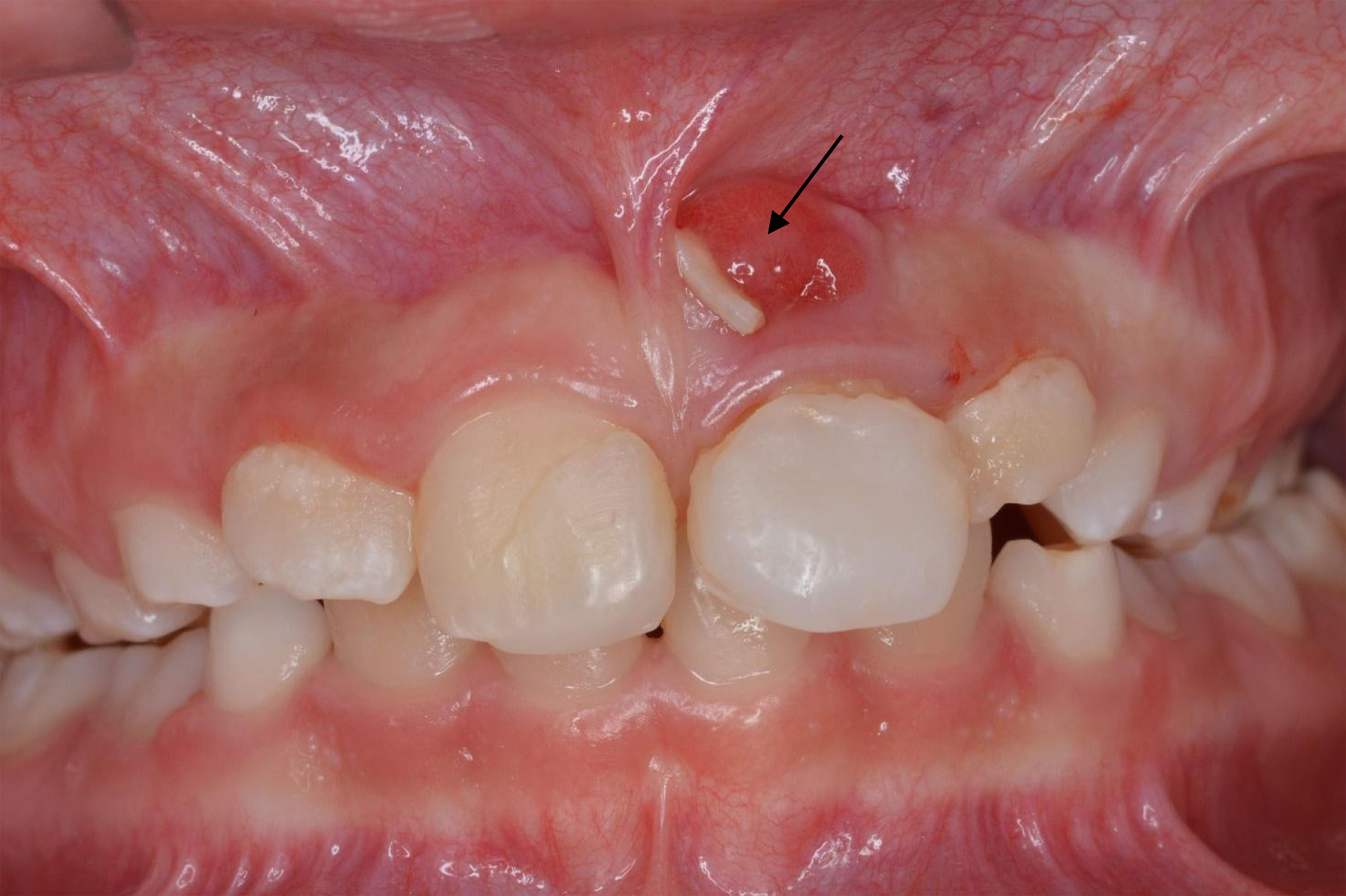 Note: Note the red color of the mucosa up to the teeth #21, and a root fragment just side by side the upper frenulum (black arrow), demonstrating an alveolysis.
 Note: A: Crown fragment; B: Root fragment.
 Note: A/B: Front view; C: Occlusal view.

