Cephalometric Evaluation of Pharyngeal Airway Space among Different Skeletal Malocclusions in United Arab Emirates Residents: A Cross-Sectional Study
ABSTRACT
Objective: To determine the relationship between skeletal malocclusion and upper pharyngeal airway space in the United Arab Emirates population using linear cephalometric measurements.
Material and Methods: A retrospective cross-sectional study was performed on lateral cephalogram radiographs acquired from the University Dental Hospital. Through convenience sampling, 70 lateral cephalograms were selected from 200, meeting the inclusion criteria for this study. Study subjects were divided into three groups: Class I (n=2S), Class II (n=2l), and Class III (n=24). The study groups were compared based on the linear upper pharyngeal airway space measurements.
Results: The three groups observed significant differences between the upper pharyngeal airway measurements. No differences in parameters were noted within the male and female study subjects. A highly significant difference (p<0.00l) in the Palatal Pharyngeal Distance was observed among the groups. Similarly, when the mean Middle Pharyngeal Distance and mean Inferior Pharyngeal Distance were compared among the three study groups, a highly significant difference (p<0.001 and p<0.004, respectively) was observed.
Conclusion: The highest variation in the linear dimensions of the upper pharyngeal airway space among the different skeletal malocclusion was observed in the Nasopharynx, Skeletal Class III having the most prominent dimensions followed by Class I and the least in Class II skeletal malocclusion.
Keywords:
Orthodontics; Cephalometry; Pharynx; Dimensional Measurement Accuracy
Introduction
Respiratory function has been extensively studied concerning craniofacial growth and has consequently played a significant role in orthodontic diagnosis and treatment planning [1]. The Skeletal system seems to respond to the influences of adjoining tissues, as demonstrated by Moss’s functional theory [2]. In particular, the mandible and cervical vertebrae grow due to functional relationships formed by all soft tissues and spaces associated with these bones. The close anatomical relationship between the mandible, cervical vertebrae, and pharyngeal airway has aroused the interest of different research groups who have examined their relationships and demonstrated a correlation between skeletal malocclusion, head posture, and pharyngeal airway space (PAS) [3,4]
Not many studies assess linear dimensions of the upper airway in the United Arab Emirates population. Because the etiology of malocclusion is multifactorial, airway study and evaluation should be added to the initial diagnosis to help diagnose possible obstructive airway syndromes, better develop a final treatment plan, and understand potential risk factors for airway-related problems. Furthermore, this knowledge would help orthodontists collaborate with the obstructive sleep apnea (OSA) team and fabricate a mandibular protruding device or prepare patients for orthognathic surgery to help them breathe better [5]. Therefore, assessing the pharyngeal structure in association with the orthodontic and orthognathic surgery diagnosis, the treatment planning process and the diagnosis and treatment of OSA might be beneficial. The null hypothesis for this study was that there is no significant difference in the linear airway dimensions among the different types of malocclusions. The alternative hypothesis is that there is a substantial difference in the linear airway dimensions among the different types of malocclusions. Therefore, this study aimed to evaluate the linear dimension of upper pharyngeal airway space among different skeletal malocclusions using lateral cephalograms for the residents of the United Arab Emirates.
Material and Methods
Study Design and Population and Sampling Technique
A retrospective study used lateral cephalograms of patients who visited the University Dental Hospital, University of Sharjah, for various dental treatments between January 2021 and January 2023. Seventy lateral cephalograms of male and female study subjects were selected for the study. Convenience sampling was used to select the 70 cephalograms from 200 cephalograms.
The estimation of sample size was carried out based on the method used by Buyukcavus and Kocakara [6]. Power analysis for upper PAS was carried out with an alpha error probability of 0.05 and a power of 80%. The assessment revealed that at least 21 study subjects were necessary for each study group. To Enhance the Power of this study, we included more study subjects than the required minimum.
Data Collection
The lateral cephalograms were acquired using the Viso7 Cephalometric unit (Planmeca, Helsinki, Finland). The radiographs were developed using the following parameters: 68kVp and 8mAs exposure time 2.0s. Patient scans were taken in the natural head position, with teeth in maximum intercuspation, and with patients in an upright position. During radiography, patients were instructed not to move their heads and to keep the tongue resting.
Head Position: The natural head position (NHP) is critical to the standard orientation of the head because it determines the craniocervical angle of the patient more accurately and facilitates radiographic cephalometric measurements. NHP is a realistic representation of the patient [7] and the most reproducible position. NHP was obtained by having the patient focus on eye level at a distant point so that the craniocervical spine angulation was in an unstrained position. The image field of focus was selected from the skull’s vertex down to nearly the 5th cervical vertebra [8].
Inclusion / Exclusion Criteria
The inclusion criteria comprised lateral cephalograms for patients of all ethnicities, sexes, races, and ages 22-65 years old who were seeking dental care at the University of Sharjah. The exclusion criteria eliminated any patient with a prior orthodontic treatment, medical history of adenoidectomy, tonsillectomy, orthognathic surgery, or trauma involving the cervical spine and head. These characteristics could interfere with airway space measurements.
Radiographs with errors affecting the region of interest (ROI) and artifacts involving ROI were excluded from the study. Scan patients undergoing (or with a history of) orthodontic treatment and a history of mid-facial skeletal trauma will be excluded from the study.
Skeletal Classification Identification
Two examiners (one orthodontist and one dental radiologist with more than 10 years of experience) conducted the radiographic measurements. They were calibrated before the commencement of the study. In case of disagreement, a third examiner with similar experience was consulted to arrive at a decision.
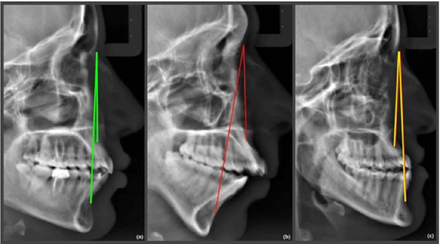
The anteroposterior skeletal malocclusion was measured by the ANB angle in degrees. The landmark identification was carried out using Steiner’s cephalometric guidelines. Point ‘A’ was marked as the most posterior point in the concavity of the anterior maxilla, Point ‘B’ was identified as the most posterior point in the concavity of the anterior mandible, and Point ‘N’ (Nasion) was marked as the most anterior point on the frontonasal suture [9]. According to the ANB angles, each study subject was classified into three groups, which determined the skeletal pattern, as shown in Figure 1.

Skeletal pattern identification using ANB angle: (a) Class I, (b) Class II, and (c) Class III.
-
1° < ANB < 4° - Class I malocclusion;
-
ANB > 4° - Class II malocclusion;
-
ANB < 0° - Class III malocclusion.
Linear Cephalometric Measurement of Pharyngeal Airway Space
The landmark identification and linear measurement were performed by two examiners on the lateral cephalogram. The measurements were added and divided by a factor [2] to obtain each parameter. The evaluation of 10% of the study subjects’ measurements was redone to ensure the reliability of the data. McNamara identified the different cephalometric landmarks for the pharyngeal airway space, and since then, numerous studies have been formulating different techniques of measurement [10].
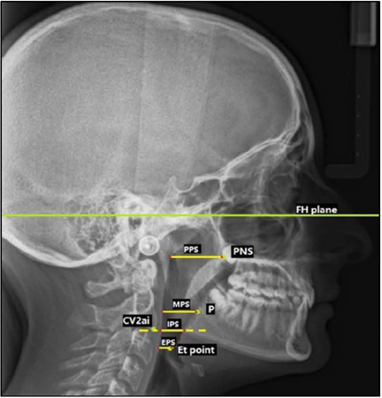
Using the guidelines from McNamara, Martin et al. defined the PAS in subjects with ideal occlusion The following linear PAS measurements were used for this present study, based on the linear cephalometric measurements by Buyukcavus and Kocakara [6], as shown in Figure 2.
Naso Pharynx
Palatal Pharyngeal Distance (PPS): Identified as the line passing from the posterior nasal spine (PNS) and parallel to FH (Frankfurt’s Horizontal) and cut out by a point on the posterior nasopharyngeal wall.
Oropharynx
Middle Pharyngeal Distance (MPS): on the line passing from a point P, parallel to FH, and the point cut by the posterior pharyngeal wall.
Hypopharynx
-
Inferior Pharyngeal Distance (IPS): on the line passing from the inferior anterior border of the second cervical vertebrae (cv2ai) and the points cut by the front and the rear pharyngeal wall.
-
Epiglottic Pharyngeal Distance (EPS): on the line passing from the extreme point on the epiglottis (Et), parallel to FH and the point cut by the posterior pharyngeal wall.
Statistical Analysis
The data was analyzed using IBM SPSS statistics, version 22 (IBM Corp, Armonk, NY, USA). At p<0.05, the results were considered statistically significant. The Pearson chi-square test was used to compare the study subjects’ gender and other variables. The one-way AN OVA was used to evaluate patients who had different malocclusion classifications. Tukey Post-Hoc tests were utilized for bilateral comparisons of significant parameters. To determine the error margin of the measurements, the same researcher repeated 50 randomly selected lateral cephalometric films after the initial measurements. For each measurement, Cronbach coefficients were calculated. The repeatability coefficients for each measurement were high (α≥876).
Results
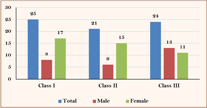
The gender distribution (n=70) revealed that 27 cephalograms were from male individuals (Figure 3). The age of the subjects ranged from 23 years to 54 years (mean = 30.5 years).

The inter-examiner and intra-examiner reliability was assessed using the Concordance Correlation Coefficient. The inter-examiner reliability varied from 0.991 (PPS) to 0.997 (IPS) (Table l). After one week, each examiner reexamined 10% (n=7) of the lateral cephalograms and measured the parameters. The intra-examiner reliability varied from 0.991 (MPS) to 0.996 (IPS) for Examiner 1, and for Examiner 2, it ranged from 0.908 to 0.995.
When the mean ANB angle of the study groups, Class I, II, and Class III, were compared using the Kruskal Wallis Test and Mann Whitney’s test, a significant difference (p<0.00l) between the study groups was observed. The highest mean ANB angle was observed in the Class II group, whereas the least mean ANB angle was observed in Class III (Table 3).
No significant difference was noted when the mean ANB angle was compared between the genders within the groups (Table 4). A highly significant difference was observed (p<0.00l) when the mean PPS of the three groups was compared using ANOVA; similarly, when the mean MPS and mean IPS were compared among the groups, a highly significant difference (p<0.001 and p<0.004, respectively). However, a significant difference was observed when the mean EPS was compared with the groups (p=0.03) (Table 5).
A significant difference was observed when a pairwise comparison of mean PPS was performed among the groups. When a pairwise comparison of mean MPS was performed among the groups, a highly significant difference (p<0.001) was observed. Similarly, when the mean MPS of Class II and Class III were compared, a highly significant difference (p=0.003) was observed. However, when the mean MPS of Class II and Class III were compared, no significant difference (p=0.68) was observed. No significant difference was observed when mean IPS was compared between Class I and Class II (p=0.14) and Class I and Class III (p=0.23). However, a significant difference was observed in Classes II and Class III (p=0.002). When mean EPS was compared between Class I and III (p=0.97) and Class II and Class III (p=0.08), no significant difference was observed; however, a substantial difference in Class I and Class II (p=04) was observed (Table 6). When the study variables (PPS, MPS, IPS, EPS) were compared between the genders, no significant difference was observed.
Discussion
There is strong evidence in the literature on the influence of orthodontic treatment on the pharyngeal airway [12,13]. Therefore, it is crucial for the orthodontist to precisely evaluate the pharyngeal airway space to formulate an efficient diagnosis and treatment plan for their patients.
Several studies have reported a relative narrowing of the PAS following surgical setback of the mandible [12,13]. In other studies, surgical advancement of the mandible was found to be associated with successful treatment of OSA [14,15].
Abundant literature reports an increase in airway space and improved breathing after rapid maxillary expansion (RME) in children. Palaisa et al. [16] reported that RME treatment increases the capacity of the nasal cavity and decreases nasal airway resistance. Furthermore, a Chinese study that used mandibular advancement devices in its patients resulted in increased PAS and reduced the distance of the hyoid bone to the mandibular plane [17].
Ozbek et al. reported that the use of functional orthopedic appliances on prepubertal Class II patients increased the sagittal dimensions of the upper pharyngeal airway. Therefore, the clinical importance of the pharyngeal airway orthodontist should not be underestimated, particularly in adolescence, since an orthodontist can influence growth and prevent any breathing conditions or complications in the future [18].
Researchers have studied the pharyngeal airway spaces in different parts of the world. In one Turkish study, the evaluation of the pharyngeal airway dimensions was compared among different skeletal patterns. Similarly, a recent Indian study evaluated the correlation between skeletal Class II skeletal malocclusion and linear dimensions of the upper pharyngeal airway [19]. Another study on the Lebanese population evaluated the dimensions of the airway using cephalometrics in different types of skeletal malocclusion [20]. They also assessed the association of airway spaces in lateral cephalograms and the gender of the study subjects [20].
To the best of our knowledge, there was a lack of literature comparing malocclusion and pharyngeal airway spaces in the Gulf Cooperation Council (GCC). We conducted a cross-sectional retrospective study using 70 lateral cephalograms from the radiology archives of the University Dental Hospital, United Arab Emirates. In the present study, lateral cephalograms were evaluated by two examiners, and the inter-rater reliability varied between 0.992 and 0.997. Habumugisha et al. [21] found interrater reliability for the pharyngeal airway measurements from 0.90 to 0.99. Another study conducted in the United States of America ranged between 0.87 to 0.90 [22]. Similarly, an Indian study also revealed excellent inter-rater reliability between the two examiners
In our study, each examiner reexamined 10% of the lateral cephalograms after one week and measured the parameters. The intra-examiner reliability varied from 0.991 to 0.996 for examiner one and from 0.908 to 0.950 for examiner 2. In the Habumugisha et al. [21] study, the inter-rater reliability for the first examiner ranged from 0.92 to 0.99, and for the second examiner, it ranged from 0.91 to 0.99. A systematic review showed that the intra-examiner reliability in most studies ranged between 0.780 and 0.999 [24].
In the past, researchers have used different landmarks and different number parameters for the evaluation of the upper airway. In Alfawazn’s [25] study, only two parameters of the upper airway (upper and lower airway width) were evaluated. A similar number of parameters were used by Indian authors [26], which also used similar parameters in their study using lateral cephalograms. However, some studies have used parameters similar to those in our research. A Brazilian study [27] and an Indian [28] study also used five upper airway parameters. In most of the studies, 5-6 parameters of the upper airway were measured [27,28].
The present study’s linear measurements were done parallel to the FH plane. A similar plane was used as a horizontal guide in a Turkish study [29] and a Lebanese study [20]. Based on the observation of the methods used in the studies, the FH plane is the most used reference plane.
The present study noted a significant difference in the mean PPS of the three study groups. The highest mean PPS was observed in Class III group, followed by Class I, and the least in Class II. Similar findings were observed in the literature [30,31]. Researchers suggest that in Class III malocclusion, the downward and forward growth of the mandible leads to increased upper airway space [32]. Clinically the knowledge of airway sizes helps the orthodontist for early management of malocclusion. Any obstruction to the airway may influence the severity of malocclusion.
Research suggests that the airway space of Class III patients is wider than that of Class I and II, possibly due to the forward position of the tongue; this theory is validated by McNamra [10]. According to his study, a rise in pharyngeal wall dimensions over 15 mm (about 0.59 in) indicates a forwardly placed tongue, which would cause a forwardly positioned mandible and, in turn, a wider airway. Similarly, Grauer et al. [33] assessed the upper pharyngeal airway space and shape and found a relationship with facial morphology. They concluded that the skeletal Class II had a forward inclination of the airway, thus reducing the space of the airway. In contrast, Class III subjects had more vertically oriented airways, implying larger airway space [33].
In the present study, there was no significant difference in the linear parameters of MPS, IPS and EPS among the different study groups. Similar results were observed in multiple studies, with no statistically significant difference being recorded in the oropharynx and hypopharynx (MPS, IPS and EPS) among study groups. However, a considerable change in the nasopharynx (PPS) was observed. Buyukcavus and Kocakara [6] also reported that the linear dimensions in the oropharynx and hypopharynx were not statistically significant when assessed with the different groups in their study.
Ozbek et al. studied the effect of functional orthopedic treatment on oropharyngeal dimensions in growing Class II malocclusion patients and found that functional treatment could increase the sagittal dimension of the upper PAS [18]. Therefore, the clinical importance of the pharyngeal airway should be considered. It is vital for adolescents when the growth and development of the maxilla and mandible are of utmost importance.
In the present study, there was no significant difference in the linear measurements of the upper PAS between male and female study subjects. Similarly, in Indian research by Dhayananth et al. [26], no significant difference was found among male and female subjects when comparing upper PAS dimensions. However, Jain et al. [32] found a significant difference in the linear dimensions of the upper airway between male and female study subjects in Class I and Class III malocclusion groups, although Class II subjects did not show any significant gender-based variation of linear upper airway dimensions.
The difference in the gender-based variation could be attributed to variation in the age group of the subjects in the two studies. One possible factor could be due to growth and development: according to literature, females’ craniofacial structures and development mature before the males’ subjects in the teenage years. A popular study by Bishara also confirms that females have an earlier onset and maturation of the anatomical structures of the face during growth compared to boys [34].
In our study, we performed a linear measurement in 2D for a 3D structure. The Pharyngeal airway space varies across all the axial planes; therefore, for the accurate evaluation of the Pharyngeal airway space, we may make use of a CBCT or CT scan for volumetric and area measurement but does the radiation dose and low reliability make it practical. Extensive literature supports the reliability and reproducibility of airway dimensions of pharyngeal airway space using lateral cephalometric radiographs when compared to CT scans. CT scans are very accurate and precise in identifying pharyngeal airway and surrounding structures, but the high radiation dose makes them impractical for routine use [35].
Although one CBCT scan may replace all conventional orthodontic radiographs, one set of these radiographs in orthodontics still entails 2-4 times less radiation than one CBCT. Depending on the scan mode, the radiation dose of a CBCT is about 3-6 times an OPG, 8-14 times a PA, and 15-26 times a lateral cephalogram [35].
Our research suggests the importance of the use of cephalometry for the assessment of the upper airway. The major advantages are the cheaper cost, ease of availability, and reduced radiation exposure as compared to the more modern advanced imaging techniques; cephalometry proves to be an important and valuable accessory for airway analysis as orthodontists already use it. The only disadvantage is that transverse dimensions cannot be assessed. Future research may be conducted to study the variation of the Pharyngeal airway space across different ethnicities and to find out what is causing the variation between different populations.
Conclusion
The most variation in the upper Pharyngeal airway Space among the different skeletal malocclusion was observed in the Nasopharynx (PPS), skeletal Class III having the most prominent dimensions followed by Class I and the least in Class II skeletal malocclusion. The variation in the lower part of the upper airway (oropharynx and hypopharynx) did not result in a difference in each of the skeletal malocclusions. Since our study population consisted of only adults who had completed growth and development, there was no difference in the Pharyngeal airway space dimensions between the genders when compared with the different skeletal malocclusion groups.
-
Financial Support
None.
-
Data Availability
The data used to support the findings of this study can be made available upon request to the corresponding author.
References
-
1 Solow B. Cranio-cervical posture: A factor in the development and function of the dentofacial structures. Eur J Orthod 2002; 24(5):447-456. https://doi.org/10.1093/ejo/24.5.447
» https://doi.org/10.1093/ejo/24.5.447 - 2 Moss ML. Functional cranial analysis and the functional matrix. Int J Orthod 1979; 17(1):21-31.
-
3 Solow B, Kreiborg S. Soft-tissue stretching: A possible control factor in craniofacial morphogenesis. Scand J Dent Res 1977; 85:505-507. https://doi.org/10.1111/j.1600-0722.1977.tb00587.x
» https://doi.org/10.1111/j.1600-0722.1977.tb00587.x -
4 Linder-Aronson S. Respiratory function in relation to facial morphology and the dentition. Br J Orthod 1979; 6(2):59-71. https://doi.org/10.1179/bjo.6.2.59
» https://doi.org/10.1179/bjo.6.2.59 -
5 Fransson AC, Tegelberg Â, Johansson A, Wenneberg B. Influence on the masticatory system in treatment of obstructive sleep apnea and snoring with a mandibular protruding device: A 2-year follow-up. Am J Orthod Dentofacial Orthop 2004; 126(6):687-693. https://doi.org/10.1016/j.ajodo.2003.10.040
» https://doi.org/10.1016/j.ajodo.2003.10.040 -
6 Buyukcavus MH, Kocakara G. Cephalometric evaluation of nasopharyngeal airway and hyoid bone position in subgroups of Class II malocclusions. Odovtos 2021; 23(1):155-167. https://doi.org/10.15517/ijds.2021.43860
» https://doi.org/10.15517/ijds.2021.43860 -
7 Lundström F, Lundström A. Natural head position as a basis for cephalometric analysis. Am J Orthod Dentofacial Orthop 1992; 101(3):244-247. https://doi.org/10.1016/0889-5406(92)70093-P
» https://doi.org/10.1016/0889-5406(92)70093-P -
8 Madsen DP, Sampson WJ, Townsend GC. Craniofacial reference plane variation and natural head position. Eur J Orthod 2008; 30(5):532-540. https://doi.org/10.1093/ejo/cjn031
» https://doi.org/10.1093/ejo/cjn031 -
9 Steiner CC. Cephalometrics for you and me. Am J Orthodontics 1953; 39:729-755. https://doi.org/10.1016/0002-9416(53)90082-7
» https://doi.org/10.1016/0002-9416(53)90082-7 - 10 McNamara JA. Influence of respiratory pattern on craniofacial growth. Angle Orthod 1981; 51(4):269-300.
-
11 Martin O, Muelas L, Vinas MJ. Nasopharyngeal cephalometric study of ideal occlusions. Am J Orthod Dentofacial Orthop 2006; 130(4):436.e1-436.e4369. https://doi.org/10.1016/j.ajodo.2006.03.022
» https://doi.org/10.1016/j.ajodo.2006.03.022 - 12 Katakura N, Umino M, Kubota Y. Morphologic airway changes after mandibular setback osteotomy for prognathism with and without cleft palate. Anesth Pain Control Dent 1993; 2(1):22-26.
-
13 Marsan G, Cura N, Emekli U. Changes in pharyngeal (airway) morphology in Class III Turkish female patients after mandibular setback surgery. J Craniomaxillofac Surg 2008; 36(6):341-345. https://doi.org/10.1016/j.jcms.2008.03.001
» https://doi.org/10.1016/j.jcms.2008.03.001 -
14 Troell RJ, Riley RW, Powell NB, Li K. Surgical management of the hypopharyngeal airway in sleep disordered breathing. Otolaryngol Clin North Am 1998; 31(6):979-1012. https://doi.org/10.1016/s0030-6665(05)70102-x
» https://doi.org/10.1016/s0030-6665(05)70102-x -
15 Prinsell JR. Maxillomandibular advancement surgery in a site-specific treatment approach for obstructive sleep apnea in 50 consecutive patients. Chest 1999; 116(6):1519-1529. https://doi.org/10.1378/chest.116.6.1519
» https://doi.org/10.1378/chest.116.6.1519 -
16 Palaisa J, Ngan P, Martin C, Razmus T. Use of conventional tomography to evaluate changes in the nasal cavity with rapid palatal expansion. Am J Orthod Dentofacial Orthop 2007; 132(4):458-466. https://doi.org/10.1016/j.ajodo.2005.10.025
» https://doi.org/10.1016/j.ajodo.2005.10.025 - 17 Poon KH, Chay SH, Chiong KF. Airway and craniofacial changes with mandibular advancement device in Chinese with obstructive sleep apnoea. Ann Acad Med Singap 2008; 37(8):637-644.
-
18 Ozbek MM, Memikoglu TU, Gögen H, Lowe AA, Baspinar E. Oropharyngeal airway dimensions and functional-orthopedic treatment in skeletal Class II cases. Angle Orthod 1998; 68(4):327-336. https://doi.org/10.1043/0003-3219(1998)068<0327:OADAFO>2.3.CO;2
» https://doi.org/10.1043/0003-3219(1998)068<0327:OADAFO>2.3.CO;2 -
19 Jadhav PJ, Sonawane SV, Mahajan N, Chavan BG, Korde SJ, Momin NM, et al. Correlation of pharyngeal airway dimensions with maxillomandibular skeletal relation and mandibular morphology in subjects with skeletal Class I and class II malocclusions and different growth patterns: A cephalometric study in selected local population. J Pharm Bioallied Sci 2021; 13(Suppl 2):S1111-S1114. https://doi.org/10.4103/jpbs.jpbs_349_21
» https://doi.org/10.4103/jpbs.jpbs_349_21 -
20 Daraze A, Delatte M, Liistro G, Zeina Majzoub. Cephalometrics of pharyngeal airway space in Lebanese adults. Int J Dent 2017; 2017:3959456. https://doi.org/10.1155/2017/3959456
» https://doi.org/10.1155/2017/3959456 -
21 Habumugisha J, Ma SY, Mohamed AS, Cheng B, Zhao MY, Bu WQ, et al. Three-dimensional evaluation of pharyngeal airway and maxillary arch in mouth and nasal breathing children with skeletal Class I and II. BMC Oral Health 2022; 22(1):320. https://doi.org/10.1186/s12903-022-02355-3
» https://doi.org/10.1186/s12903-022-02355-3 -
22 Elshebiny T, Bous R, Withana T, Morcos S, Valiathan M. Accuracy of three-dimensional upper airway prediction in orthognathic patients using dolphin three-dimensional software. J Craniofac Surg 2020; 31(4):1098-1100. https://doi.org/10.1097/SCS.0000000000006566
» https://doi.org/10.1097/SCS.0000000000006566 -
23 Diwakar R, Kochhar AS, Gupta H, Kaur H, Sidhu MS, Skountrianos H, et al. Effect of craniofacial morphology on pharyngeal airway volume measured using Cone-Beam Computed Tomography (CBCT): A retrospective pilot study. Int J Environ Res Public Health 2021; 18(9):5040. https://doi.org/10.3390/ijerph18095040
» https://doi.org/10.3390/ijerph18095040 -
24 Zimmerman JN, Lee J, Pliska BT. Reliability of upper pharyngeal airway assessment using dental CBCT: A systematic review. Eur J Orthod 2017; 39(5):489-496. https://doi.org/10.1093/ejo/cjw079
» https://doi.org/10.1093/ejo/cjw079 -
25 Alfawzan AA. Assessment of airway dimensions in skeletal Class I malocclusion patients with various vertical facial patterns: A cephalometric study in a sample of the Saudi population. J Orthod Sci 2020; 9:12. https://doi.org/10.4103/jos.JOS_10_20
» https://doi.org/10.4103/jos.JOS_10_20 -
26 Dhayananth X, Clement E, Faizee SH, Malini H, Iswarya A, Christy AS. Assessment of airway among South Indian population - A cephalometric study. Int J Cur Res Review 2021; 13(10):31-35. https://doi.org/10.31782/IJCRR.2021.SP237
» https://doi.org/10.31782/IJCRR.2021.SP237 -
27 Sprenger R, Martins LAC, Dos Santos JCB, de Menezes CC, Venezian GC, Degan VV. A retrospective cephalometric study on upper airway spaces in different facial types. Prog Orthod 2017; 18(1):25. https://doi.org/10.1186/s40510-017-0180-2
» https://doi.org/10.1186/s40510-017-0180-2 -
28 Guttal KS, Burde K. Cephalometric evaluation of upper airway in healthy adult population: A preliminary study. J Oral Maxillofac Radiol 2013; 1(2):55-60. https://doi.org/10.4103/2321-3841.120115
» https://doi.org/10.4103/2321-3841.120115 -
29 Aksu M, Gorucu-Coskuner H, Taner T. Assessment of upper airway size after orthopedic treatment for maxillary protrusion or mandibular retrusion. Am J Orthod Dentofacial Orthop 2017; 152(3):364-370. https://doi.org/10.1016/j.ajodo.2016.12.027
» https://doi.org/10.1016/j.ajodo.2016.12.027 -
30 Muto T, Yamazaki A, Takeda S. A cephalometric evaluation of the pharyngeal airway space in patients with mandibular retrognathia and prognathia, and normal subjects. Int J Oral Maxillofac Surg 2008; 37(3):228-231. https://doi.org/10.1016/j.ijom.2007.06.020
» https://doi.org/10.1016/j.ijom.2007.06.020 -
31 Iwasaki T, Hayasaki H, Takemoto Y, Kanomi R, Yamasaki Y. Oropharyngeal airway in children with Class III malocclusion evaluated by cone-beam computed tomography. Am J Orthod Dentofacial Orthop 2009; 136(3):318.e1-319. https://doi.org/10.1016/j.ajodo.2009.02.017
» https://doi.org/10.1016/j.ajodo.2009.02.017 -
32 Jain S, Raghav P, Misra V, Reddy CM, Singh S, Aggarwal S. Assessment of upper and lower pharyngeal airway width in skeletal Class I, II and III malocclusions. J Ind Orthod Soc 2014; 48(4):446-453. https://doi.org/10.1177/0974909820140702S
» https://doi.org/10.1177/0974909820140702S -
33 Grauer D, Cevidanes LS, Styner MA, Ackerman JL, Proffit WR. Pharyngeal airway volume and shape from cone-beam computed tomography: Relationship to facial morphology. Am J Orthod Dentofacial Orthop 2009; 136(6):805-814. https://doi.org/10.1016/j.ajodo.2008.01.020
» https://doi.org/10.1016/j.ajodo.2008.01.020 -
34 Bishara SE, Peterson LC, Bishara EC. Changes in facial dimensions and relationships between the ages of 5 and 25 years. Am J Orthod 1984; 85(3):238-252. https://doi.org/10.1016/0002-9416(84)90063-0
» https://doi.org/10.1016/0002-9416(84)90063-0 -
35 Signorelli L, Patcas R, Peltomäki T, Schätzle M. Radiation dose of cone-beam computed tomography compared to conventional radiographs in orthodontics. J Orofac Orthop 2016; 77(1):9-15. https://doi.org/10.1007/s00056-015-0002-4
» https://doi.org/10.1007/s00056-015-0002-4
Edited by
-
Academic Editor:
Mohammad Khursheed Alam
The data used to support the findings of this study can be made available upon request to the corresponding author.