Evaluation of Canalis Sinuosus on CBCT Images of Patients Candidate for Dental Implant Treatment in Iranian Population
ABSTRACT
Objective: To evaluate the frequency, diameter, location and path of canalis sinuosus (CS) on CBCT scans of patients candidate for dental implant treatment.
Material and Methods: In this cross-sectional study, 200 CBCT images of the maxilla were evaluated and parameters were assessed: age, gender, the canal presence and diameter, the distance between the CS and the nasal cavity floor (NC), the buccal cortical bone (BC) and the most prominent point of the alveolar ridge crest (RC). Quantitative variables were analyzed with an independent t-test, and qualititative variables were analyzed with chi-squared and McNemer tests (p<0.05).
Results: CS was detected on 100 CBCT images in the present study. There were significant relationships between the CS frequency and age and gender; however, there was no significant relationship between CS and the maxillary side. The means of BC, RC, NC and the canal diameter were 9.2±2.19, 15.15±3.13, 8.14±2.43, and 0.99±0.26 mm, respectively. There were significant relationships between the canal diameter, NC and BC with gender. However, there was no significant relationship between RC and gender.
Conclusion: Canalis sinuosus was detected, with an approximate diameter of 1 mm, in 50% of the subjects in the incisor-canine area. The use of CBCT for accurate diagnosis of canalis sinuosus is suggested before surgical procedures in the palatal aspect of the anterior maxilla.
Keywords:
Cone-Beam Computed Tomography; Dental Implants; Maxilla
Introduction
The maxillary nerve is a branch of the trigeminal nerve. The maxillary nerve in the infraorbital canal gives off the posterior superior alveolar nerve, the anterior superior alveolar nerve, and the infra orbital nerve. The infraorbital nerve travels along the infraorbital foramen, giving off a lateral branch in canal called canalis sinuosus (CS) [1,2].
Canalis sinuosus was first described by Jones, who defined CS in association with a neurovascular bundle that emerges from the posterior of the middle part of the infraorbital foramen and enters the nasal cavity laterally after traveling a tortuous path of an osseous canal measuring 2 mm in diameter. CS has been described by other researchers as a canal initiating almost 25 mm behind the infraorbital foramen, descends toward the orbital floor, circles the anterior wall of the maxillary sinus medially, and extends to the nasal cavity. The point of entry is anterior to the incisive canal, where it exhibits its anatomical variations in the anterior palate, referred to as accessory channels (ACs) [3-5].
CS contains the anterior superior alveolar nerve, vein, and artery, and its neurovascular branches form the dental neuronal plexus of the canine area. The anterior superior alveolar nerve in the CS innervates the incisors, canine, and their soft tissues, and the nasal fossa floor and the maxillary sinus [4-6].
Lack of knowledge about the position of CS is a risk factor in surgical procedures, including maxillary sinus surgeries, impacted maxillary canine surgeries, and implant placement in the canine-incisor area [4]. The canine anatomic area is very important in terms of supporting implants. The vicinity to the CS neurovascular bundle and accessory channels might endanger osseointegration, resulting in transient or permanent paresthesia, bleeding, and neurovascular injuries in the area [7-9]. Some studies have shown that CS might imitate the appearance of a periapical lesion, prompting the dentist to carry out an inappropriate endodontic treatment [10].
The CBCT technique is currently used for radiographic evaluation of the anatomic landmarks in the jaws because, in most cases, periapical and panoramic radiographs cannot depict these structures with proper details [11,12]. In addition, this radiographic technique provides more details of CS and is associated with less radiation dose than helical computed tomography (CT).
The CBCT technique is used in many cases to provide accurate data about the area in question because it significantly removes image overlapping, can provide linear and angular measurements, and reconstructs images in different planes three-dimensionally [13].
It is important to study anatomic variations to collect anthropometric data and improve clinical protocols. Therefore, it is very important for dentists to have adequate knowledge about the path and size of CS, considering anatomic variations in different populations to prevent intraoperative traumas and the relent complications and improve prognosis [14-16].
Increased application of dental implants to reconstruct the maxilla has resulted in increased postoperative complications. Evaluation of previous studies concerning hemorrhage, neurosensory disorders, absence of osseointegration, and infections in the anterior maxilla has made it is necessary to evaluate the presence of neurovascular structures before surgical procedures in the anterior maxilla [7-9].
Only a few studies have evaluated CS in different ethnic groups. Considering diversities in the position and path of CS in different ethnic groups, it is necessary to examine the CS status and position before surgical procedures in the anterior maxilla to avoid the CS neurovascular bundle. Therefore, the present study was undertaken to determine the frequency, spatial location, path and size of CS on CBCT images in a group of Iranian patients planned to receive dental implants.
Material and Methods
Study Design and Ethical Clearance
This cross-sectional study was approved by the Ethics Committee of Gilan University of Medical Sciences under the code IR.GUMS.REC.1399.560.
Data Collection
The study subjects consisted of all the CBCT scans of the maxilla of patients referring to a maxillofacial radiology center based on inclusion criteria. The samples were selected using the convenient sampling method (from newer ones to older ones) gradually from the maxillary CBCT images. All the patients signed informed consent forms before their data were collected.
The inclusion criteria consisted of the CBCT images of patients in the 20-80 age range, who were candidates to receive dental implants and were systemically healthy [17]. Patients with a history of surgery and bone grafting in the anterior maxilla, trauma (screws and plates), or pathologic entities in the anterior maxilla, images with artifacts, patient’s movement during the imaging procedure, low-quality images, syndromes and skeletal malformations, and patients under treatment with bis-phosphonates were excluded [2].
All the CBCT images were prepared using an Acteon CBCT unit (X Mind Trium, Acteon Group, Norfolk, England) under exposure conditions specific for each patient (kVp=90, FOV=8*99 inch, mA=10, and a voxel size of 100 µm. The images were processed using the On-Demand software. An oral and maxillofacial radiologist viewed the CBCT images to evaluate the canalis sinuosus. First, MPR (multiplanar reconstruction) images and panoramic series were reconstructed at 1-mm thickness to evaluate the presence or absence of canalis sinuosus. When the entire maxillary arch was present in the image field, the presence of CS was separately evaluated for each side. When the CBCT images confirmed the presence of CS, 1 mm cross-sectional images were prepared at 1 mm intervals to evaluate the study variables. When the CS was clearly visible on a cross-section, the software toolbar was used to measure the parameters of the path and diameter of the canal.
Based on MPR images, on the cross-sectional images that crossed exactly the center of the tooth (or a hypothetical position of the tooth when the tooth was missing), the following parameters were measured:
-
NC: the distance between the initiation point of CS and the nasal cavity floor;
-
BC: the distance between the initiation point of CS and the buccal cortical bone;
-
RC: the distance between the initiation point of CS and the most prominent point of the alveolar ridge crest;
-
In addition, the diameter of CS was evaluated and recorded on 1 mm cross-sections.
Data Analysis
The data were analyzed with SPSS Software, Version 26 (IBM Corp., Armonk, NY, USA). Frequencies, means, and standard deviations were used to describe data. The independent t-test was used to analyze quantitative variables. Chi-squared and McNemar tests were used to analyze qualitative variables (α=0.05).
Results
In the present study, 200 CBCT scans (400 cases of the left and right sides) were evaluated. In general, CS was detected in 50% of cases (200 of 400). Comparison of the frequencies of CS in terms of gender, age, and side are presented in Table 1. There were significant relationships between the canal frequency and age and gender, with a higher frequency of the canal in males and subjects >40 years of age. However, there was no significant relationship between the canal frequency and side.
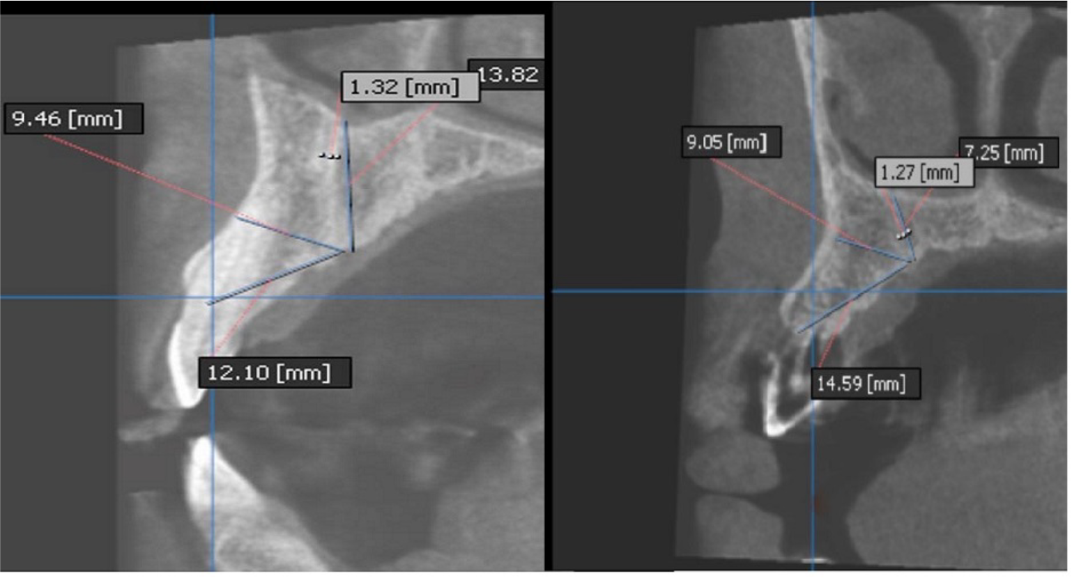
Table 2 presents the means (standard deviations) of the distance between the canal and the nasal cavity floor (NC), the distance between the canal and the buccal cortical bone (BC), the distance between the canal and the most prominent point on the alveolar crest (RC) and the canal diameter. The minimum and maximum distances recorded were 3.40 and 14.7 mm for NC, 5.60 and 19 mm for BC, 6.90 and 22 mm for RC, and 0.40 and 2 mm for the diameter, respectively (Figures 1 and 2).
Examples of CS measurements in CBCT cross sections. Case Left: NC: 13.82 mm, BC: 9.46 mm, RC: 12.10 mm, Diameter of canal: 1.32 mm. Case Right: NC: 7.25 mm, BC: 9.05 mm, RC: 14.59 mm, Diameter of canal: 1.27 mm.

Table 3 presents the relationships between the canal diameter, NC, BC, RC in the maxilla and gender, age, and side. There were no significant differences between the canal diameter and NC, BC, and RC values and age and the left and right sides. The canal diameters and NC and BC values were higher in men than in women; however, RC did not exhibit a significant relationship with gender. In addition, concerning the location of CS in the maxilla in terms of tooth position, in 100% of the cases, CS was located between the canine and incisor teeth.
Discussion
Canalis sinuosus is an anatomic variation that has not been recognized well by dentists. Since its first description by Jones in 1939, only a few reports have been published on it. Widespread acceptance of dental implant methods and improvements in three-dimensional imaging techniques have paved the way for this anatomical variation to gain the popularity it deserves. Accurate knowledge about anatomy increases clinicians’ success. Dentists should pay more attention to important structures such as the nasal cavity floor and the nasopalatine canal [18].
Before carrying out surgeries, it is very important to consider anesthetic variations precisely. Trauma to CS might result in transient or permanent paresthesia or hemorrhage in the area. Conventional imaging techniques, such as panoramic and periapical radiography, provide two-dimensional images. This canal cannot easily be identified due to the limitations of these radiographic techniques. In addition, many dentists diagnose this structure as a periapical radiolucency or a pathological lesion [19,20]. Widespread use of CBCT in dentistry has made it possible to achieve accurate 3D views of anatomic structures and high-resolution images with higher reliability and reproducibility than 2D images [18].
Therefore, the present study was undertaken to evaluate the presence or absence, the special location, and the size of CS on CBCT images of candidates for implant treatment. The results showed that 100 CBCT scans (50%) exhibited CS. In a study by Machado et al. [11] on 100 CBCT scans, the frequency of this canal was 52.1%, consistent with the present study. In studies by Anatoly et al. [21], Orhan et al. [16] and Wanzeler et al. [3], the frequencies of this canal were 67%, 70.8% and 88%, respectively. On the other hand, in studies by Manhães Júnior et al. [13] and de Oliveira-Santos et al. [2] the frequencies of this canal were 36.2% and 15.7%, respectively. The differences in the frequencies of this canal might be attributed to ethnic differences, sample sizes, and differences in voxel sizes.
In the present study, 43.2% of female and 59.8% of male subjects had CS, with a significant difference between the two genders. In a study by Aoki et al. [17] CS was detected in 133 patients (66.5%), with high reproducibility in men. In addition, Machado et al. [11] reported the highest frequency of CS in men, consistent with the present study. In studies by Gurler et al. [1] and de Oliveira-Santos et al. [2] the prevalence of CS in men was higher than in women; however, the difference was not significant statistically. Anatoly et al. [21], Orhan et al. [16] and Sekerci et al. [22] reported results different from the present study, with a higher frequency of CS in women than men, which might be attributed to ethnic differences and differences in the percentages of men and women included in these studies.
The present study showed a significant relationship between CS and age. CS was more frequent in subjects ≥40 years of age. Orhan et al. [16] reported the highest frequency of CS in the 50-59 age group. In addition, studies by Ghandourah et al. [10], Sekerci et al. [22], von Arx et al. [23] and Manhães Júnior et al. [13] showed an increase in CS frequency with aging, consistent with the present study. Aoki et al. [17], de Oliveira-Santos et al. [2], Anatoly et al. [21] and Wanzeler et al. [3] reported no relationship between CS and age, which might be attributed to differences in sample sizes and ethnicity.
In the present study, CS was slightly more frequent on the right side than on the left side, which was not statistically significant. Orhan et al. [16] reported that of 158 CBCT scans, 58 and 51 scans showed CS on the right and left sides, respectively, with no significant differences, consistent with the present study. Manhães Júnior et al. [13] and de Oliveira-Santos et al. [2] reported no significant difference in CS frequency between the left and right sides, consistent with the present study.
In the present study, the mean diameter of the canal was 1 mm. Aoki et al. [17] reported a uniform diameter throughout the canal length up to the oral cavity. Most studies have shown a diameter of 1 mm for the canal (96.6%). Ghandourah et al. [10] reported a diameter of 1 mm for CS in 82.1% of the subjects. In addition, von Arx et al. [23] and Machado et al. [11] reported a mean diameter of 1 mm for CS, consistent with the present study.
A significant relationship between the CS diameter and gender was found, with a higher mean diameter in men than in women. Machado et al. [11] and Gurler et al. [1] reported the highest diameter in mm, consistent with the present study. In contrast, Aoki et al. [17] and von Arx et al. [23] reported no effect of gender on the CS diameter, which might be attributed to differences in ethnicity, the quality and thickness of image slices, and in the participation of female and male subjects included in these studies.
The present study showed no significant relationship between the CS diameter and age. Aoki et al. [17], von Arx et al. [23] and Gurler et al. [1], too, reported no significant relationship between the CS diameter and age, consistent with the present study.
A mean of 8.14±2.43 mm for NC was found, with minimum and maximum distances of 3.40 and 14.7 mm, respectively. The mean BC was 9.24±2.19 mm, with minimum and maximum distances of 5.60 and 19 mm, respectively. The mean RC was 15.15±3.13 mm, with minimum and maximum distances of 6.90 and 22 mm, respectively. It appears that the position of CS relative to tooth positions or implant placement areas in edentulism is apical and palatal to the ridge crest. Therefore, attention to CS is more important in canine palatal impaction cases in orthodontic patents and LeFort I surgeries in patients with maxillofacial discrepancies.
Another finding of the present study was a significant relationship between NC and BC values and gender, i.e., the means of these values were higher in men than in women. However, there was no significant relationship between RC and gender. In addition, in the present study, there were no significant relationships between NC, BC, and RC values and age. No study is available that has directly evaluated the relationships between NC, BC, and RC values and patents’ age and gender. Therefore, further studies are necessary in the future.
According to the present study, there was no significant relationship between NC, BC, and RC values and the CS side in the maxilla. Aoki et al. [17] reported no significant relationship between the above values and the CS side in the maxilla, consistent with the present study. Manhães Júnior et al. [13] reported differences in the distance between CS and the alveolar ridge crest (RC) and the buccal cortical bone (BC) on both sides, with no differences in the distance between the CS and the nasal cavity floor (NC) in terms of side, which might be explained by the fact that the alveolar bone plate undergoes morphologic changes over time. In addition, Manhães Júnior et al. [13] did not evaluate issues such as trauma, edentulism, or surgery in the area under study. According to the present study, in 100% of cases, the CS was located in the canine-incisor area. Anatoly et al. [21], von Arxet et al. [23] and de Oliveira-Santos et al. [2], too, reported that CS was located near the canine-incisor area, consistent with the present study.
Conclusion
The canalis sinuosus was detected in 50% of the subjects, with a higher frequency in men and subjects >40 years of age. The CS diameter was approximately 1 mm, with a mean higher diameter in men than women. The CS was located in the canine-incisor area in all the cases. Therefore, it is suggested that the CS location be accurately determined radiographically before surgical procedures in the anterior maxilla.
Data Availability
The data used to support the findings of this study can be made available upon request to the corresponding author.
-
Financial Support
None.
References
-
[1] Gurler G, Delilbasi C, Ogut EE, Aydin K, Sakul U. Evaluation of the morphology of the canalis sinuosus using cone-beam computed tomography in patients with maxillary impacted canines. Imaging Sci Dent 2017; 47(2):69-74. https://doi.org/10.5624/isd.2017.47.2.69
» https://doi.org/10.5624/isd.2017.47.2.69 -
[2] de Oliveira-Santos C, Rubira-Bullen IR, Monteiro SA, León JE, Jacobs R. Neurovascular anatomical variations in the anterior palate observed on CBCT images. Clin Oral Implants Res 2013; 24(9):1044-1048. https://doi.org/10.1111/j.1600-0501.2012.02497.x
» https://doi.org/10.1111/j.1600-0501.2012.02497.x -
[3] Wanzeler AM, Marinho CG, Alves Junior SM, Manzi FR, Tuji FM. Anatomical study of the canalis sinuosus in 100 cone beam computed tomography examinations. Oral Maxillofac Surg 2015; 19(1):49-53. https://doi.org/10.1007/s10006-014-0450-9
» https://doi.org/10.1007/s10006-014-0450-9 -
[4] Neves FS, Crusoé-Souza M, Franco LC, Caria PH, Bonfim-Almeida P, Crusoé-Rebello I. Canalis sinuosus: A rare anatomical variation. Surg Radiol Anat 2012; 34(6):563-566. https://doi.org/10.1007/s00276-011-0907-6
» https://doi.org/10.1007/s00276-011-0907-6 -
[5] Arruda JA, Silva P, Silva L, Álvares P, Silva L, Zavanelli R, et al. Dental implant in the canalis sinuosus: A case report and review of the literature. Case Rep Dent 2017; 2017:4810123. https://doi.org/10.1155/2017/4810123
» https://doi.org/10.1155/2017/4810123 -
[6] Torres MG, de Faro Valverde L, Vidal MT, Crusoé-Rebello IM. Branch of the canalis sinuosus: A rare anatomical variation -- A case report. Surg Radiol Anat 2015; 37(7):879-881. https://doi.org/10.1007/s00276-015-1432-9
» https://doi.org/10.1007/s00276-015-1432-9 -
[7] McCrea SJJ. Aberrations causing neurovascular damage in the anterior maxilla during dental implant placement. Case Rep Dent 2017; 2017:5969643. https://doi.org/10.1155/2017/5969643
» https://doi.org/10.1155/2017/5969643 -
[8] Volberg R, Mordanov O. Canalis sinuosus damage after immediate dental implant placement in the esthetic zone. Case Rep Dent 2019; 2019:3462794. https://doi.org/10.1155/2019/3462794
» https://doi.org/10.1155/2019/3462794 -
[9] Jacobs R, Quirynen M, Bornstein MM. Neurovascular disturbances after implant surgery. Periodontol 2000 2014; 66(1):188-202. https://doi.org/10.1111/prd.12050
» https://doi.org/10.1111/prd.12050 -
[10] Ghandourah AO, Rashad A, Heiland M, Hamzi BM, Friedrich RE. Cone-beam tomographic analysis of canalis sinuosus accessory intraosseous canals in the maxilla. Ger Med Sci 2017; 15:Doc20. https://doi.org/10.3205/000261
» https://doi.org/10.3205/000261 -
[11] Machado VC, Chrcanovic BR, Felippe MB, Manhães Júnior LR, de Carvalho PS. Assessment of accessory canals of the canalis sinuosus: A study of 1000 cone beam computed tomography examinations. Int J Oral Maxillofac Surg 2016; 45(12):1586-1591. https://doi.org/10.1016/j.ijom.2016.09.007
» https://doi.org/10.1016/j.ijom.2016.09.007 -
[12] Tomrukçu DN, Köse TE. Assesment of accessory branches of canalis sinuosus on CBCT images. Med Oral Patol Oral Cir Bucal 2020; 25(1):e124-e130. https://doi.org/10.4317/medoral.23235
» https://doi.org/10.4317/medoral.23235 -
[13] Manhães Júnior LR, Villaça-Carvalho MF, Moraes ME, Lopes SL, Silva MB, Junqueira JL. Location and classification of Canalis sinuosus for cone beam computed tomography: Avoiding misdiagnosis. Braz Oral Res 2016; 30(1):e49. https://doi.org/10.1590/1807-3107BOR-2016.vol30.0049
» https://doi.org/10.1590/1807-3107BOR-2016.vol30.0049 -
[14] Ferlin R, Pagin BSC, Yaedú RYF. Evaluation of canalis sinuosus in individuals with cleft lip and palate: A cross-sectional study using cone beam computed tomography. Oral Maxillofac Surg 2021; 25(3):337-343. https://doi.org/10.1007/s10006-020-00919-7
» https://doi.org/10.1007/s10006-020-00919-7 -
[15] Ferlin R, Pagin BSC, Yaedú RYF. Canalis sinuosus: A systematic review of the literature. Oral Surg Oral Med Oral Pathol Oral Radiol 2019; 127(6):545-551. https://doi.org/10.1016/j.oooo.2018.12.017
» https://doi.org/10.1016/j.oooo.2018.12.017 -
[16] Orhan K, Gorurgoz C, Akyol M, Ozarslanturk S, Avsever H. An anatomical variant: Evaluation of accessory canals of the canalis sinuosus using cone beam computed tomography. Folia Morphol 2018; 77(3):551-557. https://doi.org/10.5603/FM.a2018.0003
» https://doi.org/10.5603/FM.a2018.0003 -
[17] Aoki R, Massuda M, Zenni LTV, Fernandes KS. Canalis sinuosus: Anatomical variation or structure? Surg Radiol Anat 2020; 42(1):69-74. https://doi.org/10.1007/s00276-019-02352-2
» https://doi.org/10.1007/s00276-019-02352-2 -
[18] Lopes-Santos G, Salzedas LMP, Bernabé DG, Ikuta CRS, Miyahara GI, Tjioe KC. Assessment of the knowledge of canalis sinuosus amongst dentists and dental students: An online-based cross-sectional study. Eur J Dent Educ 2022; 26(3):488-498. https://doi.org/10.1111/eje.12725
» https://doi.org/10.1111/eje.12725 -
[19] Shelley A, Tinning J, Yates J, Horner K. Potential neurovascular damage as a result of dental implant placement in the anterior maxilla. Br Dent J 2019; 226(9):657-661. https://doi.org/10.1038/s41415-019-0260-4
» https://doi.org/10.1038/s41415-019-0260-4 -
[20] Khalifa HM, Felemban OM. Nature and clinical significance of incidental findings in maxillofacial cone-beam computed tomography: a systematic review. Oral Radiol 2021; 37(4):547-559. https://doi.org/10.1007/s11282-020-00499-y
» https://doi.org/10.1007/s11282-020-00499-y -
[21] Anatoly A, Sedov Y, Gvozdikova E, Mordanov O, Kruchinina L, Avanesov K, et al. Radiological and morphometric features of canalis sinuosus in Russian population: Cone-beam computed tomography study. Int J Dent 2019; 2019:2453469. https://doi.org/10.1155/2019/2453469
» https://doi.org/10.1155/2019/2453469 -
[22] Sekerci AE, Cantekin K, Aydinbelge M. Cone beam computed tomographic analysis of neurovascular anatomical variations other than the nasopalatine canal in the anterior maxilla in a pediatric population. Surg Radiol Anat 2015; 37(2):181-186. https://doi.org/10.1007/s00276-014-1303-9
» https://doi.org/10.1007/s00276-014-1303-9 -
[23] von Arx T, Lozanoff S, Sendi P, Bornstein MM. Assessment of bone channels other than the nasopalatine canal in the anterior maxilla using limited cone beam computed tomography. Surg Radiol Anat 2013; 35(9):783-790. https://doi.org/10.1007/s00276-013-1110-8
» https://doi.org/10.1007/s00276-013-1110-8
Edited by
-
Academic Editor:
Emna Hidoussi Sakly