Impact of Artifacts Caused by Intraoral Dental Materials in Magnetic Resonance Imaging
ABSTRACT
Objective: To evaluate the characteristics of artifacts produced by various intraoral dental materials in magnetic resonance imaging sequences and assess their impact on the diagnostic quality of the resultant images.
Material and Methods: A clinical examination was conducted on forty-six patients who underwent magnetic resonance imaging for various brain pathologies. Parameters were recorded, including the location, span, and type of intraoral dental material. The impact of these artifacts on the diagnostic quality of the resultant image sequences was assessed. The reliability of the intraand interobserver was calculated, and Fischer's exact test was applied. P-value <0.05 was considered statistically significant.
Results: The most common signal intensity artifact observed was hypointense with hyperintense rim artifacts in FLAIR (95.5%). The most common shape of the artifacts observed was circular/void shape (84.4%). There was a statistically significant difference between the various classes of intraoral dental materials and diagnostic acceptability (p=0.017). In the present study, 28% of the study population images were graded diagnostically unacceptable in the sequences evaluated (FLAIR 37.8%, T2 3.3%, SWI 24.4%, DWI 25%).
Conclusion: Dental material artifacts interfered with the interpretation of magnetic resonance imaging at varying levels of diagnostic acceptability depending on the indication for which the imaging was performed.
Keywords:
Magnetic Resonance Imaging; Dental Materials; Artifacts
Introduction
Magnetic resonance imaging (MRI) of the head and neck region is a widely accepted imaging modality for visualizing soft tissue pathologies. It is considered one of the useful noninvasive diagnostic modalities for head and neck pathologies. It is advantageous over other three-dimensional imaging modalities because of the high spatial resolution of soft tissue images in various planes without harmful ionizing radiation. Common indications in the maxillofacial region include neurologic abnormalities, inflammatory conditions, vascular malformations, developmental anomalies, soft tissue tumors, etc. Magnetic resonance imaging uses a strong, uniform static magnetic field and radiofrequency pulses for image formation. Ferromagnetic materials have the highest magnetic susceptibility and are strongly attracted by the magnetic field [1,2]. Thus, various ferromagnetic objects may interfere with the magnetic field and cause signal distortion and artifact production during image acquisition.
Many intraoral dental materials used are ferromagnetic, which induces a magnetic field and influences the adjacent tissues. This may result in artifact production, which may or may not affect the diagnostic quality of the resultant image. Discrepancies in the strength of the magnetic field due to the presence of such dental materials and adjacent tissues lead to nonproductive signals and spatial distortions. Other unwanted effects of ferromagnetic materials reported are radiofrequency heating (physical effect) and magnetically-induced displacement of the dental material (mechanical effect). This may result in patient discomfort and instability of intra-oral dental prosthesis [3,4]. Although these effects are well documented in the literature, it does not mandate the removal of dental restorative materials before routine MRI scans. The decision to remove intraoral restorative/prosthetic material can be problematic, especially when it involves multiple teeth. No clear guidelines are available regarding the retention/removal of various dental materials and their effects. Removal of prosthetic crowns, metallic restorative materials, and orthodontic materials for imaging purposes may lead to complications like abutment teeth fracture or pulpal exposure [5].
The main concern due to the presence of intraoral dental material is artifact products, which can produce diagnostically unacceptable images [6,7]. Hubálková et al. [8], in their in vitro study, evaluated the artifacts, heating effects, and displacement forces of various dental materials on magnetic resonance imaging and stated that the presence of artifacts could influence the magnetic resonance images and the tested dental materials were safe for the patient undergoing MRI.
The type of material, span of the intraoral prosthesis, and its location influence the nature of artifact formation. In addition, the position of the dental artifact can be critical depending on the clinical region of interest under investigation [6]. There is a paucity of prospective clinical studies that address the effect of such artifacts on the diagnostic acceptability of MR images. Therefore, the present study intended to evaluate the characteristics of artifacts produced by intraoral dental materials in various MRI sequences and their correlation with the intraoral dental material. We also assessed the impact of artifacts on the diagnostic quality of the resulting magnetic resonance images.
Material and Methods
Study Design and Ethical Clearance
This prospective observational study was conducted in the Department of Oral Medicine and Radiology in collaboration with the Department of Radiodiagnosis and Imaging. Institutional Ethics Committee approval (IEC No: IEC 758/2020) and approval from the Clinical Trial Registry of India (CTRI/2021/05/033319) were obtained. Written informed consent was obtained from the recruited participants.
Sample
The study population included patients who underwent head and neck magnetic resonance imaging and had intraoral dental restorative/ prosthetic materials. Patients without dental restorations and patients with surgical plates after trauma in the maxillofacial region were excluded.
Intra-Oral Examination
Patients who underwent head and neck magnetic resonance imaging in the Department of Radiodiagnosis and Imaging were screened for dental restorations, intra-oral prostheses, and ongoing fixed orthodontic treatment. A total of 46 subjects who met the selection criteria were included in the study. A brief intraoral examination was performed under standard protocol in a well-illuminated area. Parameters such as the type of material, its location, and the span of intraoral dental materials on the teeth were noted. Patients under fixed orthodontic treatment or patients with multiple units of metallic crowns or porcelain with metal crowns were subjected to an initial MRI test scan with a scanning time of 5-8 minutes in the FLAIR sequence. No further image sequences were performed in the case of a diagnostically unacceptable image.
The intraoral location of the dental materials was divided into three parts: maxillary and mandibular right posterior region (RP), maxillary and mandibular anterior region (AA), and maxillary and mandibular left posterior region (LP). The presence of various dental materials in the oral cavity noted by the study participants was grouped into different classes based on the nature of the restorative materials and the number of teeth involved.
Intraoral dental materials were classified into four types according to the nature of restorative materials and the number of teeth involved as follows:
-
Class I: Single-tooth amalgam restorations/composite restorations/implants;
-
Class II: 1-2 single porcelain crowns/ 1-2 single metal crowns/ retention clasp of RPD/ single orthodontic bracket;
-
Class III: FPD Porcelain / Porcelain with metal backing/ 3 or more single metal crowns/fixed orthodontic retainer;
-
Class IV: Complete unit of orthodontic brackets/orthodontic brackets with wire.
MRI Parameters
Magnetic resonance images were taken for the study population using a 1.5 T Signa MRI machine (GE HealthCare Technologies, Inc., Chicago, IL, USA) with a bore of 60 cm with a maximum amplitude of 33 mT/m and a scan time of 15-20 minutes with standard parameters as follows: magnetic field strength (1.5 T), bandwidth (20-50 kHz), flip angle (180 degrees) and slice thickness (5mm). The MRI images were evaluated for intraoral dental material artifacts on four different MRI sequences, such as fluid-attenuated inversion recovery (FLAIR), T2-weighted image in axial section (T2W1), susceptibility-weighted imaging (SWI), diffusion-weighted imaging (DWI). The indications for the head and neck magnetic resonance scan, sequence, and findings were inserted in the MRI data proforma sheet.
MRI Image Examination
After the MRI scan, the images were obtained from the Insta Radiology Information System-Picture Archiving Information System (InstaRIS-PACS, Meddiff Technologies Pvt. Ltd., Karnataka, India) Image Viewer and evaluated by two trained observers after calibration. The imaging area was divided into the brain / upper face and mid-face regions for ease of evaluation. The dental material artifacts in the images were evaluated in detail for characteristics such as type, shape, location, and extent.
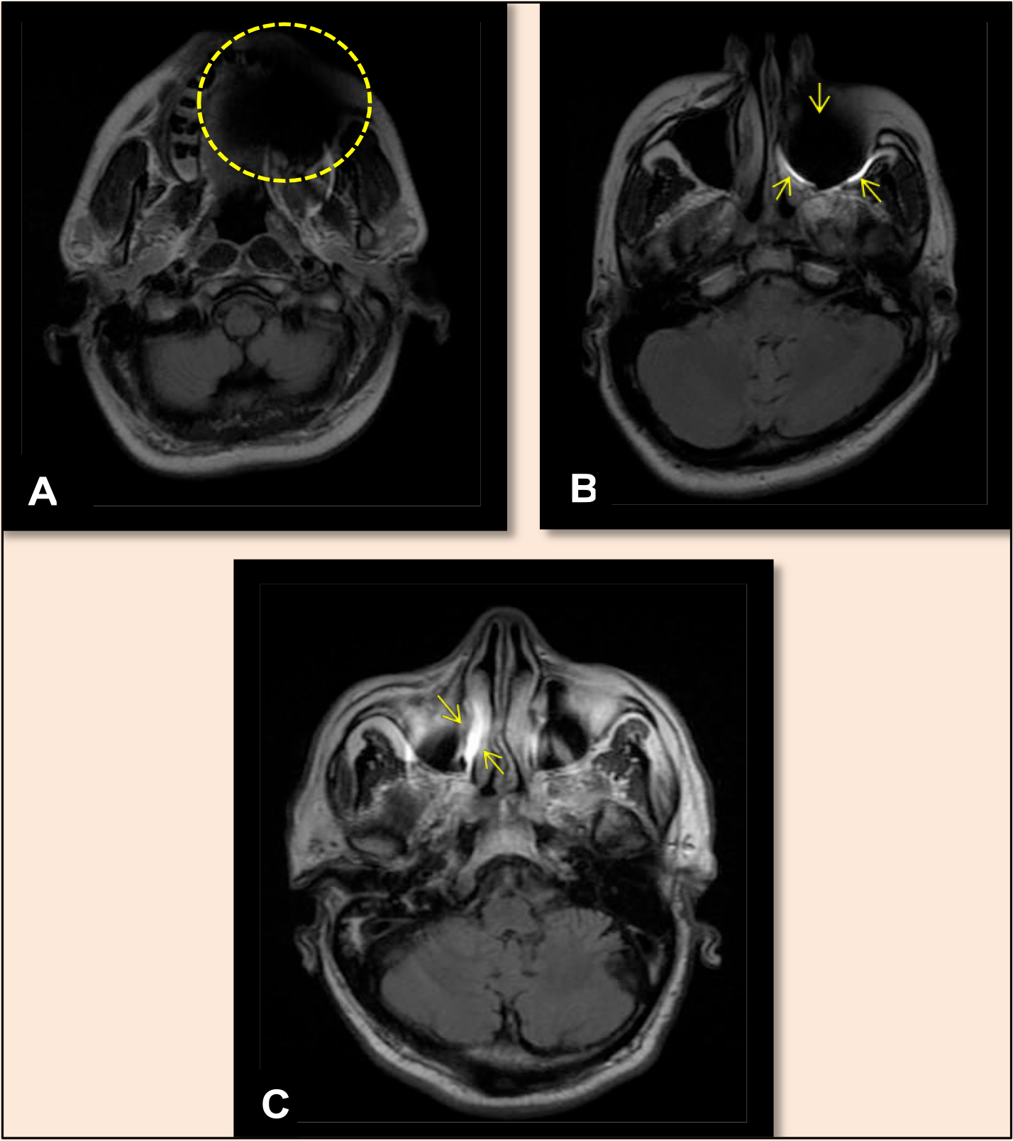
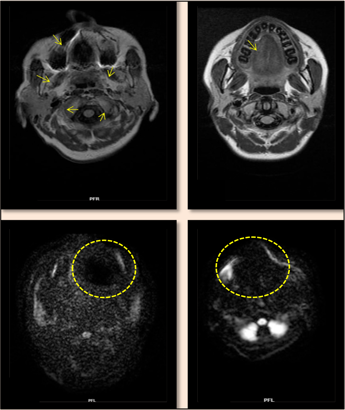
The type of artifact was noted as hypointense, hyperintense, or hypointense with hyperintense rim signal intensity (Figure 1). The shape of the artifacts was noted as void, linear, circular/void, linear, or void (Figure 2). The extent of the artifacts was reported as focal/diffuse. Furthermore, MRI artifacts were assessed and classified according to location, such as right/left/anterior/bilateral, at the brain / upper face and mid-face region level.

Showing types of the artifacts in the axial section with varying signal intensities in MR images in FLAIR sequence (A) Hypointense artifact, (B) Hypointense with hyperintense rim.

Showing the shape of the artifacts in the axial section of MR brain images in various MRI sequences. (A) Circular/Void shaped, (B) Linear shaped, (C) Void shaped, (D) Linear and Void shaped.
The diagnostic quality of the MRI images due to dental material artifacts was recorded as diagnostically acceptable or unacceptable on each of the four MRI sequences: FLAIR, T2W1, DWI, and SWI. With all four sequences in mind, the overall diagnostic acceptability of the MR images was assessed. This parameter was evaluated by the principal radiologist with more than five years of clinical experience (considered the gold standard) after considering the clinical indication of the magnetic resonance scans and the required visualization area. A total of 10% of the total MRI images were re-examined after one month to ensure interobserver and intraobserver reliability.
Statistical Analysis
Data collected were analyzed using IBM SPSS Statistics version 20 (IBM Corp., Chicago, IL, USA). Descriptive statistical methods were used to evaluate the data. Fischer's exact test was used to compare categorical variables. The reliability between and intra-observer reliability was checked using Cohen's Kappa Coefficient. A P-value <0.05 was considered statistically significant.
Results
The distribution of various classes of intraoral dental materials in the study population based on their location in the oral cavity is depicted in Table 1. Of 46 patients, 3 with Class IV materials could not continue with another image sequence after the FLAIR sequence due to the extensive artifacts that rendered the imaging unacceptable for diagnosis.
Distribution of various intra-oral dental materials based on their location in the oral cavity.
In the FLAIR sequence, the artifacts were mainly distributed in the mid-face region compared to the upper-face region. In the case of Class IV intraoral dental material, artifacts located bilaterally were seen in both the upper and middle face regions. Regardless of the class of intraoral dental materials, the T2 weighted sequence showed artifacts in the midface region in all study populations (Table 2). Class II and Class III intraoral dental materials caused most of the artifacts in the midface region in the SWI and DWI sequences (Table 3).
Distribution of the dental material artifacts in Fluid Attenuated Inversion Recovery (FLAIR) and T2 Weighted Image (T2W1) sequence concerning the location and class of the intra-oral dental materials.
Distribution of dental material artifacts in Susceptibility Weighted Imaging (SWI) and Diffusion Weighted Imaging (DWI) sequence in correlation with the location and class of the intra-oral dental materials.
The inter-observer reliability tested using Cohen’s Kappa coefficient showed substantial to almost perfect agreement (0.61-0.99) for all study variables. The intra-observer reliability in the intra-observer tested using the Cohen Kappa coefficient showed almost perfect agreement (0.81-0.99) for all study variables.
The most common type of signal intensity artifact observed in the present study was hypointense with hyperintense rim artifacts in FLAIR (95.5%), T2 weighted image (93.3%), and DWI (78.9%) sequences. Hypointense artifacts (97.4%) were commonly observed in the SWI sequence. The most common shape of the artifacts observed in our study was circular/void shaped in FLAIR (84.4%) and T2 weighted image (93.3%). However, void-shaped artifacts were commonly observed in SWI (95.1%) and DWI (85%) sequences. Also, very few images had linear-shaped artifacts in FLAIR (2.2%) and T2W1 (3.3%) sequences. The linear and void-shaped artifact was observed only on the DWI sequence (7.5%). In the present study, the typical extent of the artifact was focal in FLAIR (80%) and T2-weighted images (93.3%). Diffuse extent was observed in SWI (78%) and DWI (70%) sequences.
No significant differences in the distribution were seen between the shortand long-span intraoral dental materials in relation to the location of artifacts in the brain / upper face and midface region in any of the four MRI sequences (p>0.05) (Table 4).
Comparison of the span of the intra-oral dental materials with MRI dental artifacts at the level of skull/upper-face and mid-face in various MRI sequences.
Diagnostically acceptable images were seen in significantly higher proportion in Class II intraoral dental materials than in Class III, and diagnostically unacceptable images were observed in Class III intraoral dental materials in the FLAIR sequence. There was a statistically significant difference between the various classes of intraoral dental materials and diagnostic acceptability (p=0.017). There were no statistically significant differences seen in T2W1 (p=0.333), SWI (p=0.469), and DWI (p>0.99). The general diagnostic precision of the MRI image evaluated in various MRI sequences in the right posterior region (RP) was statistically significant (p=0.036) (Table 5).
Comparison of the diagnostic acceptability of MR images in various sequences with the class of the intra-oral dental materials in right posterior region.
In the present study, 28% of the study population images were graded diagnostically unacceptable in the sequences evaluated (FLAIR 37.8%, T2 3.3%, SWI 24.4%, DWI 25%). Images that were diagnostically acceptable even in the presence of artifacts were observed in 72% of the study population in the various sequences evaluated (FLAIR 60%, T2 96.7%, SWI 73.2%, DWI 72.5%). The MR image artifacts present in our study did not cause diagnostic problems in 2.2% of the images in the FLAIR sequence, 2.4% of SWI, and 2.5% of DWI sequences.
No significant differences were observed between the short-span and long-span intraoral dental materials concerning diagnostic accuracy in any of the four MRI sequences. The overall diagnostic precision of the MR image evaluated in various MRI sequences correlating with the span of dental materials was not statistically significant (p=0.739) (Table 6). No significant differences in distribution were observed between the short and long span of intraoral dental materials compared to the type of artifacts in the four MRI sequences (p>0.99, p>0.99, p=0.41, and p=0.687), respectively.
Comparison of diagnostic acceptability of the MR image in various sequences with the span of the intra-oral dental materials.
Discussion
Magnetic resonance imaging (MRI) of the head and neck region is a widely accepted imaging modality for visualizing soft tissue pathologies. Various ferromagnetic materials can impair MRI image acquisition, which may interfere with the magnetic field, causing signal distortion and artifact production. Many dental materials used intraorally are ferromagnetic, causing artifacts that can interfere with the diagnostic quality of the resultant image [1]. This adverse effect can be significant depending on the position, extent, type of material, and machine parameters. The present study evaluated various characteristics of artifacts caused by intraoral dental materials in commonly used MR sequences. It also correlated with the location and extent of dental materials in the oral cavity. This study also evaluated the diagnostic impedance caused by these artifacts in image interpretation.
In the present study, most of the patients (56.5%) presented dental materials of Class III, followed by Classes II (37%) and IV (6.5%). Most of the patients in the study population had long-span intraoral dental materials (63%).
Conventional MRI sequences consist of T1w, T2W1, T2 FLAIR, SWI, and DWI. In their in vitro study, Bartels et al. [9] explained that the artifacts generated in the MRI images by paramagnetic materials depend on the sequence used. The present study analyzed four magnetic resonance sequences (FLAIR, T2W1, SWI, DWI). The FLAIR sequence is commonly used in brain imaging protocols to detect subtle changes in the periphery of the hemispheres and the periventricular region close to the cerebral spinal fluid [10]. T2-weighted imaging sequences depict paramagnetic deoxyhemoglobin, methemoglobin, or hemosiderin in lesions and tissues. SWI is more sensitive to the detection of susceptibility effects and depicts significantly more small hemorrhagic lesions in diffuse axonal injury [11]. DWI is a quantitative MRI technique that can detect tissue injury outside the T2-visible lesion [12].
In the present study, dental artifacts were present in MRI images of all subjects but not necessarily in all sequences. Of the four sequences evaluated in each subject, DWI had no artifact in 5%, SWI in 4.9%, and the FLAIR sequence had no artifact in 2.2%. In contrast, all T2-weighted sequences had at least one artifact. These findings were in contrast to the in vitro study conducted by Lan et al. [13], who reported that the T2 star GRE, part of the SWI sequence, exhibited the most substantial influence on artifacts.
Camacho et al. [14] conducted an in vitro study to investigate MRI artifacts using various objects and demonstrated that the resultant signal intensity artifact, along with any susceptibility effect, depends on the object's orientation, shape, and material. The most common type of signal intensity artifact observed in the present study was hypointense with hyperintense rim artifacts in FLAIR (95.5%), T2 weighted image (93.3%), and DWI (78.9%) sequences. Krupa and Bekiesińska-Figatowska [1] reported that metallic objects within the patient’s body result in FLAIR hyperintensity due to a magnetic susceptibility artifact. Such artificial FLAIR-hyperintensity in the subarachnoid space can lead to a false diagnosis of hemorrhage in the subarachnoid space, particularly in brain MRI.
The most common shape of the artifacts observed in our study was circular/void shaped in FLAIR (84.4%) and T2 weighted image (93.3%). However, void-shaped artifacts were commonly observed in SWI (95.1%) and DWI (85%) sequences. Murakami et al., in an in-vitro study assessing artifacts caused by seven different metallic dental materials, stated that the artifact's shape depends on the scanning plane. They found that the artifacts appeared as a circular pattern in the axial plane and a 'clover-like' pattern in the sagittal plane [15]. In the present study, the typical extent of the artifact was focal in FLAIR (80%) and T2-weighted images (93.3%). Diffuse extent was observed in SWI (78%) and DWI (70%) sequences.
The present study did not show statistically significant differences between the spacing of intraoral dental materials and the location of artifacts in the brain/upper face and the midface region in any of the four MRI sequences. We could not find similar studies to compare these results.
In the present study, 28% of the study population images were graded diagnostically nonacceptable in the sequences evaluated. 72% of the images were diagnostically acceptable, even in the presence of artifacts. MR image artifacts present in our study did not cause diagnostic problems in 2.2% of the images in the FLAIR sequence, 2.4% of SWI, and 2.5% of DWI sequences. However, we could not find previous studies that assessed similar parameters.
In the present study, 6.5% with intraoral Class IV dental material had caused severe artifacts that led to the diagnostically unacceptable MR image. This was similar to the in vivo study conducted by Harris et al. [16] that evaluated patients wearing orthodontic braces who needed repeated magnetic resonance imaging for routine check-ups and concluded that metallic orthodontic appliances can distort magnetic resonance images, thus hampering the diagnostic value. Costa et al. [17] retrospectively studied the 70 MR images with artifacts and found that orthodontic appliances caused the highest artifacts (78%) but were also seen in titanium implants (18%) and metallic crowns (4%). Hinshaw et al. also evaluated artifact production using various intraoral dental materials commonly used in dentistry and reported that severe artifacts were orthodontic bands with braces, posts, and pins [18].
The present study showed major artifacts with diagnostically unacceptable quality in 23.5% and acceptable quality in 76.5% of subjects associated with Class II intraoral dental materials. At the same time, extensive metallic artifacts were found in MR images in subjects with Class III intraoral dental materials, producing diagnostically unacceptable images in 23% and diagnostically acceptable MR images in 77% (Figure 3). Mazumdar et al. [19] conducted a retrospective study evaluating 21 medical records with MRI and dental restorations. They found that patients with multiple metal-ceramic crowns with a span of more than three units demonstrated extensive artifacts. Limited artifacts were observed in patients with less than three metallic crowns. Class I materials were not assessed separately in our study due to a smaller sample size. Costa et al. [17] observed that amalgam restorations were not identified as a source of artifact generation.
In the present study, we also tried to assess the diagnostic acceptability of MRI images of patients with dental material in association with the intraoral location of the material of larger materials at this location. Diagnostic acceptability in each MRI sequence and overall grading were also compared with the span of dental materials, but this aspect was not statistically significant. A recent observational study by Ashok et al. evaluated the effect of metallic and intraoral restorations and implants on the artifacts and patient discomfort during magnetic resonance imaging. Artifacts were found in 32 out of 34 (94.1%) in the magnetic resonance imaging of the brain and 33 out of 39 (84.6%) in the MRI spine [20].
Clinical studies are sparse, and our study could be considered as a preliminary observational study of a prospective nature evaluating the span of intraoral dental materials with the location of the dental material artifact and its diagnostic acceptability. However, the results in the present study could not be conclusively interpreted due to the small sample size and uneven comparison groups due to its observational nature. This study is clinically relevant because patients undergoing head and neck magnetic resonance imaging frequently have extensive restorations or prostheses, which may cause artifacts that could impede diagnosis. In our study, the diagnostic quality of the image was unacceptable in one-fourth of the total images analyzed despite the location and span of the intraoral dental material. The present study's findings prompt the researchers to further evaluate in the same direction with a larger sample size. Further studies can help formulate a conclusive evidence-based recommendation on imaging protocols whenever head and neck MRI is performed in patients with intraoral dental materials.
The study's limitations included the small sample size and the overlapping nature of artifacts produced by various intraoral dental materials that coexist in the oral cavity. It is difficult to understand the exact type and composition of dental materials involved in intraoral clinical examination alone. The comparison of the type and location of the materials had practical limitations. Most patients with dental material artifacts had multiple types of restorations in the mouth and were close to each other. A larger sample size and stringent grouping criteria can help overcome many of these limitations.
Conclusion
Dental material artifacts interfered with the interpretation of magnetic resonance imaging at varying levels of diagnostic acceptability depending on the indication for which the imaging was performed. The artifacts in the MRI images were predominantly distributed in the midface region, and their type, shape, and extent varied according to the sequences used. Among the sequences evaluated, it was noted that diagnostically unacceptable images were demonstrated in the FLAIR sequence. More studies with a larger sample size and material distribution are warranted to obtain conclusive evidence on the effects of dental material artifacts. Further clinical research addressing novel advances and approaches in this field shall help achieve improved compliance and diagnostic accuracy.
Data Availability
The data used to support the findings of this study can be made available upon request to the corresponding author.
-
Financial Support
None.
References
-
[1] Krupa K, Bekiesińska-Figatowska M. Artifacts in magnetic resonance imaging. Polish J Radiol 2015; 80:93-106. https://doi.org/10.12659/PJR.892628
» https://doi.org/10.12659/PJR.892628 -
[2] Gaeta M, Cavallaro M, Vinci SL, Mormina E, Blandino A, Marino MA, et al. Magnetism of materials: Theory and practice in magnetic resonance imaging. Insights Imaging 2021; 179. https://doi.org/10.1186/s13244-021-01125-z
» https://doi.org/10.1186/s13244-021-01125-z -
[3] Eggers G, Rieker M, Kress B, Fiebach J, Dickhaus H, Hassfeld S. Artefacts in magnetic resonance imaging caused by dental material. MAGMA 2005; 18(2):103-111. https://doi.org/10.1007/s10334-005-0101-0
» https://doi.org/10.1007/s10334-005-0101-0 -
[4] Mathew CA, Maller S, Maheshwaran. Interactions between magnetic resonance imaging and dental material. J Pharm Bioallied Sci 2013; 5(Suppl 1):113-116. https://doi.org/10.4103/0975-7406.113309
» https://doi.org/10.4103/0975-7406.113309 - [5] Bajunaid SO. Review of techniques for the intact removal of a permanently cemented restoration. Gen Dent 2017; 65(5):48-53.
-
[6] Chockattu SJ, Suryakant DB, Thakur S. Unwanted effects due to interactions between dental materials and magnetic resonance imaging: A review of the literature. Restor Dent Endod 2018; 43(4):e39. https://doi.org/10.5395/rde.2018.43.e39
» https://doi.org/10.5395/rde.2018.43.e39 -
[7] Klinke T, Daboul A, Maron J, Gredes T, Puls R, Jaghsi A, et al. Artifacts in magnetic resonance imaging and computed tomography caused by dental materials. PLoS One 2012; 7(2):e31766. https://doi.org/10.1371/journal.pone.0031766
» https://doi.org/10.1371/journal.pone.0031766 -
[8] Hubálková H, La Serna P, Linetskiy I, Dostálová T. Dental alloys and magnetic resonance imaging. Int Dent J 2006; 56(3):135-141. https://doi.org/10.1111/j.1875-595X.2006.tb00085.x
» https://doi.org/10.1111/j.1875-595X.2006.tb00085.x -
[9] Bartels LW, Smits HF, Bakker CJ, Viergever MA. MR imaging of vascular stents: effects of susceptibility, flow, and radiofrequency eddy currents. J Vasc Interv Radiol 2001; 12(3):365-371. https://doi.org/10.1016/s1051-0443(07)61918-6
» https://doi.org/10.1016/s1051-0443(07)61918-6 -
[10] Bangerter NK, Hargreaves BA, Gold GE, Stucker DT, Nishimura DG. Fluid-attenuated inversion-recovery SSFP imaging. J Magn Reson Imaging 2006; 24(6):1426-1431. https://doi.org/10.1002/jmri.20743
» https://doi.org/10.1002/jmri.20743 -
[11] Chavhan GB, Babyn PS, Thomas B, Shroff MM, Haacke EM. Principles, techniques, and applications of T2*-based MR imaging and its special applications. Radiographics 2009; 29(5):1433-1449. https://doi.org/10.1148/rg.295095034
» https://doi.org/10.1148/rg.295095034 - [12] Nafisi-Moghadam R, Rahimdel A, Shanbehzadeh T, Fallah R. Comparison of diffuse weighted imaging and fluid attenuation inversion recovery sequences of MRI in brain multiple sclerosis plaques detection. Iran J Child Neurol 2017; 11(1):13-20.
-
[13] Lan G, Yunmin L, Pu W, Haili H. Contrastive analysis of artifacts produced by metal dental crowns in 3.0 T magnetic resonance imaging with six sequences. West China J Stomatol 2016; 34(3):277-280. https://doi.org/10.7518/hxkq.2016.03.012 [In Chinese].
» https://doi.org/10.7518/hxkq.2016.03.012 -
[14] Camacho CR, Plewes DB, Henkelman RM. Nonsusceptibility artifacts due to metallic objects in MR imaging. J Magn Reson Imaging 1995; 5(1):75-88. https://doi.org/10.1002/jmri.1880050115
» https://doi.org/10.1002/jmri.1880050115 -
[15] Murakami S, Verdonschot RG, Kataoka M, Kakimoto N, Shimamoto H, Kreiborg S. A standardized evaluation of artefacts from metallic compounds during fast MR imaging. Dentomaxillofacial Radiol 2016; 45(8):20160094. https://doi.org/10.1259/dmfr.20160094
» https://doi.org/10.1259/dmfr.20160094 -
[16] Harris TMJ, Faridrad MR, Dickson JAS. The benefits of aesthetic orthodontic brackets in patients requiring multiple MRI scanning. J Orthod 2006; 33(2):90-94. https://doi.org/10.1179/146531205225021465
» https://doi.org/10.1179/146531205225021465 - [17] Costa ALF, Appenzeller S, Yasuda C-L, Pereira FR, Zanardi VA, Cendes F. Artifacts in brain magnetic resonance imaging due to metallic dental objects. Med Oral Patol Oral Cir Bucal 2009; 14(6):E278-282.
-
[18] Hinshaw DB, Holshouser BA, Engstrom HIM, Tjan AHL, Christiansen EL, Catelli WF. Dental material artifacts on MR images. Radiology 1988; 166(3):777-779. https://doi.org/10.1148/radiology.166.3.3340777
» https://doi.org/10.1148/radiology.166.3.3340777 -
[19] Mazumdar P, Desai PD, Bhowmick S, Singh S. Artifacts in MRI due to dental restorations: A narrative review. J Dent Med Sci 2021; 20(7):47-53. https://doi.org/10.9790/0853-2007024753
» https://doi.org/10.9790/0853-2007024753 -
[20] Ashok A, Ayyadanveettil P, Devarajan E, Thavakkara V, Latha N, Saraswathy A. The effect of metallic dental restorations and implants in causing patient discomfort and artifacts during magnetic resonance imaging of the head and neck. Indian J Dent Res 2022; 33(3):258-262. https://doi.org/10.4103/ijdr.ijdr_430_21
» https://doi.org/10.4103/ijdr.ijdr_430_21
Edited by
-
Academic Editor:
Alessandro Leite Cavalcanti