Erosive Effect of Long-Term Liquid Oral Pediatric Medicines on Permanent Tooth Enamel
ABSTRACT
Objective: To evaluate in vitro the erosive effect of long-term liquid oral pediatric medicines on human enamel teeth and the preventive action of fluoride in an erosive challenge.
Material and Methods: Three commonly used medicines were selected for this study, and their endogenous pH was measured in triplicate. Thirty permanent tooth enamel specimens were prepared and divided into six groups (n=5): E1 (Zetalerg), E2 (Betamethasone), E3 (Anemifer), E4 (Anemifer+Duraphat), E5 (Coke), and E6 (artificial saliva). Specimens were immersed in 5 ml of medicine solution for 5 min, 2x/ day for 12 days, and stored in artificial saliva at 37°C between immersions. Data analysis was performed according to the enamel surface morphology using SEM.
Results: The medicines showed an acidic pH range from 2.09 to 4.14. All the specimens exposed to pediatric medicines presented some pit-like erosion pattern under SEM analysis, except for the E4 group. The degree of destruction was inversely proportional to pH formulation values. Morphology alterations could be ranked as follows: E3>E2>E1=E5. The E4 group, protected with varnish fluoride, did not present signs of surface erosion wear like E6.
Conclusion: All the pediatric medicines used promoted some enamel tooth wear, with higher severity with low pH medicines. The presence of fluoride reduced the deleterious effect of pediatric medicines on human tooth enamel.
Keywords:
Tooth Erosion; Microscopy, Electron, Scanning; Antitussive Agents; Dental Enamel
Introduction
Dental erosion is an irreversible loss of dental enamel resulting from a chemical process without bacterial involvement. It has a multifactorial etiology, with potentially erosive substances, including the consumption of food, drinks, acidic substances, and medication [1].
Oral medications for children often come in liquid form for easy swallowing [2]. Usually, they are prescribed for a short period, but in some chronic illnesses, their use may be long-term. Active ingredients in these medicines are necessary for health maintenance, but some inactive ingredients can harm teeth [3]. The oral consumption of pediatric liquid medicines with low endogenous pH levels may constitute possible etiological or aggravating factors for severe dental erosion [4-6]. Unavoidable medications may significantly contribute to erosive tooth wear [2]. Studies have raised concerns about the adverse dental effects of taking these medicines with a potential of hydrogen (pH) ≤ 5.5 [7].
Medication components can vary greatly. Most pediatric formulations are prepared with sugar, a potential risk factor for dental caries. However, to reformulate medicines without sugar, it might be necessary to add weak acidic substances that can increase the risk of dental erosion [3,7], even though acid challenges on enamel during erosion are stronger. After erosive mineral loss, a thin, partly demineralized, and softened surface layer is left, providing a structure for remineralization under favorable conditions [8]. Despite this, dental erosion has started gaining more attention from the scientific community due to its increased prevalence [2].
In vitro studies have demonstrated the erosive potential of long-term and short-term orodispersible tablet medications [7], and iron drops displayed low pH and enamel discoloration in the anterior primary teeth, as related previously [9]. Additionally, research in vitro has shown the reduced enamel microhardness capability of several medicines available in Switzerland [1], antitussive syrup [5], analgesics [6], antihistamine-containing syrups [10], and long-term medication [11]. The gravimetric method is also valid for detecting erosive enamel induced by liquid medications [12]. Moreover, a profilometer was used to evaluate the adverse effects of multivitamin syrups and effervescent tablets on the surface of restorative materials [13].
Individuals with chronic diseases must pay more attention to oral health due to their systemic conditions and the long-term use of medicines. It is also significant that dental erosion can be influenced by the contact of the liquid with a low pH and exposure time on the enamel tooth surface [1,7].
Anemia remains a condition with a high prevalence worldwide. Ferrous sulfate, an antianemic, is administered orally during the treatment of anemia [14]. Moreover, oral corticosteroid use is frequent among asthma patients, many of whom are regular users [15]. Antihistamines are frequently used in pediatric patients to treat many allergic diseases [16]. Therefore, based on the long-term use and low pH of antianemic, corticosteroid, and antiallergic medicines [4], this study aimed to evaluate in vitro the erosive effect of an antianemic, corticosteroid and antiallergic medicine on the permanent tooth enamel surface, and the preventive action of fluoride in erosive challenge.
Material and Methods
Ethical Aspects
The local ethics committee reviewed and approved the study protocol for research at Lauro Wanderley Hospital Ethical Committee (Opinion No. 6.704.653. The study was conducted at the Morphology Laboratory of the Federal University of Paraiba, Brazil. All participants provided written informed consent prior to donating their extracted teeth for research purposes.
Sample and Pediatric Medicines
Twenty human permanent teeth without cracks, dental caries, or enamel defects were evaluated for this in vitro experimental study. The teeth were stored in a 10% buffered formalin solution until the experiment was conducted.
Three commonly used pediatric liquid medicines were selected for the study. Their endogenous pH levels were measured (Table 1) using a properly calibrated microprocessor bench pH meter W3B (BEL Engineering s.r.l.., Monza, Italy). The pH measurement was performed immediately after opening the bottles to prevent the natural degradation process of medicine. These medicines were chosen based on the frequency of long-term use, as determined by a previous survey with pediatricians from a public hospital [4]. The pH values were analyzed in triplicate.
Specimen Preparation
Enamel slabs (4 x 4 x 2mm) were cut from the middle third of the buccal and palatal surfaces of each permanent tooth using a precision cutter (Labcut 1010, Extec Corp., Enfield, USA) with a diamond wheel under constant irrigation. Specimens with white spots, cracks, or any other defect were discarded. The enamel slabs underwent prophylaxis with a pumice mixture and rinsing with distilled deionized water. A double layer of nail varnish Risque (Grupo Coty, Taboão da Serra, SP, Brazil) covered half of the exposed enamel to preserve a control area of each specimen. The specimens were divided into four experimental groups (n=5): (E1) antihistaminic - Zetalerg (UCI-FARMA Indústria Farmacêutica Ltda, São Bernardo do Campo, SP, Brazil); (E2) corticosteroid - Betamethasone (EMS Sigma Pharma Ltda., Jaguariúna, SP, Brazil); (E3) antianemic - Anemifer (Pharmascience Industria Farmacêutica, Betim, MG, Brazil); and (E4) Anemifer + Duraphat (Table 1). The positive control was Coke (The Coca‑Cola Company Brasil, Jundiaí, SP, Brasil) (E5, n = 5), while the negative control was artificial saliva (E6, n = 5).
Erosive Challenge
Before the erosive challenge, the specimens were placed in a container of filtered human saliva for 24 hours. The container was constantly agitated on an orbital shaker table TE-141 (Tecnal Equipamentos Científicos, Piracicaba, SP, Brazil) to form the acquired pellicle on the enamel surface [17,18].
A commercial presentation of each liquid medicine was prepared and used according to the manufacturer’s instructions. Specimens were immersed in 5 ml of medicine solution for 5 minutes, twice a day for 12 days, and stored in artificial saliva at 37 °C between immersions. Only the E4 group was exposed to fluoride varnish Duraphat (Colgate-Palmolive Company, Hamburg, Germany) at the times T0 (day 0), T1 (day 4), T2 (day 8), and T3 (day 12) before exposure to medicine to simulate the use in the dental office. The artificial saliva was replaced daily, and the medication was renewed after each immersion. The medicines were manually shaken before each use, as prescribed on their instruction leaflets.
The 5-minute exposure time [19] was determined based on the short residence time of acidic drinks in the oral cavity and the buffering capacity of saliva. The 5 ml medicine dosage followed the prescription for children aged 7 to 12, as indicated on the instruction leaflets.
Scanning Electron Microscope (SEM) Analysis
All enamel specimens were rinsed with distilled-deionized water for analysis in a scanning electron microscope Tescan VEGA 3 LMU (Tescan Group, Kohoutovice, Czech Republic), operating at 15 kV. For morphological analysis, the specimens were sputter-coated with gold in a vacuum evaporator MED 010 Balzers Union, Liechtenstein) before microscopic examination to obtain photomicrographs of the treated specimens' surface morphology at 2000× magnification. The qualitative changes in enamel surface morphology between groups were analyzed and compared with the typical morphology of the control enamel surface (group E6), then transcribed into a standardized form. No statistical analysis was performed.
Results
The results reaffirmed the critical pH level values of the selected medicines. Zetalerg, Betamethasone, and Anemifer exhibited mean pH values of 4.14, 2.94, and 2.09, respectively. After an erosive cycle period of twelve days, clinical examination revealed that the enamel surface remained visually smooth and shiny across all groups. However, SEM analysis revealed significant variations in tooth wear among the groups that were inversely proportional to the pH levels.
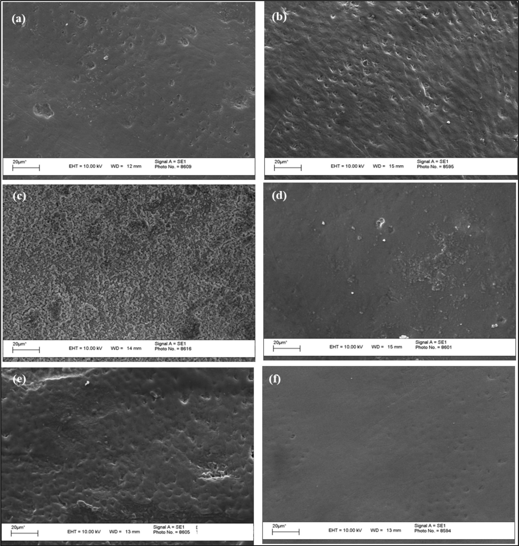
SEM observations confirmed the destructive impact of the acidic medicines on the enamel surface. Image (1a - Zetalerg) showed a relatively smooth surface with minor erosive features, while Image (1b - Betamethasone) revealed a textured surface with pronounced linear grooving consistent with moderate erosion. Image (1c - Anemifer) displayed a 'honeycomb-like' pattern with extensive enamel loss, indicating severe erosion. Image (1d - Anemifer + Duraphat) depicted a smoother surface with isolated defects, suggesting less erosion, which could be attributed to protective treatment. Image (1e - Coke) presented a rough surface with moderate erosion, less severe than Image (1c) but still significant. Image (1f - artificial saliva) showed an intact enamel surface with no signs of erosion, highlighting the contrast between the samples treated with acid and non-acidic solutions.
The morphology of the lesions varied from the most intact in Image (1f) to the severely eroded in Image (1c). The patterns of demineralization in Images (1a), (1b), and (1e) were characterized by a honeycomb-like appearance, but Image (1c) showed a more severe degree of erosion with widespread enamel destruction and areas with complete enamel loss. Specimens protected with fluoride varnish (group E4; Image 1d) resembled the negative control (group E6; Image 1f) with a smooth enamel surface and no erosion evidence.
Despite Anemifer and Coke having a very low pH level and the same type of acid in their composition, SEM images (Images 1, c, and e) demonstrated distinct demineralization patterns. This suggests that additional factors beyond pH and acid type contribute to erosion. Conversely, Zetalerg and Betamethasone, despite differences in pH levels and acid types, showed similar erosion patterns in SEM images (Images 1a and 1b), differing only in the intensity and extent of enamel surface destruction.

Scanning electron photomicrographs of the surface of permanent tooth enamel after the erosive challenge of the groups E1 (a), E2 (b), E3 (c), E4 (d), E5 (e) and E6 (f).
Discussion
The present study evaluated the in vitro erosive potential of antianemic, corticosteroid, and antiallergic medicines on human permanent teeth and the protective effect of fluoride, utilizing SEM. The low pH levels of the medications encountered in this study were identified as a potential risk factor for the development of enamel erosion, as evaluated in other studies [3,4,6,7]. However, the observations also underscore the complexity of erosion mechanisms, which differ from caries by posing a more substantial acid challenge and leading to the release of critical minerals, such as calcium, hydroxyl-, and phosphate ions, from the enamel [1].
Despite the numerous studies on dental erosion [3,6,19-24], there remains a deficiency of information regarding the contributory role of oral medicines, particularly concerning their long-term impact on permanent teeth. While numerous studies have focused on pH and other physicochemical parameters [7,19-21], the SEM analysis provides direct visual evidence of erosion, highlighting that factors beyond pH are at play, as some medications with non-critical pH levels still demonstrated erosive capabilities [22,23].
The SEM images depicted the morphological changes attributable to erosion [3,6,23,24]. For instance, the pronounced linear grooving observed on the enamel surface exposed to Betamethasone aligns with moderate erosion patterns. At the same time, the 'honeycomb-like' appearance in the Anemifer group suggests a more advanced stage of erosion. This variety in morphologies, from the relatively smooth surface seen in the Zetalerg® group to the extensive enamel loss in the Anemifer group, confirms the diverse impacts that different medicinal formulas can have on tooth enamel [3,23].
Furthermore, the present results point to the critical influence of medication administration frequency, especially for chronic conditions requiring repeated daily dosages, which may exacerbate enamel erosion over time [4,20,21]. This is of particular concern given the SEM-documented enamel damage occurring after a mere twelve-day exposure period, an exposure duration significantly shorter than the clinical timeframe for visible tooth erosion, often spanning years [3,24].
The SEM findings also corroborate previous studies reporting on the erosive effects of various oral medicines, including iron supplements [24], analgesics [3,6,23], antibiotics, antiasthmatics, and multivitamins [3,23], on primary tooth enamel. Additionally, the protective effect of topical fluoride observed in this study aligns with research suggesting that fluoride treatments can inhibit erosive demineralization and increase enamel microhardness (increased mineral density and a decrease in lesion volume, depth, and surface area), which can be observed using micro-CT and SEM [8,10].
Despite the success of fluoridated toothpaste in reducing enamel erosion caused by medical syrups [12], a systematic review indicates that the protective role of topical fluoride in dental erosion remains questionable [25]. Researchers are considering protecting or remineralizing enamel against tooth erosion [8,18]. This suggests a pressing need for further research, particularly given the promising results from varnishes in protecting enamel against erosion [26].
In vitro studies aim to replicate the oral cavity conditions, especially when using the pH cycling methodology [27]. This approach allows for data collection in a controlled environment, eliminating the influence of external variables [3,5,6,10,11]. However, it's important to note the limitations of these studies. They do not fully capture the complexity of the oral environment, including the salivary pellicle's protective effects, saliva's buffering capacity, and the impact of individual dietary habits. Furthermore, the research did not evaluate key chemical factors such as titratable acidity, viscosity, acid type, and mineral content, which are crucial for understanding erosion potential [6,11,20-22,24]. Considering the limitations of in vitro studies, protocols that use pH cycling are considered the closest to the natural process of demineralization and remineralization, as it occurs clinically [27].
Lastly, the implications for permanent teeth should be cautiously interpreted. Pediatric medicines likely have a less pronounced erosive effect on permanent teeth when compared to deciduous teeth due to the latter's lower mineralization levels [12,23]. Regardless of the absence of clinical signs, it is imperative to raise patient awareness about the potential for morphological changes and to implement preventive measures. This includes consulting with healthcare providers to seek medications that minimize the impact on saliva secretion or provide alternative formulations with diminished erosive potential [1].
Conclusion
All pediatric medicines evaluated in this study contributed to some degree of enamel tooth wear. Medicines with low pH values exacerbate the severity of wear. The application of fluoride on the enamel surface reduced the deleterious effects of pediatric medicines on permanent tooth enamel, as observed with SEM.
-
Financial Support
None.
Data Availability
The data used to support the findings of this study can be made available upon request to the corresponding author.
Acknowledgments
The authors would like to express their sincere gratitude to the Federal University of Paraíba (UFPB) and the National Council for Research and Development (CNPq) for their financial support through the Scientific Initiation Scholarship.
References
-
[1] Lussi A, Megert B, Shellis P. The erosive effect of various drinks, foods, stimulants, medications and mouthwashes on human tooth enamel. Swiss Dent J 2023; 133(7-8):440-455. https://doi.org/10.61872/sdj-2023-07-08-01
» https://doi.org/10.61872/sdj-2023-07-08-01 -
[2] Yip K, Lam PPY, Yiu CKY. Prevalence and associated factors of erosive tooth wear among preschool children-A systematic review and meta-analysis. Healthcare 2022; 10(3):491. https://doi.org/10.3390/healthcare10030491
» https://doi.org/10.3390/healthcare10030491 -
[3] Babu KLG, Rai K, Hedge AM. Pediatric liquid medicaments - Do they erode the teeth surface? An in vitro study: Part I. J Clin Pediatr Dent 2008; 32(3):189-194. https://doi.org/10.17796/jcpd.32.3.j22m7t8163739820
» https://doi.org/10.17796/jcpd.32.3.j22m7t8163739820 -
[4] Passos IA, Sampaio FC, Martínez CR, Freitas CHSM. Sucrose concentration and pH in liquid oral pediatric medicines of long-term use for children. Rev Panam Salud Publica 2010; 27(2):132-137. https://doi.org/10.1590/s1020-49892010000200007
» https://doi.org/10.1590/s1020-49892010000200007 -
[5] Kulkarni P, Anand A, Bansal A, Jain A, Tiwari U, Agrawal S. Erosive effects of pediatric liquid medicinal syrups on primary enamel: An in vitro comparative study. Indian J Dent 2016; 7(3):131-133. https://doi.org/10.4103/0975-962X.189338
» https://doi.org/10.4103/0975-962X.189338 -
[6] Rocha CT, Nogueira ASB, Almeida JA, Rodrigues LKA, Neves BG. Erosive effect of analgesics on primary tooth enamel - An in vitro study. Pesqui Bras Odontopediatria Clín Integr 2022; 22:e210149. https://doi.org/10.1590/pboci.2022.066
» https://doi.org/10.1590/pboci.2022.066 -
[7] Anand L, Pentapati K, Shenoy R, Yelleti G, Kumar S. Sugar content and erosive potential of commonly prescribed Orodispersible tablets - An in vitro study. F1000 Res 2023; 12:228. https://doi.org/10.12688/f1000research.130786.2
» https://doi.org/10.12688/f1000research.130786.2 -
[8] Akküç S, Duruk G, Keleş A. Remineralization effect of three different agents on initial caries and erosive lesions: A micro-computed tomography and scanning electron microscopy analysis. BMC Oral Health 2023; 23(1):106. https://doi.org/10.1186/s12903-023-02805-6
» https://doi.org/10.1186/s12903-023-02805-6 - [9] Babaei N, Molaei T, Belyad S, Hekmatfar S. Relationship of pH and the viscosity of five different iron supplements with the absorption of iron ions and enamel discoloration in the anterior primary teeth (an in vitro study). Dent Res J 2021; 18:7.
-
[10] Kufiyah AK, Bagasi AMH, Nawlalili SM, Bazaid DS, Marghalani AA, Fathi A. Effect of Zamzam water on microhardness of primary tooth enamel after erosion induced by claritin syrup: An in-vitro study. J Int Soc Prev Community Dent 2021; 11(2):173-178. https://doi.org/10.4103/jispcd.JISPCD_420_20
» https://doi.org/10.4103/jispcd.JISPCD_420_20 -
[11] Yılmaz N, Baygin O, Cakıroglu TN, Tüzüner T, Deger O. In vitro evaluation of the effects of frequently prescribed pediatric drugs on the microhardness of permanent tooth enamel. Dent Med Probl 2019; 56(2):131-136. https://doi.org/10.17219/dmp/105562
» https://doi.org/10.17219/dmp/105562 -
[12] Qasim AA, Alani BW, Al Qassar SSS. Effects of fluoridated tooth paste on medically erosive enamel in bonded primary teeth during maxillary arch expansion in cleft palate patient: An in vitro study. J Orthod Sci 2021; 10:17. https://doi.org/10.4103/jos.JOS_67_20
» https://doi.org/10.4103/jos.JOS_67_20 -
[13] Guler EBG, Bayrak GD, Unsal M, Selvi Kuvvetli S. Effect of pediatric multivitamin syrups and effervescent tablets on the surface microhardness and roughness of restorative materials: An in vitro study. J Dent Sci 2021; 16(1):311-317. https://doi.org/10.1016/j.jds.2020.03.017
» https://doi.org/10.1016/j.jds.2020.03.017 -
[14] Paulino CTDS, Nishijima M, Sarti FM. Association of iron supplementation programs with iron-deficiency anemia outcomes among children in Brazil. Nutrients 2021; 13(5):1524. https://doi.org/10.3390/nu13051524
» https://doi.org/10.3390/nu13051524 -
[15] Ekström M, Nwaru BI, Hasvold P, Wiklund F, Telg G, Janson C. Oral corticosteroid use, morbidity and mortality in asthma: A nationwide prospective cohort study in Sweden. Allergy 2019; 74(11):2181-2190. https://doi.org/10.1111/all.13874
» https://doi.org/10.1111/all.13874 -
[16] Parisi GF, Licari A, Papale M, Manti S, Salpietro C, Marseglia GL, et al. Antihistamines: ABC for the pediatricians. Pediatr Allergy Immunol 2020; 31(Suppl 24):34-36. https://doi.org/10.1111/pai.13152
» https://doi.org/10.1111/pai.13152 -
[17] Hara AT, Ando M, González-Cabezas C, Cury JA, Serra MC, Zero DT. Protective effect of the dental pellicle against erosive challenges in situ. J Dent Res 2006; 85(7):612-616. https://doi.org/10.1177/154405910608500706
» https://doi.org/10.1177/154405910608500706 -
[18] Vieira A, Ruben JL, Huysmans MCDNJM. Effect of titanium tetrafluoride, amine fluoride and fluoride varnish on enamel erosion in vitro. Caries Res 2005; 39(5):371-379. https://doi.org/10.1159/000086843
» https://doi.org/10.1159/000086843 -
[19] Gowdar IM, Aldamigh SA, Alnafisah AM, Wabran MS, Althwaini AS, Alothman TA. Acidogenic evaluation of pediatric medications in Saudi Arabia. J Pharm Bioallied Sci 2020; 12(Suppl 1):S146-S150. https://doi.org/10.4103/jpbs.JPBS_46_20
» https://doi.org/10.4103/jpbs.JPBS_46_20 -
[20] Singana T, Suma NK. An in vitro assessment of cariogenic and erosive potential of pediatric liquid medicaments on primary teeth: A comparative study. Int J Clin Pediatr Dent 2020; 13(6):595-599. https://doi.org/10.5005/jp-journals-10005-1824
» https://doi.org/10.5005/jp-journals-10005-1824 -
[21] Xavier AF, Moura EF, Azevedo WF, Vieira FF, Abreu MH, Cavalcanti AL. Erosive and cariogenicity potential of pediatric drugs: Study of physicochemical parameters. BMC Oral Health 2013; 13:71. https://doi.org/10.1186/1472-6831-13-71
» https://doi.org/10.1186/1472-6831-13-71 -
[22] Lussi A, João-Souza SH, Megert B, Carvalho TS, Baumann T. The erosive potential of different drinks, foodstuffs and medicines - A vade mecum. Swiss Dent J 2019; 129(6):479-487. https://doi.org/10.61872/sdj-2019-06-02 [In German].
» https://doi.org/10.61872/sdj-2019-06-02 -
[23] Mahmoud EF, Omar OM. Erosive and cariogenic potential of various pediatric liquid medicaments on primary tooth enamel: A SEM study. Dent Med Probl 2018; 55(3):247-254. https://doi.org/10.17219/dmp/91539
» https://doi.org/10.17219/dmp/91539 -
[24] Pasdar N, Alaghehmand H, Mottaghi F, Tavassoli M. Experimental study of iron and multivitamin drops on enamel microhardness of primary tooth. J Int Soc Prev Community Dent 2015; 5(6):518-524. https://doi.org/10.4103/2231-0762.170524
» https://doi.org/10.4103/2231-0762.170524 -
[25] Né YGS, Souza-Monteiro D, Frazão DR, Alvarenga MOP, Aragão WAB, Fagundes NF, et al. Treatment for dental erosion: A systematic review of in vitro studies. PeerJ 2022; 10:e13864. https://doi.org/10.7717/peerj.13864
» https://doi.org/10.7717/peerj.13864 -
[26] Otel I, Dias K, Pereira R, Fonseca M, Jesus AP, Mata A, et al. Investigation of the protective suitability of a dental fluorinated varnish by means of X ray fluorescence and raman spectroscopy. J Trace Elem Med Biol 2022; 71:126938. https://doi.org/10.1016/j.jtemb.2022.126938
» https://doi.org/10.1016/j.jtemb.2022.126938 -
[27] Yu OY, Zhao IS, Mei ML, Lo EC, Chu CH. A review of the common models used in mechanistic studies on demineralization-remineralization for cariology research. Dent J 2017; 5(2):20. https://doi.org/10.3390/dj5020020
» https://doi.org/10.3390/dj5020020
Edited by
-
Academic Editor:
Wilton Wilney Nascimento Padilha