
 Radiographic Evaluation of the Lower First Permanent Molars of Children from Southeastern Brazil
ABSTRACT
Objective: To evaluate the health condition of the lower first permanent molars of children aged 6 to 12 years from the Southeast states in Brazil (Espírito Santo, Minas Gerais, Rio de Janeiro, and São Paulo).
Material and Methods: 1300 digital panoramic radiographs (2600 lower first permanent molars) from a database were analyzed by a calibrated examiner for the presence of carious lesions in dentin, pulp involvement, presence of restorative and/or filling material, and periapical lesion.
Results: Data were analyzed using the R software (p<0.05; 95% IC). The right and left sides were compared using the McNemar's test. 51.1% of the sample were male, and 48.9% were female (mean of 9.2 years; SD = 1.9). 6.4% of the teeth had carious lesions, 7.3% had restorative material, and 0.3% were absent. The mixed linear regression model showed that age advance was proportional to the increase in the number of missing teeth (p=0.012), decayed, restored, and periapical lesions (p<0.001). There was a higher incidence of restored teeth in females (8.5%) than in 6.1% of males (p=0.019).
Conclusion: There was a low incidence of dentin dental caries lesions and a low rate of loss of the permanent lower molar in children from Southeastern Brazil. As the individual ages, alterations in the lower first molars increase. There was no association with sex, except for the presence of restorations.
Keywords:
Dental Caries; Prevalence; Radiography; Tooth Loss
Introduction
The first permanent molar is derived from the primary dental lamina, and its formation usually begins around the 17th week of gestation. Its hard tissue begins to form at birth, and its coronal development is completed in the third year of life [1]. The eruption of the first permanent molars occurs around 6 to 7, but this tooth will only be fully formed between 9 and 10 years of age. It is usually the first permanent tooth to erupt, exposed for a longer time to the risk factors for caries in mixed dentition, especially in children with poor hygiene habits and a cariogenic diet [2]. In addition to its masticatory function, this tooth is essential for establishing the occlusion key, lifting the vertical dimension, and developing the dental arch and the temporomandibular joint. It also acts as a guide for the eruption and positioning of the other molars [3].
Dental caries harms the quality of life, is considered a public health problem in Brazil, and is one of the significant challenges of the Unified Health System [4]. Approximately 50% of preschoolers in different countries have caries experience [5]. This estimate is confirmed in studies conducted in Brazil, where prevalence rates range from 20.3% [6] to 53.6% [7]. Caries are the result of mineral loss from hard dental tissues. It is considered a dynamic, multifactorial, non-communicable disease mediated by biofilm and modulated by a cariogenic diet. Biological, behavioral, psychosocial, and environmental factors are also determinants of its development [8].
Some factors favor the development of carious lesions in the first permanent molar, such as occlusal anatomy with several grooves that facilitate food retention, poor hygiene, asymptomatic eruption, and, in some cases, defects in enamel development, such as Molar-Incisor Hypomineralization (MIH), where the dental structure appears porous and susceptible to fractures due to a failure in enamel mineralization and maturation [9,10]. Furthermore, teeth are vulnerable to caries within the first 2 to 4 years after eruption, as the enamel is not fully mineralized. Maturation and hardening continue in the oral cavity after the incorporation of fluoride, calcium, and other ions into the dental structure [11].
The early loss of this tooth is closely related to caries disease, and due to its importance in facial development, its absence can cause numerous damages to oral health [12]. Among these damages are compromised chewing function and dental misalignment due to adjacent and antagonistic teeth migration. This can result in alterations in the curve of Spee and affect the temporomandibular joint (TMJ), potentially triggering temporomandibular disorders (TMD). Additionally, the absence of the first permanent molar can impact facial development and occlusal key, compromising the proper distribution of occlusal and chewing forces [3,12]. Previous studies conducted in Iran and Saudi Arabia analyzed panoramic radiographs to verify the rate of loss of the first permanent molar in children aged 7 to 15 years, confirming that 36.9% and 40% had an absence of this dental element, respectively [12,13].
Panoramic radiography is commonly used as a primary diagnostic radiographic technique, a screening method for the entire maxillary and mandibular dentition [14]. This exam emits a low radiation dose, and its execution technique is simple, fast, and comfortable for the patient. Such characteristics make it widely used to approach child patients [15], whether in the evaluation of the stages of dental development (Nolla's stages), periapical and periodontal lesions, surrounding bone features, traumas, and other pathological changes [16].
The radiographic evaluation of the first permanent molars and the respective incidence rates of carious lesions and other alterations associated with these teeth is essential to assess the magnitude of this health problem. Considering the scarcity of studies that radiographically evaluated the molars of Brazilian children, the present study aimed to assess radiographically the health condition of the first lower permanent molars of children aged 6 to 12 years from the Southeastern states of Brazil.
Material and Methods
Study Design and Ethical Clearance
This descriptive cross-sectional observational study was approved by a public university's Human Research Ethics Committee (Opinion No. 5.560.433). The radiographic examinations used in this project were performed for diagnostic purposes, independent of this research, so the patients were not exposed to radiation without proper indication. The identification of the individuals was kept confidential, and data were not disclosed along with the results.
Pilot Study
A pilot study was conducted, and the proposed methodology was faithfully reproduced to calibrate the examiner. An Oral Radiologist (F.S.V.) with more than ten years of experience provided detailed explanations regarding the conditions under investigation: dental absence, dentin caries lesion, dentin and pulp caries lesion, restorative material, filling material, and periapical lesion. During the assessments, a lesion was classified as dentin caries if the image displayed a radiolucent area within the tooth crown involving dentin layers. If this radiolucent spot extended to the dental pulp, it was categorized as a dentin and pulp caries lesion. Conversely, a radiopaque area filling the crown structure and contrasting with the surrounding dental layers indicated the presence of restorative material. Visualizing a a radiopaque area within the root canals, contrasting with dental tissues and adjacent bone, was deemed necessary to confirm the presence of filling material. Periapical lesions were assessed as radiolucent areas in the periapical region, potentially exhibiting thickening of the periodontal ligament space, rupture of the alveolar cortex, and bone rarefactions. Dental absences were identified when the anatomical region corresponding to the first permanent lower molars lacked teeth.
The pilot study analyzed two hundred panoramic radiographs that were not included in the final sample. The image analysis was conducted under standardized conditions, and the data were meticulously recorded on a form containing information about age, sex, and the presence or absence of the following first molar features: dental absence; dentin caries lesion; dentin and pulp caries lesion; restorative material; filling material, and periapical lesion.
After 15 days, 20% of the pilot study sample was reassessed to calculate intra-rater agreement. The examiner was only allowed to begin the evaluations of the final sample after obtaining Kappa test values higher than 0.75. Thus, it was ensured that the evaluator could analyze the images without compromising the research results.
Sample Selection
The sample was obtained by convenience sampling, including all panoramic radiographs of children aged 6 to 12, available in a database. The images were acquired between the years 2020 and 2023 in different cities of the Brazilian states of Minas Gerais, Rio de Janeiro, São Paulo, and Espírito Santo, using the device OP 300 Kavo (Instrumentarium Corp., Helsinki, Finland).
Also, to be included in the study, the images should present medium contrast and density, maximum sharpness and detail, no technical error of execution, and only the inherent distortion and magnification determined by the manufacturer (25%). Radiographs in which it was impossible to complete visualization of the region of the lower first molars were excluded.
Image Evaluation
A dentist specialized in Pediatric Dentistry (L.A.M.P.) and with more than five years of experience, previously calibrated in the pilot study, evaluated the radiographs in a room with standardized lighting conditions, using a 21.5-inch LCD monitor and high resolution (1920 x 1080) (Dell S2240L - Dell Computadores do Brazil Ltda., Eldorado do Sul, RS, Brazil).
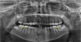
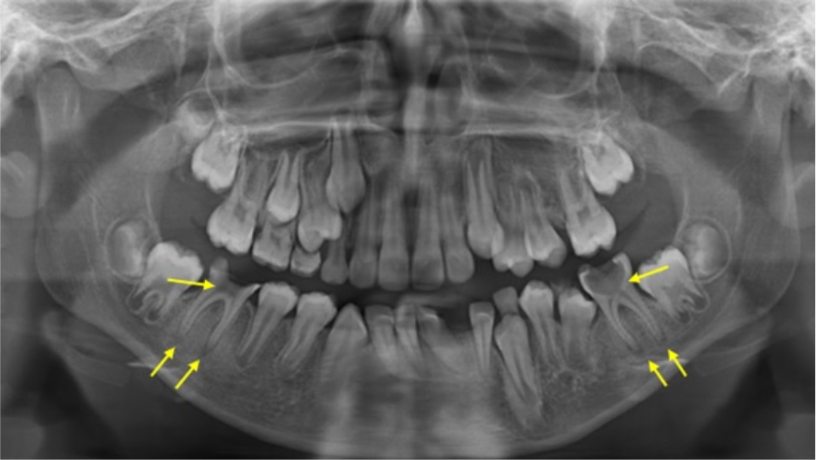
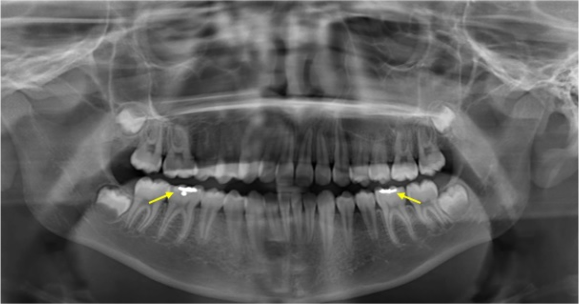
A maximum of 20 images per day were evaluated to avoid visual fatigue and consequent impairment of the assessments. The zoom, brightness, and contrast tools could have been used at the evaluator's discretion. Figures 1 to 4 are panoramic radiographs of the sample and show some examples of the alterations evaluated.

Male patient, 6 years old. The presence of a carious dentin lesion in tooth 36 is observed.

Male patient, 11 years old. There is a carious lesion in dentin with pulp and periodontal involvement in teeth 36 and 46.
Female patient, 12 years old. The presence of restorative material is observed in teeth 36 and 46.

Statistical Analysis
Data were evaluated through descriptive statistics using the R software, version 4.1.2. Continuous variables were described by means and standard deviations, and categorical variables were defined by their absolute and relative frequencies. The comparison between the sides was made using the McNemar hypothesis test for dependent samples due to the dependence on the side of the tooth for the same individual. The evaluation of the effect of sex and age considered the tooth (and no longer the individual, as in the comparison between the sides) as the sample unit. In these models, we considered the side of the tooth as a random effect. The effect of sex and age on dental manifestations was evaluated using a generalized linear model of the binomial family with a random effect of the side. The chi-square test compared the associations between the dental conditions studied. The level of significance was considered at 5%.
Results
The Kappa value, calculated for intra-examiner agreement, was 0.914, indicating an almost perfect degree of agreement. Data from 1,300 children were analyzed, with 51.1% of the sample being male and a mean age of 9.2 years (± 1.9 years). The distribution of all ages and sexes of the sample is shown in Table 1.
No statistical evidence of differences was identified for the conditions studied, comparing the left and right sides (Table 2).
Table 3 shows the distribution of dental conditions according to gender. Note that female individuals had a higher frequency of molars restored in males (p=0.019). For the other conditions, there was no difference between sexes (p>0.05).
The means and standard deviations of age according to the presence or absence of a dental condition are shown in Table 4. It was noted that the mean age among participants with missing lower first molar was 11.6 years. To test the effect of age on the presence of the tooth, a mixed linear regression model was adjusted to control for the effect of the tooth side and thus obtained a p-value of 0.012, indicating that age had an impact on tooth loss. It was also observed in the following variables: dentin and pulp caries (average age of 10.4 years), restoration (10.2 years), and periapical lesion (10.5 years) (Table 4).
Means and standard deviations of age according to the presence/absence of dental conditions.
The box plot graphs of the ages according to the presence or absence of tooth condition are represented in Figure 5. The median age was higher when the following conditions occurred: caries in the dentin + pulp, restoration, filling, and periapical lesion.

Table 5 shows the two two-by-two associations between the dental conditions studied. In the first block of variables, there is an association between the conditions and carious lesion in the dentin. It was impossible to observe significant associations between carious lesions only in dentin and carious lesions in dentin + pulp, restoration, filling, or periapical lesions. In the group of teeth with carious lesions in the dentin and pulp (n=33), it was observed that 18.2% presented restoration, 6.1% filling, and 66.7% periapical lesion. All differences were significant. There was also an association between the presence of restoration and filling and periapical lesion and between the presence of filling and periapical lesion.
Association between a carious lesion in dentin and pulp with restoration, filling, and periapical lesion.
Discussion
This study evaluated the radiographic condition of the first lower permanent molars of children aged 6 to 12 years from the Southeast region of Brazil. Epidemiological surveys are essential to characterizing the profile of the population and identifying the primary diseases and injuries affecting it. Through these data, it becomes possible to elaborate public policies, actions, and strategies for health promotion and prevention [17].
The last survey of the Brazilian population's oral health conditions showed an improvement in the experience of caries in permanent teeth of the age group of 12 years, with a decrease of 26% between 2003 and 2010. The DMFT index (number of decayed, missing, and filled permanent teeth) for this same age group also showed a regression over the years, being 6.7 (very high prevalence) in 1986, 2.8 (moderate prevalence) in 2003 and 2.1 (low prevalence) in 2010 [18].
Teixeira et al. [19] reported that this regression observed in the DMFT index has been occurring in response to the implementation of Brazilian government projects aimed at prevention, specialized treatments, and rehabilitation. Crescente et al. [20] corroborate, citing that the National Oral Health Policy (PNSB) came into force in 2004 and made oral health care more accessible through the insertion of Oral Health Teams (ESB) in the Family Health Strategies (ESF) and also expanded water fluoridation in Brazilian cities. These facts may explain the low prevalence of alterations in the first permanent molar found in the present study since 6.4% of the teeth evaluated were decayed and 7.3% restored.
This research included participants from the Southeast region of Brazil. According to Kazeminia et al. [21], geographical, cultural, nutritional, social, and structural factors should be considered when discussing the prevalence of oral conditions, especially caries. Costa et al. [22] showed that the Southeast and South regions had lower DMFT indices when compared to other areas in Brazil.
According to Pontigo-Loyola et al. [23], the first permanent molars are the teeth most affected by caries lesions due to some features such as their anatomy, posterior location that makes hygiene difficult and often be confused with deciduous teeth, receiving less attention from those responsible. In the study by Teixeira et al. [19], a higher frequency of involvement was observed in tooth 46 (first right lower permanent molar). In contrast, in the present study, there was no statistical difference between the right and left sides in the occurrence of the studied conditions.
Another factor that justifies susceptibility to caries lesions in the first permanent molars is the presence of enamel developmental defects, such as Molar Incisor Hypomineralization. This is a qualitative defect that occurs during the maturation or mineralization phase of enamel. Clinically, it presents as a friable structure prone to fractures, becoming a decisive risk factor for the development of caries lesions. The adhesion of restorative materials is also compromised, contributing to the rapid progression of cavities, hypersensitivity, and the need for invasive treatments in a short period [24].
According to Sfreddo et al. [25], there is evidence indicating that some risk factors provide a sex bias, putting women at higher risk of developing caries lesions than men. Martinez-Mier and Zandona [26] cited factors such as different compositions and salivary flow rates, hormonal fluctuations, eating habits, genetic variations, and specific social roles between families. Zhu et al. [27] showed that girls between 6 and 8 had more caries than boys. However, the present study did not show differences in the occurrence of caries disease between the sexes, although it evidenced that females had a higher frequency of restorations in the first permanent molars.
This study's results showed an association between the increase in age and the incidence of alterations. The older the individual, the higher the index of the absence of the first molar and the presence of deep caries lesion, restoration, and filling material, corroborating the findings of Urvasizoglu et al. [28], who found an incidence rate of caries of 7.2% in 7-year-olds and 23.3% in 10-year-olds. According to Martignon et al. [29], this fact can be justified by the longer period of exposure to caries risk factors in older individuals. In addition, the independence in choices and the behavioral and social changes faced by children and adolescents favor harmful oral health habits, such as frequent sucrose consumption, inadequate oral hygiene, and lack of preventive dental care.
The apparent decline in caries at an early age does not mean it is no longer a public health problem. Pizzo et al. [30] and Zhu et al. [27] stated that the experience of caries in childhood presents an increased risk for adulthood. According to the Ministry of Health [18], the DMFT index of the age group of 35 to 44 years fell 19%, from 20.1 to 16.3 in seven years, but it is still considered a very high degree of severity. Therefore, understanding what may be occurring differently in the younger population is necessary to guide future prevention and rehabilitation policies in oral health.
The methodology of this study used panoramic radiographs to detect the conditions of the molars. Jeon et al. [14] showed that this radiographic technique may present low resolution and image distortions. However, it is a widely used method to monitor dental development in children, evaluate facial growth structures, and is usually used as a primary diagnostic exam. The ease of performing the evaluations from an expressive database and the care not to submit individuals to radiation unnecessarily justify the choice of this test to carry out the present study.
Considering the maxilla, the overlap of anatomical structures, such as the maxillary sinus, maxillary zygomatic process, and adjacent permanent tooth germs, hinders visualization and compromises the interpretation of the variables analyzed in this study. Therefore, this study analysis focused on the lower molars, where there are fewer anatomical interferences, thus ensuring a more accurate and reliable assessment.
The DMFT index is widely used in epidemiological studies on oral health. The literature presents few studies that performed epidemiological surveys through radiographic examinations in children. With this, the information on the depth of caries lesion and periapical involvement of the first permanent molars is limited, constituting a differential of this work. Significant associations were found between the presence of a deep carious lesion and periapical lesion (p< 0.001), in addition to the deep carious lesion and the presence of filling material (p=0.003). Therefore, the absence of adequate and early treatment of carious lesions and the treatment performed incompletely or unsatisfactorily can evolve and worsen the disease, which in the future may lead to tooth loss [29].
The study of the conditions of the first permanent molar is essential to assist in the future planning of preventive and educational programs and early dental interventions, aiming to avoid complex treatments and, consequently, improving the population's quality of life. Therefore, this study contributes by providing necessary evidence regarding the studied population. Through these epidemiological data, it is possible to monitor trends over time and evaluate the effectiveness of interventions and public policies.
The limitations of this study are inherent to cross-sectional investigations, which do not allow establishing a cause-and-effect relationship. However, it should be emphasized that the sample is within acceptable limits concerning the generalization of the findings. In addition, future studies that evaluate and compare populations from other regions of the country and with associated clinical data may provide more comprehensive results.
Conclusion
There was a low incidence of carious lesions in dentin and a low rate of lower permanent molar loss in children from Southeastern Brazil. As the individual ages, alterations in the first molars increase. There was no association with sex, except for the presence of restorations, which were more common in females. Epidemiological studies are essential to encourage the planning of preventive measures to maintain children's oral health and development.
-
Financial Support
None.
Data Availability
The data used to support the findings of this study can be made available upon request to the corresponding author.
References
-
[1] Cobourne MT, Williams A, Harrison M. National clinical guidelines for the extraction of first permanent molars in children. Br Dent J 2014; 217(11):643-648. https://doi.org/10.1038/sj.bdj.2014.1053
» https://doi.org/10.1038/sj.bdj.2014.1053 - [2] Nordeen KA, Kharouf JG, Mabry TR, Dahlke WO, Beiraghi S, Tasca AW. Radiographic evaluation of permanent second molar substitution after extraction of permanent first molar: Identifying predictors for spontaneous space closure. Pediatr Dent 2022; 44(2):123-130.
- [3] Botelho K, Cavalho L, Maciel R, Franca C, Colares V. Clinical aspects of first permanents molars - In children between 6 and 8 years old. Odontol Clín-Cient 2011; 10(2):167-171.
-
[4] Corrêa-Faria P, Paixão-Gonçalves S, Paiva SM, Pordeus IA. Incidence of dental caries in primary dentition and risk factors: A longitudinal study. Braz Oral Res 2016; 30(1). https://doi.org/10.1590/1807-3107BOR-2016.vol30.0059
» https://doi.org/10.1590/1807-3107BOR-2016.vol30.0059 -
[5] Baelum V, Van Palenstein Hw, Hugoson A, Yee R, Fejerskov O. A global perspective on changes in the burden of caries and periodontitis: Implications for dentistry. J Oral Rehabil 2007; 34(12):872-906. https://doi.org/10.1111/j.1365-2842.2007.01799.x
» https://doi.org/10.1111/j.1365-2842.2007.01799.x -
[6] Abanto J, Tsakos G, Paiva SM, Carvalho TS, Raggio DP, Bönecker M. Impact of dental caries and trauma on quality of life among 5- to 6-year-old children: Perceptions of parents and children. Community Dent Oral Epidemiol 2014; 42(5):385-394. https://doi.org/10.1111/cdoe.12099
» https://doi.org/10.1111/cdoe.12099 -
[7] Corrêa-Faria P, Martins-Júnior PA, Vieira-Andrade RG, Marques LS, Ramos-Jorge ML. Factors associated with the development of early childhood caries among Brazilian preschoolers. Braz Oral Res 2013; 27(4):356-362. https://doi.org/10.1590/S1806-83242013005000021
» https://doi.org/10.1590/S1806-83242013005000021 -
[8] Machiulskiene V, Campus G, Carvalho JC, Dige I, Ekstrand KR, Jablonski-Momeni A, et al. Terminology of dental caries and dental caries management: Consensus report of a workshop organized by ORCA and cariology research group of IADR. Caries Res 2020; 54(1):7-14. https://doi.org/10.1159/000503309
» https://doi.org/10.1159/000503309 - [9] Nogueira AJ. Comprometimento do primeiro molar após 1 ano de sua erupção. Rev Odontoped 1995; 4(3):135-145. [In Portuguese].
- [10] Petrik, JA, Zanchin C, Silva E, Carvalho L, Souza J, Lazzarin H. Evaluation of the condition of the first permanent molars in children and adolescents assisted in a social project. Arq Mudi 2022; 24(1):1-11.
-
[11] Lynch RJ. The primary and mixed dentition, post-eruptive enamel maturation and dental caries: A review. Int Dent J 2013; 63(2):3-13. https://doi.org/10.1111/idj.12076
» https://doi.org/10.1111/idj.12076 -
[12] Rezaie M, Ghapanchi J, Haghnegahdar A, Khojastehpour L, Khorshidi H, Heidari H. A radiographic evaluation of missing of permanent first molars in a group of Iranian children and adults: A retrospective study. Int J Dent 2018; 2018:5253965. https://doi.org/10.1155/2018/5253965
» https://doi.org/10.1155/2018/5253965 -
[13] Almugla YM. Prevalence of Missing first permanent molars in a selected population in a university dental clinic setting: A retrospective radiographic study. Int J ClinPediatr Dent 2021; 14(2):269-272. https://doi.org/10.5005/jp-journals-10005-1941
» https://doi.org/10.5005/jp-journals-10005-1941 -
[14] Jeon KJ, Han SS, Lee C, Choi YJ, Jung HI, Kim YH. Application of panoramic radiography with a multilayer imaging program for detecting proximal caries: A preliminary clinical study. DentomaxillofacRadiol 2020; 49(8):20190467. https://doi.org/10.1259/dmfr.20190467
» https://doi.org/10.1259/dmfr.20190467 -
[15] Akkaya N, Kansu O, Kansu H, Cagirankaya LB, Arslan U. Comparing the accuracy of panoramic and intraoral radiography in the diagnosis of proximal caries. DentomaxillofacRadiol 2006; 35(3):170-174. https://doi.org/10.1259/dmfr/26750940
» https://doi.org/10.1259/dmfr/26750940 -
[16] Kweon HH, Lee JH, Youk TM, Lee BA, Kim YT. Panoramic radiography can be an effective diagnostic tool adjunctive to oral examinations in the national health checkup program. J Periodontal Implant Sci 2018; 48(5):317-325. https://doi.org/10.5051/jpis.2018.48.5.317
» https://doi.org/10.5051/jpis.2018.48.5.317 -
[17] Gimenez T, Bispo BA, Souza DP, Viganó ME, Wanderley MT, Mendes FM, et al. Does the decline in caries prevalence of Latin American and Caribbean children continue in the new century? Evidence from systematic review with meta-analysis. PLoS One 2016; 11(10):e0164903. https://doi.org/10.1371/journal.pone.0164903
» https://doi.org/10.1371/journal.pone.0164903 - [18] Brasil. Ministério da Saúde. Secretaria de Atenção Primária à Saúde. Departamento de Saúde da Família. SB Brasil 2020: Pesquisa Nacional de Saúde Bucal: Projeto técnico. Brasília, 2022. [In Portuguese].
- [19] Teixeira MK, Antunes LAA, de Abreu FV, Gomes CC, dos Santos Antunes L. Primeiro molar permanente: Estudo da prevalência de cárie em crianças. Int J Dent 2011; 10(4):223-227. [In Portuguese].
-
[20] Crescente LG, Gehrke GH, Santos CMD. Changes in the prevalence of decayed permanent teeth in Brazil and upper-middle income countries in the years 1990 and 2017. Cien Saude Colet 2022; 27(3):1181-1190. https://doi.org/10.1590/1413-81232022273.46812020
» https://doi.org/10.1590/1413-81232022273.46812020 -
[21] Kazeminia M, Abdi A, Shohaimi S, Jalali R, Vaisi-Raygani A, Salari N, et al. Dental caries in primary and permanent teeth in children's worldwide, 1995 to 2019: A systematic review and meta-analysis. Head Face Med 2020; 16(1):22. https://doi.org/10.1186/s13005-020-00237-z
» https://doi.org/10.1186/s13005-020-00237-z -
[22] Costa SM, Abreu MH, Vasconcelos M, Lima RC, Verdi M, Ferreira EF. Inequalities in the distribution of dental caries in Brazil: a bioethical approach. Cien Saude Colet 2013; 18(2):461-470. https://doi.org/0.1590/s1413-81232013000200017
» https://doi.org/0.1590/s1413-81232013000200017 -
[23] Pontigo-Loyola AP, Márquez-Corona ML, Minaya-Sánchez M, Lucas-Rincón SE, Casanova-Rosado JF, Robles-Minaya JL, et al. Correlation between the caries status of the first permanent molars and the overall DMFT Index: A cross-sectional study. Medicine 2020; 99(5):e19061. https://doi.org/10.1097/MD.0000000000019061
» https://doi.org/10.1097/MD.0000000000019061 -
[24] Lopes LB, Machado V, Botelho J, Haubek D. Molar-incisor hypomineralization: An umbrella review. Acta Odontol Scand 2021; 79(5):359-369. https://doi.org/10.1080/00016357.2020.1863461
» https://doi.org/10.1080/00016357.2020.1863461 -
[25] Sfreddo CS, Moreira CHC, Nicolau B, Ortiz FR, Ardenghi TM. Socioeconomic inequalities in oral health-related quality of life in adolescents: A cohort study. Qual Life Res 2019; 28(9):2491-2500. https://doi.org/10.1007/s11136-019-02229-2
» https://doi.org/10.1007/s11136-019-02229-2 -
[26] Martinez-Mier EA, Zandona AF. The impact of gender on caries prevalence and risk assessment. Dent Clin North Am 2013; 57(2):301-315. https://doi.org/10.1016/j.cden.2013.01.001
» https://doi.org/10.1016/j.cden.2013.01.001 -
[27] Zhu F, Chen Y, Yu Y, Xie Y, Zhu H, Wang H. Caries prevalence of the first permanent molars in 6-8 years old children. PLoS One 2021; 16(1):e0245345. https://doi.org/10.1371/journal.pone.0245345
» https://doi.org/10.1371/journal.pone.0245345 -
[28] Urvasizoglu G, Bas A, Sarac F, Celikel P, Sengul F, Derelioglu S. Assessment of permanent first molars in children aged 7 to 10 years old. Children 2022; 10(1):61. https://doi.org/10.3390/children10010061
» https://doi.org/10.3390/children10010061 -
[29] Martignon S, Roncalli AG, Alvarez E, Aránguiz V, Feldens CA, Buzalaf MAR. Risk factors for dental caries in Latin American and Caribbean countries. Braz Oral Res 2021; 35(Suppl 01):e053. https://doi.org/10.1590/1807-3107bor-2021.vol35.0053
» https://doi.org/10.1590/1807-3107bor-2021.vol35.0053 -
[30] Pizzo G, Matranga D, Maniscalco L, Buttacavoli F, Campus G, Giuliana G. Caries severity, decayed first permanent molars and associated factors in 6-7 years old schoolchildren living in Palermo (Southern Italy). J Clin Med 2023; 12(13):4343. https://doi.org/10.3390/jcm12134343
» https://doi.org/10.3390/jcm12134343
Edited by
-
Academic Editor:
Wilton Wilney Nascimento Padilha



 *Indicates that the median age was higher in the presence of the evaluated condition.