 Alginate wound dressing containing calendula extract: sterile and non-sterile properties evaluation
Abstract
This study evaluated the effects of gamma sterilization on a novel wound dressing composed of polyester-viscose gauze impregnated with an alginate hydrogel containing 10% v/v calendula glycolic extract. The investigation examined the impact of 15 kGy gamma radiation on the morphology and properties of the alginate-calendula wound dressing, including swelling, biodegradation, water vapor permeability, and pH. The HPLC fingerprint of gamma-irradiated calendula glycolic extract was also analyzed. Physicochemical properties of sterile and non-sterile dressings were characterized using scanning electron microscopy (SEM-FEG), X-ray diffraction (XRD), and thermal analysis. Both the non-irradiated and irradiated calendula glycolic extracts had similar chromatographic fingerprint profiles. Hydrogel coverage on the gauze fabric was observed in SEM images, along with slight surface modifications in the sterile dressing. XRD and thermal analyses presented peaks associated with the textile substrate and reduced weight loss due to water evaporation caused by sterilization. After irradiation with gamma rays, swelling (500-600%), biodegradation (70-80%), water vapor permeability (70-85 g/m.day.Pa), and pH (6.4) of dressings did not show any significant changes. Overall, the findings show that gamma radiation maintains the flavonoid content, moisture level, low biodegradation rate, and slightly acidic pH, which makes it an appropriate method for sterilizing the alginate-calendula wound dressing.
Keywords:
Wound dressing; Biomaterial; Gamma irradiation; Sterilization; Pot marigold.
INTRODUCTION
Alginate is a natural polysaccharide composed of (1→4) linked β-D-mannuronic acid (M) and α-L- guluronic acid (G) residues randomly arranged along the chains commonly extracted from brown marine algae (Aderibigbe, Buyana, 2018). It has been investigated for various biomedical purposes and the development of wound dressings because it has been proven to be biocompatible, biodegradable, non-toxic, and have moderate gelation properties induced by the addition of divalent cations such as Ca2+, Ba2+, Cu2+, and Sr2+ (Donati, Christensen, 2023). Alginate-based wound dressings can be formed into different forms, such as hydrogels, films, membranes, foams, nanofibers, and sponges.
When preparing wound dressings, taking precautions to prevent microbial contamination is important. Furthermore, if the product is intended for use on an open wound, it must be sterile. Different sterilization techniques can be employed, such as radiation (gamma, X-ray, and e-beam), chemical sterilization (ethylene oxide, hydrogen peroxide, and ozone), and wet or dry heat (IAEA, 2008; Tao et al., 2021). Each of these methods offers distinct benefits; however, the choice of sterilization technique must account for its potential impact on the physical and biological properties of the wound dressing materials.
Gamma-ionizing radiation is one of the most widespread sterilization techniques in the medical field. It is applied to eliminate microbial contaminants from various materials, including implants, surgical sutures, cannulas, syringes, gloves, needles, and bone grafts (IAEA, 2018). Gamma rays are generated by radioactive isotopes (60Co or 137Cs), and a standard radiation dose of 25 kGy has been recommended for sterilization of medical devices. Sterilization by γ-irradiation occurs through thedirectionization of essential cellular molecules, such as DNA, and indirectly through the reaction of free radicals (e.g., hydroxyl radicals) generated in the cellular fluid, which damage cell membranes and enzymes involved in DNA repair (IAEA, 2018).
Gamma radiation offers advantages over other sterilization methods, including superior penetration, enhanced sterility assurance, effectiveness independent of temperature and pressure, and the absence of residues (da Silva et al., 2021; Singh et al., 2016). However, gamma irradiation can also alter the physical and chemical characteristics of polymers and biomaterials. For instance, it can cause modifications such as the formation of free radicals, crosslinking, and chain scissions, which may impact properties like biodegradation or bioactivity (Ashfaq et al., 2020; Ferry, Ngono, 2021; Naikwadi et al., 2022; Türker et al., 2014). Although such modifications may enhance certain properties of biomolecules, several studies have shown that gamma radiation negatively affects sterilized materials, particularly the mechanical integrity and biocompatibility of wound dressing (Beh, 2023; Rutala et al., 2023; S. A. Bento et al., 2023).
Gamma radiation can break the glycosidic bonds of alginate chains, reduce molecular weight, and impair its capacity to support a suitable environment for wound healing (Chang et al., 2022; Lee et al., 2003). Baldos et al. (2021) developed an alginate wound dressing impregnated with honey, whichretained its physicochemical properties after electron beam sterilization. However, the specific interactions between radiation, polymers, and bioactive compounds must be fully understood to preserve their therapeutic efficacy.
Calendula officinalis, known as calendula or pot marigold, is a medicinal plant widely used for several pharmacological activities. Topical preparations of calendula are indicated as an aid in treating oral and oropharyngeal mucosal inflammation, mild skin inflammations (such as sunburn), and minor wounds (Brazil, 2021). Calendula glycolic extract contains flavonoid and terpenoid glycosides, polar carotenoids, phenolic acids, tannins, amino acids, and polysaccharides (Andersen et al., 2010). Among these compounds, flavonoids, terpenoids, and coumarins exhibited antiinflammatory, antimicrobial, and collagen-promoting activities (Shahane et al., 2023).
Previously, it was developed an alginate hydrogel containing calendula glycolic extract, which demonstrated low cytotoxicity against 3T3 cells anda significant improvement in wound closure, with reduced inflammation, increased macrophage activity, and enhanced collagen deposition (Possa et al., 2024). In addition, Kumar et al. (2020) showed that gamma radiation leads to modifications in the structure and antioxidant activity of flavonoids. These alterations may reduce their ability to neutralize free radicals or modulate inflammation, potentially diminishing therapeutic effects.
The impact of gamma radiation on the bioactive compounds of calendula and the properties of alginate wound dressings, such as swelling capacity and biodegradability, remain poorly understood. Therefore, this study aims to investigate the effects of gamma ionizing radiation on a novel wound dressing composed of a polyester-viscose gauze impregnated with a sodium alginate hydrogel containing calendula glycolic extract.
EXPERIMENTAL
Plant material identification
The Calendula officinalis Linn. flowers (Chamel Produtos Naturais, Paraná, Brazil) were purchased from the local market (batch number 7044). The plant material was authenticated according to the guidelines of the Brazilian Pharmacopoeia (Brasil, 2019). Briefly, the petals were separated from the floral capitula and sectioned to a thickness of 60 µm using a microtome. The sections were then immersed in a 2% (v/v) sodium hypochlorite aqueous solution for clarification and stained with a 10 mg/mL ethanolic safranin solution diluted 1:1 in ultrapure water. Prepared slides were examined under an Olympus CX31 optical microscope, and images were captured using an Olympus C-7070 camera at 400x magnification. Additionally, the abaxial and adaxial surfaces of the petals were also examined using a Tescan Vega 3 field emission scanning electron microscope (FEG-SEM). Energy Dispersive Spectroscopy (EDS) analysis was performed at 15 kV and 102 mA.
Calendula glycolic extract
In an amber glass flask, 10 grams of powdered calendula petals (250-500 microns) were macerated in 100 mL of a polyethylene glycol-water (9:1, v/v) solution for 14 days at 25 °C with intermittent stirring. The extract was then vacuum-filtered using a Büchner funnel, collected, and stored in an amber flask.
Alginate-calendula hydrogel dressing preparation
A 1.5% (w/v) aqueous low-viscosity sodium alginate solution (mannuronic acid to guluronic acid (M/G ratio) of 1.56; MW 120,000-190,000 g/mol; Sigma-Aldrich) was prepared by dissolving the alginate in deionized water under magnetic stirring at room temperature. Glycolic calendula extract (10%, v/v) was then incorporated into the solution. The textile substrate, sterile viscose-polyester gauze (70% viscose and 30% polyester non-woven, Cicatrisan®, Sanfarma; 12x12 cm), was placed on an acrylic plate of matching size. The alginate hydrogel solution containing calendula extract was poured and distributed uniformly over the plate's fabric surface. Subsequently, the plate underwent oven drying with air circulation (Nova Ética, 410/ND) at 40°C for 4 hours. After cooling, the wound dressings were individually packed in surgical-grade paper and polyester/polypropylene laminated film (Hospflex, São Paulo, Brazil) and sealed for sterilization.
Sterilization of dressings by gamma ionizing radiation
The dressings (n=15) were sterilized by gamma radiation at a dose of 15 kGy using a 60Co source at the Radiation Technology Center (RTC) of the Institute of Nuclear and Energy Research (Instituto de Pesquisas Energéticas e Nucleares, IPEN, São Paulo, Brazil). Irradiation occurred in a multipurpose facility with a radiation rate of 5 kGy/h under ambient temperature and atmosphere. The glycolic calendula extract (5.0 mL, n=5) was also subjected to gamma radiation under the same conditions.
High-performance chromatography (HPLC) fingerprint
HPLC analysis of sterile and non-sterile glycolic calendula extracts was performed on a Nexera-i MT Shimadzu chromatographic system equipped with an SPD-M10A diode-array UV detector. A Synergi™ Polar-RP 4 µm, 80 Å, 250 x 4.6 mm column was used. The separation was carried out using a gradient elution mode with mobile phase A (water) and B (methanol containing 0.1% formic acid) changed as follows: 0-3 min (30-50% B), 3-19 min (50-50% B), and 19-20 min (30-30% B). The flow rate was set at 0.8 mL/ min. Before analysis, the calendula glycolic extract was diluted with ultrapure water (1:10) and filtered through 0.45 µm nylon membranes. Then, 10 μL aliquots of the resulting solution were injected into the system, and the chromatograms were recorded at 255 and 355 nm wavelengths. Rutin standard (Sigma-Aldrich) was also injected in the same conditions.
Field emission gun scanning electron microscopy (FEG-SEM)
The microstructural evaluation of the produced dressings was performed using a MIRA 3 model (LMH, Shimadzu, Kyoto, Japan) scanning electron microscope. Samples were cut, mounted onto stubs, and vacuumcoated with gold using a SC7620 Mini Sputter Coater/ Glow Discharge System. Micrographs were obtained at acceleration voltages of 8kV and magnifications of 500x and 1000x.
X-ray diffraction
X-ray diffraction patterns of dressings and control gauze were obtained using an Ultima IV model (Rigaku, Tokyo, Japan) X-ray diffractometer. Measurements were performed under the following conditions: CuKα radiation (λ = 1.5418 °A), 40 mA current, 40 kV voltage, 2θ angle ranging from 5° to 50°, and 2°/min scan rate.
Thermal analysis
Thermograms were obtained using an STA 600 instrument (Perkin Elmer, Waltham, MA, USA) under nitrogen flow (50 mL/min) at a constant heating rate of 10°C/min. The temperature range used was from 20°C to 550°C. Thermogravimetric analysis (TGA) and differential scanning calorimetry (DSC) were performed to assess the thermal stability and potential presence of crystalline/amorphous phases in the dressings.
Biodegradation
Sterile and non-sterile dressings were characterized for enzymatic degradation using lysozyme (from chicken egg white, 70,000 U/mg; Sigma-Aldrich®) following a modified version of the procedure described by Kim et al. (2007). Dressings (2x2 cm) were pre-dried in desiccators (0% relative humidity) for 48 hours. Subsequently, they were incubated in vials containing 1.5 mg/mL lysozyme dissolved in phosphate buffer solution (PBS, pH 7.4; prepared using analytical grade Na2HPO4 and KH2PO4 Sigma-Aldrich). Vials were maintained at 37°C and weighed at 2, 6, 8, 12, and 14 days. The lysozyme solution was replaced every two days to prevent medium saturation (verified by enzymatic activity assay).
Swelling degree
Liquid absorption (swelling) was determined according to a modified version of the procedure previously described (Azam et al., 2023; Mao et al., 2023). Dressings (2x2 cm) were pre-dried in desiccators (0% relative humidity) for 48 hours and weighed. Samples were then incubated in vials containing 5 mL of simulated exudate fluid (pH 7.4, composition: PBS plus 0.0736 g CaCl2, 1.6596 g NaCl, 200 mL deionized water). Vials were kept in a water bath at 37°C, and weights were recorded at 30, 60, 90, and 120 minutes to capture the initial rapid swelling phase. The swelling degree (SD%) was calculated as
Where Wi is the initial mass, and Wf is the final mass after swelling.
Water vapor permeability (WVP)
WVP tests were conducted using a modified ASTM E96-95 method (da Silva et al., 2021). For one week, sterile and non-sterile dressings were pre-dried in desiccators (0% relative humidity). Dressings (4.6 cm diameter) were glued to the surface of cells containing anhydrous calcium chloride (CaCl2; Sigma-Aldrich®). Cells were placed in a desiccator with 75% relative humidity at 25°C and weighed at 1, 2, 4, 6, 8, 12, and24 hours. Weight changes were plotted as a function of time. After the test, the water vapor transmission rate (WVTR) was calculated, and the film thickness of the dressings was measured. WVP (g/m.day.Pa) was then calculated as
where: S is the water vapor saturation pressure (Pa) at 25°C, R1 is the relative humidity (R.H.) in the desiccator (75%), R2 is the R.H. in the permeability cell (25%), d is the film thickness of the dressing (cm). Under these conditions, the driving force [S(R1 - R2)] was 1753.55 Pa.
pH
Sterile and non-sterile dressings (5x5 cm) were immersed in 10 mL of distilled water for 5 minutes at 25°C. The pH was measured using a calibrated pH meter previously calibrated with pH 4.01 and pH 7.01 buffer solutions.
Statistical analysis
A one-way ANOVA analysis of variance followed by post-hoc Tukey tests was performed to find statistically significant differences between groups, with confidence of 95% (p<0.05). The mean values are presented in graphs, and the error bars correspond to the standard deviation.
RESULTS AND DISCUSSION
Calendula characterization
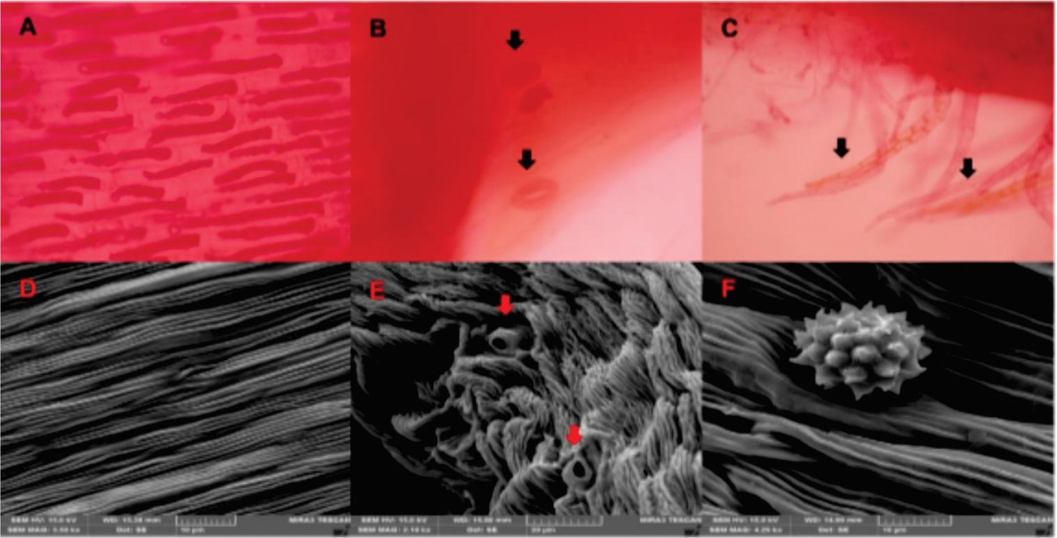
fThe flowers are part of the calendula plant used to extract bioactive compounds. The petals were analyzed using optical and scanning electron microscopy (FEGSEM) to identify anatomical structures (Figure 1). The adaxial surface exhibited elongated, rectangular epidermal cells with straight to slightly sinuous walls and no stomata (Figures 1A and 1D), consistent with descriptions in the Brazilian Pharmacopoeia. Similar epidermal cells were observed on the abaxial surface, accompanied by anomocytic stomata (Figures 1C and 1E). Additionally, multicellular glandular trichomes with oval, biseriate heads were identified on the abaxial surface (Figure 1C), and spherical pollen grains featuring short spines on the anther (Figure 1F).

Images obtained with optical microscopy (4000x) and FEG-SEM from histological sections of calendula petals. Optical microscopy: (A) Epidermal cells on the adaxial surface. (B) Anomocytic stomata on the abaxial surface. (C) Glandular trichomes. FEG-SEM: (D) Epidermal cells on the adaxial surface. (E) Stomata on the abaxial surface. (F) Pollen on the abaxial surface.
The observed anatomical structures are consistent with those described in the Calendula officinalis L. monograph (Brasil, 2019). Microscopic markers are helpful for accurate identification, quality control, efficacy, and safety of herbal drugs. Authenticating the raw drugs is crucial to distinguish them from adulterants, mainly because some plant species with similar morphological flowers can be substitutes or adulterants for C. officinalis (De Souza et al., 2010).
HPLC fingerprint of calendula extract
The chromatographic fingerprint of the sterile and non-sterile calendula glycolic extract was analyzed using a diode-array detector (DAD) at 255 nm and 355 nm. Chromatograms and UV-DAD spectra are shown in Figure 2. The compounds corresponding to the three main peaks highlighted in the Figure exhibited characteristic flavonoid absorptionbands: maximum absorption (λmax) in the range of 352-355 nm (designated as band I) and at 255 nm (band II). More polar compounds primarily showed absorption in the band I, with peak values between 325 and 330 nm, indicating the presence of phenolic acids. Under the same chromatographic conditions, the rutin standard overlapped with compound 2, indicating the presence of this flavonoid in the calendula glycolic extract in both non-sterile and sterile samples.

HPLC chromatograms of sterile and non-sterile glycolic extracts of calendula and rutin standard recorded at 255 nm, and characteristic UV-Vis spectra of compounds highlighted in peaks 1, 2, and 3.
Figure 2 also demonstrates the similar fingerprints of the sterile and non-sterile calendula glycolic extracts, indicating that the main peaks corresponding to phenolic and flavonoid compounds, including rutin, were preserved after sterilization.
Rutin (quercetin-3-O-rutinoside) is known for its wound-healing properties. Numerous investigations have reported that flavonoids exhibit bioactivity through their anti-inflammatory, angiogenic, reepithelialization, and antioxidant effects. These effects support wound healing by modulating biomarker expression across multiple pathways (Zulkefli et al., 2023). The preservation of the flavonoids in the extract after the gamma radiation is promising, suggesting that the sterile alginate-calendula dressing could accelerate the wound-healing process.
Alginate-calendula hydrogel dressing
Alginates have been widely used in wound healing due to their beneficial properties, such as biocompatibility, non-toxicity, and high absorption ability, which can absorb excess wound fluid, provide a moist environment for cell migration, and minimize bacterial infections at the wound site (Zhang, Zhao, 2020). In order to produce hydrogel dressings, Calendula glycolic extract was added to the alginate dispersion. The hydrogel containing the extract had a yellow hue due to the carotenoid presence in the calendula extract. A thin film was formed by the C. officinalis extract-based alginate hydrogel, which fills the pores of the gauze, as shown in Figure 3A. It is an appropriate material for medical applications and makes administration easy. Incorporating hydrogel in textile structures allows the production of a functionalized textile base with the ability to respond to environmental stimuli, improving users' quality of life by producing more comfortable materials (Alves et al., 2022).

Macroscopy and FEG-SEM images of the dressings. (A) Packaged dressing. FEG-SEM: (B) Control gauze; (C) Non-sterile dressing; (D) Sterile dressing. Magnification of 500x and scale 100 µm.
After sterilization, no macroscopic changes were observed in the calendula extract and hydrogel dressing. Gamma ionizing radiation is a rapid, convenient, and extensive sterilization process commonly used for medical devices, including polysaccharides (Kim et al., 2007; Li et al., 2009; Mazor, Zilberman, 2017; Zimoch- Korzycka et al., 2016). Because ionizing energy rapidly penetrates through the polysaccharide, destroying the DNA or RNA of pathogen agents (Mollah et al., 2021).
However, it is reported that high irradiation doses lead to the degradation of polysaccharides, such as sodium alginate, by cleavage of the glycosidic bonds involving a radical scission mechanism (El-Mohdy, 2017; Huq et al., 2012). The degradation rate is related to the polymer concentration and irradiation dose (Mollah et al., 2021; Wasikiewicz et al., 2005). Gamma irradiation induces glycosidic bond cleavage on the alginate chain, which is the primary reaction. The cleavage is accompanied by C=O and COOH groups forming at the reducing end (Chang et al., 2022). Studies have revealed that gamma irradiation has a more pronounced depolymerization effect in aqueous media than in solid forms, like dressings, which are less affected. The free radicals generated during the radiolysis of water promote chain scission of alginate and enhance mobility in the aqueous medium, reducing the recombination possibility of the chains (Chang et al., 2022).
The physical network structure of linear anionic polysaccharide alginate can be reduced by depolymerization, which can impact the wound-healing properties of alginate, such as gelling behavior, liquid absorption ability, and drug release. Thus, the physical properties of alginate hydrogel dressings submitted to gamma irradiation were evaluated.
Field emission gun scanning electron microscopy (FEG-SEM)
The images provided by FEG-SEM can confirm the hydrogel's deposition and distribution on textile gauze. The dressing has a uniform alginate hydrogel filling throughout its entire surface, as shown in the micrographs (Figure 3B-D). The control sample, which is the gauze, has fibers with surface depression, as shown in Figure 3B. Some surface depressions remain on the non-sterile dressing surface, as shown in Figure 3C. The presence of depressions can aid in water absorption, dressing permeability, and active compound diffusion through wound dressing.
Figure 3D shows the surface of the sterile dressing after being exposed to ionizing radiation. Surface depression is also present; also, the micrography shows physical alterations distributed across the dressing surface, which may be a result of alginate depolymerization by gamma ionizing radiation.
X-ray diffraction
XRD patterns of unmodified gauze (control) show the peaks at 2θ = 17.5°, 22.4°, and 25.5° that corresponded to the cellulose crystalline structure (Bains et al., 2019; Said et al., 2021) (Figure 4).

The pattern of XRD was similar for both sterile and non-sterile dressings, which may be because of the lower hydrogel content compared to the textile substrate, which does not indicate any crystallization in the dressing caused by radiation.
Thermal analysis
The DSC and TGA curves are shown in Figure 5. As shown in all the samples, three peaks are present in the DSC analysis for the textile substrate's water evaporation, polyester melting, and cellulose decomposition. The gauze substrate is made up of 70% viscose and 30% polyester. The endothermic peak at 250°C is related to the polyester melting (Alves et al., 2022), and at 350°C is related to the cellulose degradation (Said et al., 2021), as also observed by the exothermic peak at 400°C in the dressing samples. In TGA, the mass loss of 60% could be explained by the breakdown of the cellulose chain into smaller chains, which liberate CO2 and H2O.

TGA thermogram of non-sterile dressing presents a mass loss of 12% related to the loss of trapped water molecules interacting with -OH and -COO- polar groups in the alginate chain by hydrogen bonding, while sterile dressing has a slight weight loss of 2%. Gamma irradiation may have reduced the interaction between water molecules and alginate. The endothermic event in DSC related to water evaporation for dressing samples was lower than 120°C. Sterile dressings showed a significant reduction in this event, peaking at 68°C, as opposed to control and non-sterile samples at 85°C. Some alginate depolymerization could have occurred with the sterilization because it was reported that shorter alginate chains cause lower heat resistance and bond energy (Chang et al., 2022). Moreover, the region of weight loss at 150-250°C is attributed to the glycosidic bond fracture, dehydration, decarboxylation, and decarbonylation of alginate in dressings (Prabhu et al., 2021).
Biodegradation
The biodegradation test revealed an average remaining mass of approximately 70% and 80 % for the non-sterile and sterile dressing, respectively, with no statistical difference after 14 days (p>0.05; Figure 6A). Knowing hydrogel's enzymatic biodegradation for wound dressing in vivo applications is necessary.

Comparison of the characteristics of the sterile and non-sterile dressings against the control gauze. (A) Biodegradation with lysozyme in PBS pH 7.4; (B) Swelling degree in simulated exudate fluid (pH=7,4). Bars represent mean ± S.D. (n=3).
To mimic the in vivo degradation performance, the degradation of dressing was examined at pH 7.4 in phosphate buffer saline (PBS) with lysozyme. This enzyme is highly present in human serum and is used to evaluate the polymer's biodegradation due to hydrolyzing the glycosidic bonds of polysaccharides (Kim et al., 2007). While the enzyme degradation process is complex, it is primarily due to protein adsorption on the material's surface. Alginate contains functional groups such as hydroxyl and carboxyl, which provide several possibilities for enzyme adsorption (Li et al., 2009). Alginate is linked through 1-4 linkage that lysozyme cannot identify, and it is stable in the presence of this enzyme (Zimoch-Korzycka et al., 2016). Mass losses can be caused by either alginate degradation by lysozyme or polymer dissolution in the PBS solution. The enzyme solution has been renewed every two days to prevent saturation, indicating low polymer degradation.
Additionally, the alginate slows degradation rate has been reported and hypothesized to occur by periodate oxidation, which cleaves the carbon-carbon bond of the cis-diol group in the uronate residue and alters the chain conformation, resulting in hydrolysis of alginate in aqueous solutions (Bouhadir et al., 2001). The slow biodegradation and maintenance of the structural features of the dressing can reduce its change time and avoid pain to removal, besides promoting sustained drug delivery (Abourehab et al., 2022).
Swelling degree
Figure 6B displays the results of the swelling tests. The dressings exhibited a swelling degree of approximately 500 and 600% in the initial 30 minutes, maintaining equilibrium during the experiment. The gauze is a control sample made of fibers that take up significant amounts of water, which increases over time. In dressing production, the alginate hydrogel fills these fibers, thereby decreasing the surface area in contact with water. Although the hydrogel does not totally cover some fibers, as seen in FEG-SEM, the swelling ability of the dressings is similar to the control gauze at the first 30 min; however, it remains stable even though it significantly increases in the control gauze (p<0.05). The difference indicates that the control gauze is saturated with water absorption. The swelling rate for the sterile dressing was 500% lower than that of the nonsterile dressing, which was close to 600%. However, the degree of swelling for both sterile and non-sterile dressings is not statistically different (p>0.05). After 120 minutes of testing, the samples showed stability in their water retention capacity.
Thethree-dimensionalnetworkofalginate hydrogel can expand and hold the liquid when immersed in water. Although sterile dressings experienced a slight decrease in their swelling ability, the hydrogel structure maintained by the physical crosslinking of the polymer chains remained after sterilization, boosting its ability to absorb water. The dressing's exposure to radiation did not affect the polymer-water interaction, preserving the hydrogel's swelling behavior. The ability to absorb and retain large amounts of water is the main feature of alginate hydrogel as dressing, controlling the level of wound exudate and preventing tissue maceration by effectively absorbing excess fluid, facilitating various cellular processes like proliferation and differentiation (Ribeiro et al., 2024).
Water vapor permeability (WVP)
According to the WVP test (Table I), there were no statistical differences between sterile and non-sterile dressings, even though sterile dressings had a higher water vapor permeability (85.76±0.51 g/m.day.Pa) than non-sterile ones (71.72±0.40 g/m.day.Pa). It can be inferred from this result that the alginate hydrogel retains its ability to transport water vapor after being exposed to radiation. The ideal moisture rate must be maintained and regulated to help wound dressings heal faster, promote cell growth, and prevent skin vitality loss.
Proper wound treatment dressings should prevent the wound from drying out and facilitate autolytic debridement, thus preventing excessive water loss and drying. In addition, avoid excessive accumulation of exudate and hindering the healing process. The sterile dressing promotes a moist environment in the affected area (~500% swelling and ~85 g/m.day.Pa WVP), preventing the dressing from adhering to the regenerated tissue and facilitating the autolytic debridement and dressing renewal, being adequate for wound treatment (Alves et al., 2022).
pH
The gamma radiation did not influence the pH, i.e., the pH values of the non-sterile and sterile dressings were 6.3 and 6.4, respectively. The slight rise in pH values indicates that the gamma irradiation dose (15 kGy) did not cause significant changes. Many different endogenous and exogenous factors influence the pH in wounds (Schneider et al., 2007). In the wound, an acidic pH supports the natural healing process by suppressing bacterial growth, reducing proteolytic activity, enhancing fibroblast growth, leading to more oxygen supply, contributing to the process of angiogenesis, collagen formation, and an increase in the immune response with the recruitment of macrophages (Percival et al., 2014). However, chronic wounds have a more alkaline pH; thus, topical acidic preparations support natural wound healing by normalizing the skin pH and restoring the skin barrier (Baron et al., 2020).
CONCLUSION
Alginate hydrogel dressings containing Calendula officinalis extract were developed and have the potential to be widely used for wound treatment. Flavonoids and phenolic acids are still present in calendula extract after being sterilized using a dose of 15 kGy using gamma ionizing. Sterilization of dressings containing C. officinalis extract led to microscopic changes; however, gamma radiation did not significantly impact the evaluated dressing properties, including swelling, biodegradation, pH, and water vapor permeability. The desirable properties for promoting efficient wound healing were observed, including maintaining and regulating the ideal moisture rate. The sterile dressing exhibits low biodegradation, which is crucial for carrying on the active compounds of the calendula extract, reducing the time required to change the dressing and not affecting the cell regeneration process. Further studies to identify extract composition are required to confirm the viability of this sterilization method, enabling the production of an innovative wound dressing composed of a polyester-viscose compress based on calendula glycolic extractsodium alginate hydrogel.
-
ACKNOWLEDGMENTS
We acknowledge the Institute of Nuclear and Energy Research (Instituto de Pesquisas Energéticas e Nucleares, IPEN) for the gamma radiation sterilization, the CNPq for the scholarship, the Araucaria Foundation for the financial support, and the c-LABMU/UEPG for the support.
REFERENCES
-
Abourehab MAS, Rajendran RR, Singh A, Pramanik S, Shrivastav P, Ansari MJ, et al. Alginate as a Promising Biopolymer in Drug Delivery and Wound Healing: A Review of the State-of-the-Art. Int J Mol Sci. 2022;23. https://doi.org/10.3390/ijms23169035
» https://doi.org/10.3390/ijms23169035 -
Aderibigbe BA, Buyana B. Alginate in wound dressings. Pharmaceutics. 2018;10. https://doi.org/10.3390/pharmaceutics10020042
» https://doi.org/10.3390/pharmaceutics10020042 -
Alves C, Ribeiro A, Pinto E, Santos J, Soares G. Exploring Z-Tyr-Phe-OH-based hydrogels loaded with curcumin for the development of dressings for wound healing. J Drug Deliv Sci Technol. 2022;73. https://doi.org/10.1016/j.jddst.2022.103484
» https://doi.org/10.1016/j.jddst.2022.103484 -
Andersen FA, Bergfeld WF, Belsito D V., Hill RA, Klaassen CD, Liebler DC, et al. Final Report of the Cosmetic Ingredient Review Expert Panel Amended Safety Assessment of Calendula officinalis-Derived Cosmetic Ingredients. Int J Toxicol. 2010;29:221S-243S. https://doi.org/10.1177/1091581810384883
» https://doi.org/10.1177/1091581810384883 -
Ashfaq A, Clochard M-C, Coqueret X, Dispenza C, Driscoll MS, Ulański P, et al. Polymerization Reactions and Modifications of Polymers by Ionizing Radiation. Polymers (Basel). 2020;12. https://doi.org/10.3390/polym12122877
» https://doi.org/10.3390/polym12122877 -
Azam F, Ahmad F, Ahmad S, Zafar MS, Ulker Z. Synthesis and characterization of natural fibers reinforced alginate hydrogel fibers loaded with diclofenac sodium for wound dressings. Int J Biol Macromol. 2023;241:124623. https://doi.org/https://doi.org/10.1016/j.ijbiomac.2023.124623
» https://doi.org/https://doi.org/10.1016/j.ijbiomac.2023.124623 -
Bains D, Singh G, Kaur N, Singh N. Development of Biological Self-Cleaning Wound-Dressing Gauze for the Treatment of Bacterial Infection. ACS Sustain Chem Eng. 2019;7:969-78. https://doi.org/10.1021/acssuschemeng.8b04608
» https://doi.org/10.1021/acssuschemeng.8b04608 - Baldos DT, Puno JM, Tolentino MM, Montefalcon DR V, Diano GT, Asaad CO. Radiation sterilization of Honey and Honey-Alginate Wound Dressing from Stingless Bee (Tetragonula biroi) Collected from Sta. Maria, Laguna, Philippines. J Trop Life Sci. 2021;11.
-
Baron JM, Glatz M, Proksch E. Optimal Support of Wound Healing: New Insights. Dermatology. 2020;236:593-600. https://doi.org/10.1159/000505291
» https://doi.org/10.1159/000505291 -
Beh CC. Sterilization Techniques of Biomaterials (Implants and Medical Devices) BT - Biomaterials and Biopolymers. In: Domb A, Mizrahi B, Farah S, editors., Cham: Springer International Publishing; 2023, p. 255- 69. https://doi.org/10.1007/978-3-031-36135-7_12
» https://doi.org/10.1007/978-3-031-36135-7_12 - Bouhadir KH, Lee KY, Alsberg E, Damm KL, Anderson KW, Mooney DJ. Degradation of partially oxidized alginate and its potential application for tissue engineering. Biotechnol Prog. 2001;17:945-50.
- Brasil. Agência Nacional de Vigilância Sanitária - ANVISA. Farmacopeia Brasileira. Farm Bras 6a Ediçãoa Edição 2019;2:1-744.
- Brazil. Herbal Medicines Formulary. 2nd editio. Brasília: 2021.
-
Chang KA, Chew LY, Law KP, Ng JF, Wong CS, Wong CL, et al. Effect of gamma irradiation on the physicochemical properties of sodium alginate solution and internally crosslinked film made thereof. Radiat Phys Chem. 2022;193:109963. https://doi.org/https://doi.org/10.1016/j.radphyschem.2022.109963
» https://doi.org/https://doi.org/10.1016/j.radphyschem.2022.109963 -
Donati I, Christensen BE. Alginate-metal cation interactions: Macromolecular approach. Carbohydr Polym. 2023;321:121280. https://doi.org/10.1016/j.carbpol.2023.121280
» https://doi.org/10.1016/j.carbpol.2023.121280 -
El-Mohdy HLA. Radiation-induced degradation of sodium alginate and its plant growth promotion effect. Arab J Chem. 2017;10:S431-8. https://doi.org/https://doi.org/10.1016/j.arabjc.2012.10.003
» https://doi.org/https://doi.org/10.1016/j.arabjc.2012.10.003 -
Ferry M, Ngono Y. Energy transfer in polymers submitted to ionizing radiation: A review. Radiat Phys Chem. 2021;180:109320. https://doi.org/https://doi.org/10.1016/j.radphyschem.2020.109320
» https://doi.org/https://doi.org/10.1016/j.radphyschem.2020.109320 -
Huq T, Khan A, Dussault D, Salmieri S, Khan RA, Lacroix M. Effect of gamma radiation on the physicochemical properties of alginate-based films and beads. Radiat Phys Chem. 2012;81:945-8. https://doi.org/10.1016/j.radphyschem.2011.11.055
» https://doi.org/10.1016/j.radphyschem.2011.11.055 - IAEA. Trends in Sterilization of Health Care. October 2008.
-
Kim IY, Yoo MK, Seo JH, Park SS, Na HS, Lee HC, et al. Evaluation of semi-interpenetrating polymer networks composed of chitosan and poloxamer for wound dressing application. Int J Pharm. 2007;341:35- 43. https://doi.org/10.1016/j.ijpharm.2007.03.042
» https://doi.org/10.1016/j.ijpharm.2007.03.042 -
Kumar AAA, Sonwani S, Bakkiam D, Ramanathan N, Rao T. Gamma radiation induced changes in antioxidant and radioprotective activities of flavonoids differing mainly with respect to -OH substitution pattern in B and C rings. J Radioanal Nucl Chem. 2020;326:801-11. https://doi.org/10.1007/s10967-020-07366-4
» https://doi.org/10.1007/s10967-020-07366-4 -
Lee DW, Choi WS, Byun MW, Park HJ, Yu Y-M, Lee CM. Effect of γ-Irradiation on Degradation of Alginate. J Agric Food Chem. 2003;51:4819-23. https://doi.org/10.1021/jf021053y
» https://doi.org/10.1021/jf021053y - Li X, Xie H, Lin J, Xie W, Ma X. Characterization and biodegradation of chitosan-alginate polyelectrolyte complexes. Polym Degrad Stab. 2009;94:1-6.
-
Mao G, Tian S, Shi Y, Yang J, Li H, Tang H, et al. Preparation and evaluation of a novel alginatearginine-zinc ion hydrogel film for skin wound healing. Carbohydr Polym. 2023;311:120757. https://doi.org/ https://doi.org/10.1016/j.carbpol.2023.120757
» https://doi.org/» https://doi.org/10.1016/j.carbpol.2023.120757 - Mazor E, Zilberman M. Effect of gamma-irradiation sterilization on the physical and mechanical properties of a hybrid wound dressing. Polym Adv Technol. 2017;28:41-52.
- Mollah MZI, Rahaman MS, Faruque MRI, Khandaker MU, Osman H, Alamri S, et al. Effects of Radiation Sterilization Dose on the Molecular Weight and Gelling Properties of Commercial Alginate Samples. Front Mater. 2021;8:761995.
- Naikwadi AT, Sharma BK, Bhatt KD, Mahanwar PA. Gamma Radiation Processed Polymeric Materials for High Performance Applications: A Review. Front Chem. 2022;10.
-
Percival SL, McCarty S, Hunt JA, Woods EJ. The effects of pH on wound healing, biofilms, and antimicrobial efficacy. Wound Repair Regen Off Publ Wound Heal Soc [and] Eur Tissue Repair Soc. 2014;22:174-86. https://doi.org/10.1111/wrr.12125
» https://doi.org/10.1111/wrr.12125 -
Possa GDOK, Chopek S, Pereira AV, Koga AY, Oliveira MRP, Costa MDM. Calendula glycolic extract enhances wound healing of alginate hydrogel. Acta Cir Bras. 2024;39:99724. https://doi.org/https://doi.org/10.1590/acb399724
» https://doi.org/https://doi.org/10.1590/acb399724 -
Prabhu S, S. G. B, Gudennavar SB. Sodium alginate/ bismuth (III) oxide composites for γ-ray shielding applications. J Appl Polym Sci. 2021;138:50369. https://doi.org/https://doi.org/10.1002/app.50369
» https://doi.org/https://doi.org/10.1002/app.50369 -
Ribeiro M, Simões M, Vitorino C, Mascarenhas-Melo F. Hydrogels in Cutaneous Wound Healing: Insights into Characterization, Properties, Formulation and Therapeutic Potential. Gels (Basel, Switzerland). 2024;10. https://doi.org/10.3390/gels10030188
» https://doi.org/10.3390/gels10030188 -
Rutala WA, Donskey CJ, Weber DJ. Disinfection and sterilization: New technologies. Am J Infect Control. 2023;51:A13-21. https://doi.org/10.1016/j.ajic.2023.01.004
» https://doi.org/10.1016/j.ajic.2023.01.004 -
S. A. Bento C, Gaspar MC, Coimbra P, de Sousa HC, E. M. Braga M. A review of conventional and emerging technologies for hydrogels sterilization. Int J Pharm. 2023;634:122671. https://doi.org/10.1016/j.ijpharm.2023.122671
» https://doi.org/10.1016/j.ijpharm.2023.122671 -
Said MM, Rehan M, El-Sheikh SM, Zahran MK, Abdel-Aziz MS, Bechelany M, et al. Multifunctional Hydroxyapatite/Silver Nanoparticles/Cotton Gauze for Antimicrobial and Biomedical Applications. Nanomater (Basel, Switzerland). 2021;11. https://doi.org/10.3390/nano11020429
» https://doi.org/10.3390/nano11020429 -
Schneider LA, Korber A, Grabbe S, Dissemond J. Influence of pH on wound-healing: a new perspective for wound-therapy? Arch Dermatol Res. 2007;298:413- 20. https://doi.org/10.1007/s00403-006-0713-x
» https://doi.org/10.1007/s00403-006-0713-x -
Shahane K, Kshirsagar M, Tambe S, Jain D, Rout S, Ferreira MKM, et al. An Updated Review on the Multifaceted Therapeutic Potential of Calendula officinalis L. Pharmaceuticals. 2023;16:611. https://doi.org/10.3390/ph16040611
» https://doi.org/10.3390/ph16040611 -
da Silva GLP, Morais LC de A, Olivato JB, Marini J, Ferrari PC. Antimicrobial dressing of silver sulfadiazine-loaded halloysite/cassava starch-based (bio)nanocomposites. J Biomater Appl. 2021;35:1096- 108. https://doi.org/10.1177/0885328221995920
» https://doi.org/10.1177/0885328221995920 -
Singh R, Singh D, Singh A. Radiation sterilization of tissue allografts: A review. World J Radiol. 2016;8:355. https://doi.org/10.4329/wjr.v8.i4.355
» https://doi.org/10.4329/wjr.v8.i4.355 - De Souza APTB, Barni ST, Ferreira RA, Couto AG. Desenvolvimento tecnológico de soluções extrativas hidroetanólicas das flores de Calendula officinalis L. empregando planejamento fatorial. Lat Am J Pharm. 2010;29:13-21.
-
Tao M, Ao T, Mao X, Yan X, Javed R, Hou W, et al. Sterilization and disinfection methods for decellularized matrix materials: Review, consideration and proposal. Bioact Mater. 2021;6:2927-45. https://doi.org/10.1016/j.bioactmat.2021.02.010
» https://doi.org/10.1016/j.bioactmat.2021.02.010 -
Türker NS, Özer AY, Kutlu B, Nohutcu R, Sungur A, Bilgili H, et al. The effect of gamma radiation sterilization on dental biomaterials. Tissue Eng Regen Med. 2014;11:341-9. https://doi.org/10.1007/s13770-014-0016-9
» https://doi.org/10.1007/s13770-014-0016-9 -
Wasikiewicz JM, Yoshii F, Nagasawa N, Wach RA, Mitomo H. Degradation of chitosan and sodium alginate by gamma radiation, sonochemical and ultraviolet methods. Radiat Phys Chem. 2005;73:287-95. https://doi.org/https://doi.org/10.1016/j.radphyschem.2004.09.021
» https://doi.org/https://doi.org/10.1016/j.radphyschem.2004.09.021 -
Zhang M, Zhao X. Alginate hydrogel dressings for advanced wound management. Int J Biol Macromol. 2020;162:1414-28. https://doi.org/10.1016/j.ijbiomac.2020.07.311
» https://doi.org/10.1016/j.ijbiomac.2020.07.311 - Zimoch-Korzycka A, Kulig D, Jarmoluk A, Marycz K, Matuszczak W. Study of enzymatically treated alginate/ chitosan hydrosols in sponges formation process. Polymers (Basel). 2016;8:8.
-
Zulkefli N, Che Zahari CNM, Sayuti NH, Kamarudin AA, Saad N, Hamezah HS, et al. Flavonoids as Potential Wound-Healing Molecules: Emphasis on Pathways Perspective. Int J Mol Sci. 2023;24. https://doi.org/10.3390/ijms24054607
» https://doi.org/10.3390/ijms24054607
Edited by
-
Associated Editor:
Carlota Rangel-Yagui