 Can idebenone protect gastric tissue against oxidative stress, inflammation, and necrosis?
Abstract
This study aims to investigate the antiulcer effects of Idebenone (IDE) in Wistar rats with indomethacin-induced gastric ulcer and compare its effectiveness with famotidine (FAM). 36 rats were randomly divided into six groups: healthy; healthy+IDE200; ulcer (U); U+FAM; U+IDE100; U+IDE200. Six hours after the administration of 25 mg/kg of indomethacin, the antiulcer effects of FAM (40 mg/kg) and two different doses of IDE (100 and 200 mg/kg) in the gastric tissue of rats were examined through macroscopic, biochemical, molecular, and histopathological analyses. Comparative assessments were conducted between the healthy control group and the group treated with indomethacin alone. IDE effectively suppressed the indomethacin-induced increase in malondialdehyde (MDA) and mitigated the reduction in glutathione (GSH) levels. Furthermore, IDE treatment led to a significant decrease in mRNA expressions of interleukin 1 beta (IL-1β), tumor necrosis factor-alpha (TNF-α), and nuclear factor kappa beta (NF-κB)-p65 in ulcerative rats. Histopathological examinations revealed that both IDE doses improved the ulcer damage caused by indomethacin in the rats' stomach tissues. This study demonstrates that IDE administration exhibits a protective effect against indomethacin-induced gastric ulcers by reducing oxidative stress and inflammatory processes. IDE’s potential to prevent ulcer formation in the gastric tissue of rats suggests that it could be a valuable therapeutic agent in clinical practice.
Keywords:
Anti-inflammatory; Antioxidant; Gastric ulcer; Idebenone; Indomethacin
INTRODUCTION
Gastric ulcer, a prevalent gastrointestinal ailment, manifests through abnormalities in the mucosal and submucosal layers (Narayanan, Reddy, Marsicano, 2018). The pathogenic mechanisms driving gastric ulcers involve a complex interplay of factors, including heightened generation of reactive oxygen species (ROS) (Eraslan et al., 2020), diminished cell proliferation, and intensified inflammation (Alatawi et al., 2023; Mahmoud et al., 2021). The production of ROS activates the nuclear factor kappa beta (NF-κB) signaling pathway, a pivotal component of cellular inflammatory cascades. NF-κB activation leads to elevated levels of various pro-inflammatory cytokines, such as interleukin 1 beta (IL-1β) and tumor necrosis factor-alpha (TNF-α), which significantly contributes to the pathogenesis of gastric ulcers (El-Gendy et al., 2023). Moreover, the overexpression of proinflammatory cytokines gives rise to the generation of ROS, lipid mediators, and secondary cytokines (Kumar et al., 2018). Consequently, it becomes imperative to regulate oxidative stress-associated parameters, such as glutathione (GSH) and malondialdehyde (MDA), and promote an anti-inflammatory response to effectively mitigate the development of gastric ulceration. Current therapeutic approaches for the treatment of gastric ulcers underscore the importance of comprehensive strategies that target both oxidative stress and inflammation. However, the prolonged use of commonly prescribed synthetic anti-ulcer medications often leads to significant adverse effects. In particular, the long-term administration of non-steroidal anti- inflammatory drugs, such as indomethacin, can result in serious gastric ulcers, highlighting the critical need for the development of safer and more effective gastroprotective agents. An ideal treatment option should provide comprehensive protection with minimal side effects, thereby reducing the necessity for polypharmacy. In this context, new therapeutic options with antioxidant and anti-inflammatory properties could fill this treatment gap, improving patient adherence and overall treatment efficacy.
The synthetic short-chain analogue of CoQ10, idebenone (hydroxydecylubiquinone, IDE), was introduced by Japanese Takeda Chemical Industries in 1986 to address cognitive decline/dementia (Gueven et al., 2021). IDE plays a pivotal role as a crucial antioxidant for the cell membrane. Integrated into the mitochondrial electron transport chain, IDE facilitates the production of adenosine triphosphate (Avci et al., 2021). Notably, previous research indicated IDE's efficacy in guarding against oxidative stress and reducing the levels of various proinflammatory cytokines in diverse tissue damage scenarios, including the colon (Shastri et al., 2020) and liver (Fadda et al., 2018). However, the extent of IDE's protective effect on gastric ulcers remains undiscovered and warrants further investigation. This knowledge gap emphasizes the need for a comprehensive investigation of the interrelationship of IDE with gastric ulcers.
Based on this information, the present study aims to investigate the gastroprotective effects of IDE in a rat model of indomethacin-induced gastric ulcers. This investigation will employ a comprehensive approach, incorporating biochemical analyses, molecular evaluations, and histopathological examinations.
MATERIAL AND METHODS
Ethical approval
This study, along with all animal protocols, received approval from the Animal Experiments Local Ethics Committee (HADYEK) of Atatürk University on 05.05.2022, documented under number E-42190979- 050.01.04-2200137204.
Animals
For this study, 36 female Wistar rats, aged 3-4 months and weighing 220-260 grams, were procured from the Atatürk University Medical Experimental Application and Research Center. Prior to the experiment, the rats were subjected to a one-week adaptation period. Standard plastic cages were utilized to house all animals under consistent conditions (temperature: 22 ± 1 °C, relative humidity: 40-80%, 12 h light-dark cycle). Throughout the experiment, the rats had unrestricted access to the standard rat water and food (ad libitum). All experimental procedures were conducted in accordance with national guidelines for the ethical use and care of laboratory animals.
Chemicals
The IDE used in this study, specifically Idebex 90 mg in the form of 30 film-coated tablets, was procured from Neutec, Turkey. Indomethacin, obtained as 25 mg capsules (Endol), was purchased from Deva, Turkey, while Famotidine (FAM) in the form of Famodin 40 mg tablets (30 tablets) was acquired from Sandoz, Turkey. Thiopental sodium was sourced from Ibrahim Ethem Ulagay AS (Istanbul, Turkey), and all other chemical substances required for laboratory tests were obtained from Sigma and Merck, Germany.
Experimental design
The 36 rats were randomly divided into 6 groups (six rats in each group). Experimental groups are shown in Table I.
The dosage of IDE (100 and 200 mg/kg) was established based on prior research (Gou, Jin, Xia, 2022; Akpinar et al., 2022; Fadda et al., 2018). 200 mg/ kg IDE was used to determine the effect of IDE when administered alone on the stomach tissue of healthy rats.
Indomethacin-induced gastric ulcer model
An indomethacin-induced gastric ulcer model was established following the methodology outlined in the previous studies (Ugan et al., 2020). Experimental design is shown in Table II. With the exception of the healthy group, all rats underwent a 24-hour fasting period, during which they had unrestricted access to water. Following this fasting period, IDE and FAM (Kucukler, Kandemir, Yildirim, 2022) were orally administered to their respective experimental groups. 5 minutes later, indomethacin (Calik et al., 2020) was administered through the gastric gavage to groups 3 - 6. Six hours after indomethacin administration, all rats received an intraperitoneal injection of 50 mg/kg thiopental. The abdomen was excised, and the gastric tissue was removed for macroscopic examination of ulcer areas on the gastric surface. Following this assessment, half of the gastric tissues were preserved at -80 °C for biochemical and molecular analyses, while the other half was stored in 10% formalin for histopathological investigations.
Biochemical investigations
Gastric tissue samples from all rats were processed as follows: they were cleaned, fixed with liquid nitrogen, and preserved at -80°C. Subsequently, 1 ml of PBS was administered to 100 mg of all samples, and the samples were ground in liquid nitrogen using a Tissue Lyser II (Qiagen). After the grinding process, each sample was centrifuged, and the supernatants obtained from centrifugation were utilized as samples. GSH (Sedlak, Lindsay, 1968) and MDA levels (Ohkawa, Ohishi, Yagi, 1979) in each sample supernatant and standard were determined using an enzyme-linked immunosorbent assay (ELISA) reader. GSH and MDA levels were expressed as nmol/mg protein. The mean and standard deviation for each dataset were presented per mg of protein.
Protein determination
The protein concentrations were determined using the commercial protein standards (Sigma Aldrich, Total protein kit-TP0300-1KT-(USA)), and the Lowry technique was employed for this purpose.
Molecular investigations
Gene expression analyses
A quantitative reverse transcription polymerase chain reaction (qRT-PCR) was designed to assess the mRNA expression levels of IL-1β, TNF-±, and NF-κB-p65. This process involved homogenizing gastric tissues, isolating RNA, synthesizing complementary DNA (cDNA), and quantitatively evaluating the expression levels of the target mRNAs (Kose et al., 2021).
RNA extraction from gastric tissue
Gastric tissue samples, each weighing 20 mg, were individually weighed and stabilized in RNAlater RNA Stabilization Reagent (Qiagen). After stabilization, the tissues were homogenized using the Tissue Lyser II (Qiagen). Total RNA was then purified using the RNeasy Mini Kit (Qiagen), following the manufacturer's instructions, and processed in QIAcube (Qiagen, Hilden, Germany). The total mRNA content was determined using NanoDrop spectrophotometry (All Sheng) at 260 nm.
Reversed transcriptase reaction and cDNA synthesis:
The production of cDNA from total RNA was performed using a High-Capacity cDNA Reverse Transcription Kit (Applied Biosystems, Foster City, CA, USA). Each reaction comprised 10μl of RNA, and cDNA synthesis was carried out using a T100 Thermal Cycler (BIO-RAD) following specified temperature conditions. The quantity of cDNA was assessed with Nanodrop spectrophotometry (All Sheng), and the resulting cDNA was stored at -20 °C. The following components were used for the cDNA synthesis reaction: total RNA (10 μl), 25 X dNTP mix (0.8 μl), 10X RT random primers (2 μl), reverse transcription 10X buffer (2 μl), diethylpyrocarbonate H2O (4.2 μl), and MultiScribe reverse transcriptase (1 μl). Take3 Plate and Epoch Spectrophotometer System (Biotek) were utilized to evaluate and measure the cDNA concentrations.
Quantitative determination of IL-1β, TNF-±, and NF-κB-p65 mRNA gene expressions by qRT - PCR:
The relative expression analyses of IL-1β, TNF-±, and NF-κB-p65 from rat RNA-derived cDNA were conducted using the StepOnePlus Real-Time PCR System technology (Applied Biosystems, USA). For the real-time polymerase chain reaction, β-actin (housekeeping gene) (Rn00667869_m1) expression results were employed as the reference gene. The details of TaqMan® (Thermo Fisher Scientific) gene primers and probes used in the qRT-PCR study are shown in Table III. The amplification and quantification processes were conducted on a Corbett Rotor-Gene device (Thermo Fisher Scientific). TaqMan Gene Expression Assays were carried out with 100 ng cDNA, 10 μl TaqMan Master Mix (2X), and 1 μl Assay (20X), completed to 20 μl with RNase-free H2O, for 40 cycles. The cycle threshold (Ct) represents the cycle at which the fluorescent signal observed in qRT-PCR experiments surpasses the designated threshold value. The results were subjected to statistical analysis after the Ct values were automatically transformed into delta delta Ct (2-ΔΔCt).
Histopathological procedures
The stomach samples were evaluated both macroscopically and microscopically for ulcer foci on the gastric surface. Samples taken from each experimental animal were stretched on a wax plate and ulcer areas on the stomach surface were counted. The histopathological evaluation followed the established procedures outlined in previous studies (Toktay et al., 2022). The stomach tissue blocks from the corpus region for all experimental groups were fixed in a 3.7% formaldehyde (10% formalin) solution for 72 hours. After fixation, each tissue sample underwent routine processing. Dehydration was achieved through a series of increasing alcohol concentrations (50%, 70%, 80%, 96%, and 99%), with each preservation step lasting 1 hour. Subsequently, the tissues were cleaned in a xylene solution for three to fifteen minutes and then preserved in molten liquid paraffin for infiltration. After processing, the tissues were meticulously blocked in paraffin and each paraffin block was sectioned to a thickness of 5 micrometers for histopathological analysis. The paraffin sections were cut on adhesive- covered slides and stained with Harris Hematoxylin and Eosin Y. Evaluation of stained slides was conducted at ×100 magnification, with a minimum of five areas assessed for each tissue slide.
The Harris Hematoxylin and Eosin Y staining findings were semi-quantitatively scored based on the degree of density, regularity of the gastric layer, depth of thickening, the presence of hemorrhage, and necrotic cells. To elucidate the significant differences in terms of histopathological changes among the experimental groups, a scoring system was employed. This system consisted of categories such as '- (no), -/+ (none or almost no +, minor damage), ++ (moderate damage), and +++ (severe damage).' The intention behind this scoring approach was to was to explicitly demonstrate meaningful distinctions in observed damage among the experimental groups (Karaoglan et al., 2020).
Statistical analysis
The statistical analysis of the molecular, biochemical and histopathological results was conducted using GraphPad Prism, version 5.0. The data are presented as means ± standard deviation (SD). The normal distribution of continuous variables was assessed using the Shapiro-Wilk test and the Kolmogorov-Smirnov test. Since the normality assumption was met for comparisons of three or more independent groups, ANOVA was preferred. Following the ANOVA test, post-hoc tests were conducted using the Tukey test when variances were homogeneous. The biochemical, molecular and histopathological datas showed a normal distribution and homogeneous variance. Therefore, for all datas a one-way analysis of variance and Tukey's test were applied for comparisons between groups. The statistical significance level was taken as p<0.05.
To facilitate interpretation, means sharing the same letter within the same column are not significantly different, while means with different letters in the same column indicate statistically significant differences between the groups. This approach ensures clarity in distinguishing between the experimental groups and enhances the robustness of the statistical comparisons.
RESULTS
Impacts of IDE on oxidant and antioxidant parameters in gastric tissue
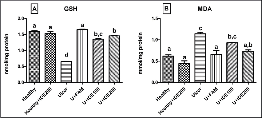
The administration of 200 mg/kg IDE alone did not elicit a statistically significant effect on the GSH and MDA levels compared to the healthy group (p>0.05). Notably, the GSH levels, indicative of the antioxidant system, were significantly lower in the ulcer group than in the healthy group (p<0.05). IDE administration (U+IDE100 and U+IDE200) dose-dependently and significantly attenuated the decrease in GSH levels induced by indomethacin (p<0.05). Conversely, the MDA levels, indicative of the oxidant status, increased in the ulcer groups compared to the healthy group (p<0.05). When compared with the ulcer group, IDE administration (U+IDE100 and U+IDE200) significantly and dose-dependently reduced indomethacin-induced increase in MDA levels (p<0.05). Moreover, the MDA levels in the group administered 200 mg/kg IDE were the most similar to those of the healthy and FAM groups (Figure 1).

The effects of IDE on GSH (A) (nmol/mg protein) and MDA (B) levels (nmol/mg protein) in indomethacin-induced gastric ulcer in rats (FAM: Famotidine; GSH: Glutathione; IDE100: 100 mg/kg Idebenone; IDE200: 200 mg/kg Idebenone, MDA: Malondialdehyde; U: Ulcer) The levels of MDA and GSH were measured according to the modified methods with an ELISA reader. a, b, c, and d: There is no statistically significant difference between the groups in the columns symbolized with the same letter. There is a statistically significant difference between groups for columns symbolized by different letters.
Impacts of IDE on anti-inflammatory parameters in gastric tissue
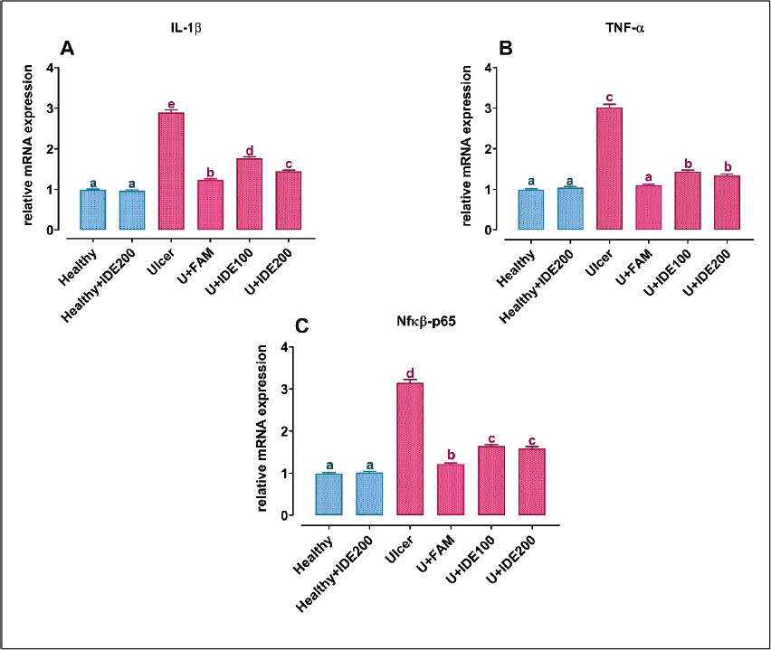
Administering only 200 mg/kg IDE to rats did not result in a statistically significant effect on IL-1β, TNF-±, and NF-κB-p65 mRNA expression levels compared to the healthy group (p>0.05). Notably, IL- 1β mRNA expression levels significantly increased in the ulcer groups compared to the healthy group (p<0.05). However, this induced elevation in IL-1β mRNA expression levels was significantly and dose-dependently reduced in the groups treated with IDE (U+IDE100 and U+IDE200) compared to the ulcer group (p<0.05). Similarly, TNF-± and NF-κB -p65 mRNA expression levels significantly increased in the ulcer groups compared to the healthy group (p<0.05). IDE administration (U+IDE100 and U+IDE200) significantly and dose-dependently reduced the indomethacin-induced increases in TNF-± and NF-κB-p65 mRNA expression levels compared to the ulcer group (p<0.05) (Figure 2).

The effects of IDE on IL-1β (A), TNF-± (B), and NFκB-p65 (C) mRNA expression levels in indomethacin-induced gastric ulcer in rats (FAM: Famotidine; IDE100: 100 mg/kg Idebenone; IDE200: 200 mg/kg Idebenone, IL-1β: interleukin 1 beta; NFκB-p65: nuclear factor kappa B-p65; TNF-±: tumor necrosis factor-alpha; U: Ulcer) The expression of mRNAs was detected using quantitative Real-Time PCR analysis. β-actin was used as the reference gene. a, b, c, d and e: There is no statistically significant difference between the groups in the columns symbolized with the same letter. There is a statistically significant difference between groups for columns symbolized by different letters.
Impacts of IDE on histopathological changes in gastric tissue
The histopathological effects of IDE were evaluated by counting ulcer foci on the stomach surface and analyzing light microscopy findings from Harris Hematoxylin and Eosin Y staining, as shown in Figures 3A-B and Figure 4. Additionally, the total histopathological damage findings, scored semi- quantitatively based on Harris Hematoxylin and Eosin Y results, are presented in Table IV.

Macroscopic and microscopic results of histopathological effects of IDE on indomethacin-induced gastric ulcer in rats (FAM: Famotidine; IDE100: 100 mg/kg Idebenone; IDE200: 200 mg/kg Idebenone; U: Ulcer. Arrowhead (>): Superficial epithelial losses, Circle (O): Macroscopik ulcer area; Star (*): Necrotic ulcer area;)

Counting results of ulcer foci in indomethacin-induced gastric ulcer in rats (FAM: Famotidine; IDE100: 100 mg/kg Idebenone; IDE200: 200 mg/kg Idebenone; U: Ulcer. a, b, c, and d: There is no statistically significant difference between the groups in the columns symbolized with the same letter. There is a statistically significant difference between groups for columns symbolized by different letters.
A clear correlation emerged between the macroscopic and microscopic findings. No gastric damage was observed in the healthy and healthy+IDE200 groups. Conversely, the gastric tissues of the ulcer group exhibited visible ulcer foci macroscopically, with numerous areas of the mucosa displaying bleeding in the form of linear or focal black spots of varying sizes. The regular structure of gastric pits was lost, and deterioration was evident in the ulcer group, with necrotic cells observed on the mucosal surface and in the hemorrhagic areas extending deep into the mucosa. Inflammatory cells were also present in these areas. No ulcer foci were noted in the U+FAM group, where the histopathological appearance resembled that of the healthy group, with regular epithelial and connective tissue structures observed. The IDE-administered groups exhibited a dose-dependent increase in the anti- ulcerative effect. In both the U+IDE100 and U+IDE200 groups, the stomach tissue returned to its normal structure. Visible black ulcer foci of varying sizes significantly decreased in many areas of the gastric mucosa in these groups. Inflammatory cells, necrotic areas, and edema were reduced compared to the ulcer groups. Notably, the histopathological appearance of the U+IDE200 group surpassed that of both the ulcer and U+IDE100 groups. Moreover, the histopathological appearance of the 200 mg/kg IDE-administered group most closely resembled that of the healthy and FAM groups.
DISCUSSION
Indomethacin, recognized as one of the most potent nonsteroidal anti-inflammatory drugs, is known for causing more severe damage to the gastric mucosa in rats compared to other drugs in the same class. Therefore, the indomethacin-induced gastric ulcer model has been extensively utilized to explore gastroprotective effects in experimental animal studies such as rats (Isnain et al., 2022) and mice (Da Silva et al., 2018). Research on indomethacin-induced gastric ulcers has explored various mechanisms. Some proposed mechanisms explaining the induction of indomethacin- induced gastric ulcers include the suppression of cyclooxygenase, disturbance of the gastric mucosal antioxidant system, dysregulation of prostaglandins, and the provocation of inflammation in the gastric mucosa (Balaha et al., 2022). Consequently, targeting antioxidant and anti-inflammatory systems has become a prominent strategy in the development of new anti- ulcer agents. In light of this information, the presented study evaluated the anti-ulcer effects of IDE in a gastric ulcer model induced by indomethacin in Wistar rats. IDE, known for its potent antioxidant and anti- inflammatory properties, was assessed biochemically, molecularly, and histopathologically.
The pathogenesis of gastric ulcers is intricately linked to the roles of ROS and oxidative stress (Barboza et al., 2018). Oxidative stress, characterized by a significant imbalance between free radical formation and the protective actions of antioxidants, is a pivotal factor contributing to tissue injury (Chandra, Sachan, 2022). GSH, a crucial antioxidant, plays a key role in counteracting the damaging effects of oxidative stress. Given the strong connection between oxidative stress and gastric ulcers, the assessment of key parameters such as GSH and MDA levels is instrumental in understanding the extent of cell and tissue damage associated with this condition (Mahmoud et al., 2023). Evaluating these markers provides valuable insights into the oxidative stress-induced alterations in cellular integrity and the efficacy of endogenous defense mechanisms. Thus, investigating the GSH and MDA levels emerges as a key method in unraveling the complexities of cell and tissue damage in the context of gastric ulcers. In our study, we specifically explored oxidative stress factors associated with gastric ulcers, focusing on GSH and MDA. Consistent with previous research (AlKreathy, Alghamdi, Esmat, 2020), our findings revealed an increase in MDA levels and a decrease in GSH levels following gastric ulcer injury. These observations underscore the pivotal role of oxidative stress in the pathophysiology of gastric ulcers, highlighting the potential of GSH and MDA as crucial biomarkers in assessing the extent of cellular damage in this context (Bayir et al., 2023). In our research, we meticulously assessed the levels of the GSH and MDA in the gastric tissue. Our results revealed a significant increase in the MDA levels and a concurrent decrease in the GSH levels in the ulcer group compared to the healthy group. This finding suggests an exacerbation of oxidative stress reactions in the gastric tissues following the occurrence of gastric ulcers. Conversely, in the groups administered with IDE, we observed a notable dose-dependent increase in the GSH levels and a decline in the MDA levels, dose-dependently. The observed changes in GSH and MDA levels indicate a protective effect of IDE, emphasizing its strong antioxidant properties. These findings align with prior research that establishes IDE's protective impact by regulating oxidative stress. The modulation of oxidative stress markers in gastric tissues further supports the potential of IDE as a therapeutic agent with gastroprotective properties, offering insights into its mechanism of action against oxidative damage associated with gastric ulcers (Clementi et al., 2022). Akpinar et al. (2022) reported that IDE significantly alleviated oxidative stress markers in a rat model of sepsis-induced lung damage by increasing the GSH levels and decreasing the MDA levels. Similarly, Similarly, IDE was shown to significantly modulate GSH and MDA levels in the liver tissue of rats experiencing sepsis (Gou, Jin, Xia, 2022). Azim et al. (2015) demonstrated in their study on micelles that administering 200 mg/kg of IDE exerted hepatoprotective effects by modulating changes in GSH and MDA levels associated with liver damage. These effects appear to be mediated by IDE's antioxidant, anti- inflammatory, and anti-apoptotic properties. Choi et al. (2024) further explored IDE's potential in LPS-induced systemic inflammatory diseases, showing that IDE administration protected against tissue damage, reduced the expression of inflammatory enzymes and cytokines, suppressed inflammatory responses in macrophages, inhibited the NF-κB signaling pathway, reduced reactive oxygen species and lipid peroxidation, and normalized antioxidant enzyme activities. Additionally, Al-Rasheed et al. (2013) demonstrated in a rat study that 200 mg/kg of IDE regulated TNF-± levels and restored GSH levels associated with oxidative renal damage, suggesting that IDE's protective effects are likely related to its antioxidant and anti-inflammatory actions. These findings collectively support the potential of IDE as a protective agent against oxidative stress. Consistent with these findings from the literature, our study suggests that IDE administration effectively reduces the generation of oxidant parameters while concurrently increasing the production of antioxidant parameters. This protective action contributes to shielding the gastric mucosa from ulcer damage by controlling oxidative stress and inflammatory markers. Our results align with the notion that IDE, known for its antioxidant properties, exerts a protective effect against indomethacin-induced gastric ulcers by regulating oxidative stress. By corroborating the established literature, these findings strengthen the evidence supporting the potential therapeutic role of IDE in mitigating oxidative damage and inflammation, offering valuable insights into its mechanisms of gastroprotection.
Indomethacin-induced ulcers, associated with the inhibition of protective factors and an inflammatory response marked by increased neutrophil infiltration (Ugan, Un, 2020), further underscore the significance of suppressing pro-inflammatory cytokines, including IL-1β, TNF-±, and NF-κB, in gastric ulcer treatment (Akanda, Park, 2017). In this study, we assessed the mRNA expression of IL-1β, TNF-±, and NF-κB p65 in the gastric tissues of rats to elucidate the potential anti-ulcer effect of IDE in indomethacin-induced gastric ulcers. Our observations revealed the increased expression levels of IL-1β, TNF-±, and NF-κB in the ulcer group, consistent with previous studies associating gastric ulcers with elevated inflammatory cytokines (Ma et al., 2022; Yi et al., 2022). Notably, IDE administration reduced these inflammatory markers, indicating its potent anti-inflammatory characteristics. These findings align with existing literature, showing IDE's efficacy in modulating inflammatory markers in various animal models, such as testicular torsion/ detorsion (Abdelzaher et al., 2022), diabetic wounds (Jintao, 2022), renal damage (Blanco et al., 2020), and liver injury (Jiang et al., 2021). Valduga et al. (2023) further demonstrated, in an experimental model of Duchenne muscular dystrophy using mdx mice, that IDE treatment protected dystrophic muscle cells by reducing inflammatory molecules like NF-κB and TNF. This collective evidences supports the argument that IDE exerts a protective effect against gastric ulcers by suppressing the inflammatory response, highlighting its potential therapeutic value in mitigating inflammation- associated tissue injury.
ROS, functioning as a redox signaling messenger at normal intracellular concentrations, plays a pivotal role in various essential cellular processes, including cell differentiation, proliferation, and growth (Pizzino et al., 2017). Indomethacin exacerbates this damage by causing vascular damage and gastric cell necrosis (Neamatallah, 2023). To examine the microscopic damages and complement our macroscopic examination, histopathological analyses were conducted. Macroscopic analysis of the gastric mucosa revealed that indomethacin induced the appearance of lesions, with areas presenting hemorrhagic ulcers and an increased ulceration index, consistent with previous reports (Shahzad et al., 2023). In our investigation, the histological total damage score was notably high in the ulcer group, whereas IDE administration significantly reduced this score. These histopathological findings were consistent with our macroscopic observations. The ulcer group exhibited necrotic cells, inflammatory cell infiltration, and epithelial cell loss. Conversely, IDE treatment ameliorated these histopathological abnormalities, with the greatest protection achieved using 200 mg/kg IDE. These results demonstrate that IDE is protective against histopathological damage to gastric tissue induced by indomethacin-induced gastric ulcers. Importantly, our histopathological findings align with our biochemical and molecular findings, providing a comprehensive picture of IDE's therapeutic efficacy in mitigating both macroscopic and microscopic aspects of gastric ulceration. This holistic approach strengthens the credibility of IDE as a potential therapeutic agent for gastric ulcers by addressing multiple facets of the pathological process.
The clinical relevance of our study lies in its potential implications for the treatment and management of gastrointestinal disorders, particularly those associated with oxidative stress and inflammation. IDE, as investigated in our study, demonstrates promising effects in reducing oxidative stress, necrosis, and inflammation in the gastric tissues of experimental animals. These findings suggest that IDE could potentially serve as a therapeutic agent for conditions characterized by gastric mucosal damage, such as indomethacin-induced ulcers. Furthermore, IDE's mechanism of action, which includes its antioxidant and anti-inflammatory properties, aligns with current therapeutic strategies aimed at protecting the gastric mucosa and preventing ulcer formation. By elucidating these mechanisms, our study contributes to the growing body of literature exploring novel treatments for gastrointestinal diseases. Moreover, the use of animal models in our study provides a platform for further translational research to evaluate IDE's efficacy and safety in clinical trials. Given its antioxidant properties and potential to mitigate gastric damage, IDE may offer a new avenue for therapeutic intervention in patients with gastrointestinal disorders.
With its antioxidant and anti-inflammatory properties, IDE has the potential to alleviate various symptoms observed in patients, thereby improving the patient experience by preventing the need for multiple medications (Zhang et al., 2024). IDE, which has significant potential in clinical applications, stands out due to its various benefits, especially in neurological disorders such as dementia (Qi et al., 2020), along with its effects on the central nervous system (Kosa et al., 2020). Dementia is a complex neurological condition that often requires long-term treatment plans. The antioxidant and anti-inflammatory properties of IDE may reduce the risk of ulcer formation, which is common in dementia patients. Therefore, as an alternative to multidrug treatment strategies, the use of IDE alone or in minimal combinations can simplify treatment processes and improve the quality of life for patients. This highlights another clinical significance of our study.
Studies have shown that autoimmune disorders, such as rheumatoid arthritis, disproportionately affect women due to hormonal and genetic factors, making female animal models more reflective of clinical conditions. Additionally, adverse reactions to non- steroidal anti-inflammatory drugs, such as indomethacin, are more pronounced in women (Fischer et al., 2024; Hambardzumyan et al., 2019). The use of female rats in our study aligns with these clinical realities. We accounted for the influence of sex hormones in our study design, and this choice is consistent with findings regarding sex-based differences in inflammation and drug responses.
The limitations of this study include the inability to investigate certain biochemical markers such as superoxide dismutase. Additionally, a pylorus-ligation model is required for pH evaluation. However, in the indomethacin-induced ulcer model, the decrease in the protective layer plays a role rather than an increase in acid. Therefore, pH measurement was not conducted in our study. Despite these limitations, our study holds particular value as the first to unveil the antiulcer effect of IDE on indomethacin-induced gastric ulcer. We anticipate that this study will serve as a foundational piece, guiding and inspiring further in-depth research in this domain.
Our results suggest that IDE may be beneficial in ulcers induced by indomethacin, one of the most potent and widely used cyclooxygenase inhibitors. However, based on our findings study, further research is needed to investigate the effects of IDE in ulcers caused by various non-steroidal anti-inflammatory drugs. Non- steroidal anti-inflammatory drugs-induced ulcers are multifactorial, and their severity and response to treatment can vary significantly. Factors such as the type and dose of the non-steroidal anti-inflammatory drugs, duration of use, patient characteristics, and underlying health conditions can influence the development of these ulcers and their response to therapy.
Our study provides a foundation for further exploration of IDE's potential as a therapeutic agent in indomethacin-induced ulcers. Future research should include rigorous clinical trials to assess the efficacy, safety, and optimal dosage of IDE in patients with indomethacin-induced ulcers. In conclusion, these mechanistic insights contribute to the understanding of the protective effects of IDE on stomach ulcers caused by indomethacin and may pave the way for future clinical trials to evaluate its therapeutic potential in human patients.
DATA AVAILABILITY STATEMENT
Not Informed.
ACKNOWLEDGEMENTS
We would like to thank the Dean of Faculty of Medicine of Tekirdag Namik Kemal University.
-
Financial Interest
This declaration is "not applicable". The authors declare that no funding, grants, or other support was received for this study.
-
Conflict of interest statement
The authors declare that they have no conflict of interest of financial or personal nature.
REFERENCES
- Abdelzaher WY, Mostafa-Hedeab G, Sayed Abobakr Ali AH, Fawzy MA, Ahmed AF, Bahaa El-Deen MA, et al. Idebenone regulates sirt1/Nrf2/TNF-alpha pathway with inhibition of oxidative stress, inflammation, and apoptosis in testicular torsion/detorsion in juvenile rats. Hum Exp Toxicol. 2022;41:1-11.
- Akanda MR, Park BY. Involvement of MAPK/NF-κB signal transduction pathways: camellia japonica mitigates inflammation and gastric ulcer. Biomed Pharmacother. 2017;95:1139-1146.
- Akpinar E, Kutlu Z, Kose D, Aydin P, Tavaci T, Bayraktutan Z, et al. Protective effects of idebenone against sepsis induced acute lung damage. J Invest Surg. 2022;35(3):560-568.
- Al-Rasheed NM, Faddah LM, Mohamed AM, Abdel Baky NA, Al-Rasheed NM, Mohammad RA. Potential impact of quercetin and idebenone against immuno- inflammatory and oxidative renal damage induced in rats by titanium dioxide nanoparticles toxicity. J Oleo Sci. 2013;62(11):961-971.
- Alatawi YF, Alhablani MA, Al-Rashidi FA, Khubrani WS, Alqaisi SA, Hassan HM, et al. Garcinol-attenuated gastric ulcer (GU) experimentally induced in rats via affecting inflammation, cell proliferation, and DNA polymerization. Cureus. 2023 (in press).
- Alkreathy HM, Alghamdi MK, Esmat A. Tetramethylpyrazine ameliorates indomethacin- induced gastric ulcer in rats: impact on oxidative, inflammatory, and angiogenic machineries. Saudi Pharm J. 2020;28(8):916-926.
- Avci B, Gunaydin C, Guvenc T, Yavuz CK, Kuruca N, Bilge SS. Idebenone ameliorates rotenone-induced parkinson's disease in rats through decreasing lipid peroxidation. Neurochem Res. 2021;46(3):513-522.
- Azim SA, Darwish HA, Rizk MZ, Ali SA, Kadry MO. Amelioration of titanium dioxide nanoparticles-induced liver injury in mice: possible role of some antioxidants. Exp Toxicol Pathol. 2015;67(4):305-314.
- Balaha MF, Almalki ZS, Alahmari AK, Ahmed NJ, Balaha MF. AMPK/mTOR-driven autophagy & Nrf2/HO-1 cascade modulation by amentoflavone ameliorates indomethacin-induced gastric ulcer. Biomed Pharmacother. 2022 (in press).
- Barboza KRM, Coco LZ, Alves GM, Peters B, Vasquez EC, Pereira TMC, et al. Gastroprotective effect of oral kefir on indomethacin-induced acute gastric lesions in mice: impact on oxidative stress. Life Sci. 2018;209:370-376.
- Bayir Y, Tagiyeva N, Albayrak A, Ismayilov A, Akpinar E, Toktay E, et al. Effects of polygonum cognatum meissn extract on indomethacin induced gastric damage in rats. Biotech Histochem, 2023;98(6):1-8.
- Blanco LP, Pedersen H L, Wang X, Lightfoot YL, Seto N, Carmona?Rivera C, et al. Improved mitochondrial metabolism and reduced inflammation following attenuation of murine lupus with coenzyme Q10 analog idebenone. Arthritis Rheumatol. 2020;72(3):454-464.
- Calik I, Yayla M, Cinar I, Cadirci E, Albayrak A, Sirin B, et al. LP44 (4-[2 (methylthio)phenyl]-N-(1,2,3,4- tetrahydronaphthalen-1-yl)-1-piperazinehexanamide) exerts anti-ulcer effects via 5-hydroxytryptamine receptor 7 activation on indomethacin-induced gastric ulcers in rats. Inflammopharmacology. 2020;28:893- 902.
- Chandra P, Sachan N. Evaluation of mechanism(s) of action underlying the antioxidant and antiulcer activity of Sesamum indicum leaves extract in experimental rats. Indian J Pharmacol. 2022;54(6):423-430.
- Clementi ME, Pizzoferrato M, Bianchetti G, Brancato A, Sampaolese B, Maulucci G, et al. Cytoprotective effect of idebenone through modulation of the intrinsic mitochondrial pathway of apoptosis in human retinal pigment epithelial cells exposed to oxidative stress induced by hydrogen peroxide. Biomedicines. 2022 (in press).
- Choi Y, Cho YL, Park S, Park M, Hong KS, Park YJ, et al. Anti-Inflammatory Effects of Idebenone Attenuate LPS-Induced Systemic Inflammatory Diseases by Suppressing NF-κB Activation. Antioxidants (Basel). 2024 (in press).
- Da Silva DM, Martins JLR, De Oliveira DR, Florentino IF, Da Silva DPB, Dos Santos FCA, et al. Effect of allantoin on experimentally induced gastric ulcers: pathways of gastroprotection. Eur J Pharmacol. 2018;821:68-78.
- El-Gendy ZA, Taher RF, Elgamal AM, Serag A, Hassan A, Jaleel GAA, et al. Metabolites profiling and bioassays reveal bassia indica ethanol extract protective effect against stomach ulcers development via HMGB1/TLR- 4/NF-κB pathway. Antioxidants. 2023 (in press).
- Eraslan E, Tanyeli A, Guler MC, Kurt N, Yetim Z. Agomelatine prevents indomethacin-induced gastric ulcer in rats. Pharmacol Rep. 2020;72:984-991.
- Fadda LM, Hagar H, Mohamed AM, Ali HM. Quercetin and idebenone ameliorate oxidative stress, inflammation, DNA damage, and apoptosis induced by titanium dioxide nanoparticles in rat liver. Dose Response. 2018 (in press).
- Fischer S, Neun O, Rüsseler M, Herrmann E, Schippers P, Münzberg M, et al. Female sex as a negative predictor of outcomes of ankle arthrodesis: a retrospective comparative monocentric study. J Orthop Surg Res. 2024 (in press).
- Gou T, Jin X, Xia J. Idebenone reduces sepsis-induced oxidative stress and apoptosis in hepatocytes via RAGE/ p38 signaling. Ann Transl Med. 2022;10(24):1363.
- Gueven N, Ravishankar P, Eri R, Rybalka E. Idebenone: when an antioxidant is not an antioxidant. Redox Biol. 2021 (in press).
- Hambardzumyan K, Hermanrud C, Marits P, Vivar N, Ernestam S, Wallman JK, et al. Association of female sex and positive rheumatoid factor with low serum infliximab and anti-drug antibodies, related to treatment failure in early rheumatoid arthritis: results from the SWEFOT trial population. Scand J Rheumatol. 2019 (in press).
- Isnain FS, Liao NC, Tsai HY, Zhao YJ, Huang CH, Hsu JL, et al. Freshwater clam extract attenuates indomethacin-induced gastric damage in vitro and in vivo. Foods. 2022 (in press).
- Jiang JX, Tomilov A, Montgomery C, Hui CK, Török NJ, Cortopassi G. Shc inhibitor idebenone ameliorates liver injury and fibrosis in dietary NASH in mice. J Biochem Mol Toxicol. 2021 (in press).
- Jintao Y. Idebenone-loaded wound dressings promote diabetic wound healing through downregulation of IL1b, Nfkb genes and upregulation of Fgf2 gene. Res Vet Sci. 2022;151:128-137.
- Karaoglan ES, Bayir Y, Albayrak A, Toktay E, Ozgen U, Kazaz C, et al. Isolation of major compounds and gastroprotective activity of alchemilla caucasica on indomethacin induced gastric ulcers in rats. Eurasian J Med. 2020;52(3):249-253.
- Kosa P, Wu T, Phillips J, Leinonen M, Masvekar R, Komori M, et al. Idebenone does not inhibit disability progression in primary progressive MS. Mult Scler Relat Disord. 2020 (in press).
- Kose D, Kose A, Halici Z, Cadirci E, Tavac? T, Gürbüz MA, et al. Bosentan, a drug used in the treatment of pulmonary hypertension, can prevent development of osteoporosis. Iran J Basic Med Sci. 2021;24(7):922- 927.
- Kucukler S, Kandemir FM, Yildirim S. Protective effect of chrysin on indomethacin induced gastric ulcer in rats: role of multi-pathway regulation. Biotech & Histochem. 2022;97(7):490-503.
- Kumar S, Gupta E, Kaushik S, Kumar Srivastava V, Mehta SK, Jyoti A. Evaluation of oxidative stress and antioxidant status: Correlation with the severity of sepsis. Scand J Immunol. 2018 (in press).
- Ma N, Sun Y, Yi J, Zhou L, Cai S. Chinese sumac (Rhus chinensis Mill.) fruits alleviate indomethacin-induced gastric ulcer in mice by improving oxidative stress, inflammation and apoptosis. J Ethnopharmacol. 2022 (in press).
- Mahmoud MF, Abdo W, Nabil M, Drissi B, El-Shazly AM, Abdelfattah MAO, et al. Apple (Malus domestica Borkh) leaves attenuate indomethacin-induced gastric ulcer in rats. Biomed Pharmacother. 2023 (in press).
- Mahmoud MF, Nabil M, Abdo W, Abdelfattah MAO, El-Shazly AM, El Kharrassi Y, et al. Syzygium samarangense leaf extract mitigates indomethacin-induced gastropathy via the NF-κB signaling pathway in rats. Biomed Pharmacother. 2021 (in press).
- Narayanan M, Reddy KM, Marsicano E. Peptic ulcer disease and helicobacter pylori infection. Mo Med, 2018;115(3):219-224.
- Neamatallah T. Caffeic acid phenethyl ester attenuates indomethacin-induced gastric ulcer in rats, Naunyn Schmiedebergs Arch Pharmacol, 2024;397(3):1791- 1801.
- Ohkawa H, Ohishi N, Yagi K. Assay for lipid peroxides in animal tissues by thiobarbituric acid reaction. Anal Biochem. 1979;95(2):351-358.
- Pizzino G, Irrera N, Cucinotta M, Pallio G, Mannino F, Arcoraci V, et al. Oxidative stress: harms and benefits for human health. Oxid Med Cell Longev. 2017 (in press).
- Qi FX, Hu Y, Kang LJ, Li P, Gao TC, Zhang X. Effects of butyphthalide combined with idebenone on inflammatory cytokines and vascular endothelial functions of patients with vascular dementia. J Coll Physicians Surg Pak. 2020;30(1): 23-27.
- Sedlak J, Lindsay RH. Estimation of total, protein- bound, and nonprotein sulfhydryl groups in tissue with ellman's reagent. Anal Biochem. 1968;25:192-205.
- Shahzad N, Anjum I, Ahsan H, Alamgeer Syed SK, Mushtaq MN. Gastroprotective potential and mechanisms of action of Hedera nepalensis. Braz J Pharm Sci 2023 (in press).
- Shastri S, Shinde T, Sohal SS, Gueven N, Eri R. Idebenone protects against acute murine colitis via antioxidant and anti-inflammatory mechanisms. Int J Mol Sci. 2020;21(2):484.
- Toktay E, Yayla M, Sahin L, Harmankaya A, Ozic C, Aksu Kilicle P, et al. The effects of dragon fruit (Hylocereus polyrhizus) extract on indomethacin- induced stomach ulcer in rats. J Food Biochem. 2022 (in press).
- Ugan RA, Un H. The protective roles of butein on indomethacin induced gastric ulcer in mice. Eurasian J Med. 2020;52(3):265-270.
- Ugan RA, Un H, Kose D, Cadirci E, Bal Tastan T, Yayla M, et al. Can aprepitant used for nausea and vomiting be good gastrointestinal complaints? Naunyn Schmiedebergs Arch Pharmacol. 2020;393(12):2463- 2472.
- Valduga AH, Mizobuti DS, Moraes FDSR, Mâncio RD, Moraes LHR, Hermes TA, et al. Protection of dystrophic muscle cells using Idebenone correlates with the interplay between calcium, oxidative stress and inflammation. Int J Exp Pathol. 2023;104(1):4-12.
- Yi L, Lu Y, Yu S, Cheng Q, Yi L. Formononetin inhibits inflammation and promotes gastric mucosal angiogenesis in gastric ulcer rats through regulating NF-κB signaling pathway. J Recept Signal Transduct Res. 2022;42(1):16-22.
- Zhang H, Wu H, Qi X, Wu F, Zhang D. Effect of butylphthalide combined with idebenone on vascular dementia: A retrospective observational analysis. Medicine (Baltimore). 2024 (in press).
Edited by
-
Associated Editor:
Camila Manoel Crnkovic