 Taxonomic updates of Cyathus berkeleyanus (Tul. & C.Tul.) Lloyd (Nidulariaceae, Basidiomycota) with nomenclatural notes
ABSTRACT
Cyathus berkeleyanus (Tul. & C.Tul.) Lloyd was originally collected by Charles Darwin in Rio de Janeiro, Brazil. The type material has been lost over the years, and the designation of a new type with a modern description has become necessary. Through an extensive literature review, the illustration present in the Lloyd work was considered as the lectotype of the species. Furthermore, due to the new morphological characters recognized for the genus in recent years, it has also become necessary to delimit an epitype for the species. For this, materials from Brazilian fungi collections were requested on loan and analyzed using the proposed methodology for the group. An updated description, photo plate, taxonomic notes, and geographic distribution of the species are provided.
Keywords:
biodiversity; bird’s nest fungi; gasteroid fungi; systematic; typification
RESUMO
Cyathus berkeleyanus (Tul. & C.Tul.) Lloyd foi originalmente coletado por Charles Darwin no Rio de Janeiro, Brasil. O material tipo foi perdido ao longo dos anos e a designação de um tipo moderno tornou-se necessário. Através de uma extensa revisão de literatura, a ilustração presente no trabalho do Lloyd foi considerada como o lectótipo da espécie. Além disso, devido às novas características morfológicas reconhecidas para o gênero nos últimos anos, também se tornou necessário delimitar um epítipo para a espécie. Para isso, materiais de coleções fúngicas brasileiras foram solicitados em empréstimo e analisadas utilizando a metodologia proposta para o grupo. Uma descrição atualizada, pranchas fotográficas, notas taxonômicas e a distribuição geográfica da espécie é fornecida.
Palavras-chave:
biodiversidade; fungos gasteróides; fungos ninho-de-pássaro; sistemática; tipificação
Introduction
Cyathus Haller is a genus of gasteroid fungi characterized by small basidiomata in the form of an inverted bell or cone, with tiny circular structures in the interior, called peridioles (Brodie 1975). Distributed worldwide, Cyathus species are saprobic and grow mainly on decaying wood, although they can also occur on soil, manure, litter, or seeds (Brodie 1975, Blenis & Chow 2005). The group was divided into seven groups by Brodie (1975) according to the plication pattern on the peridium, basidiomata color, and presence of tunica. Subsequently, Zhao et al. (2007) divided the genus into three major groups (ollum, pallidum and striatum), based on the striae plication and peridium tomentum. The last major study with all genus of Nidulariaceae fungi (Cyathus, Crucibulum Tul. & C.Tul., Nidula V.S. White, Nidularia Fr., Mycocalia J.T. Palmer and Retiperidiolia Kraisit. Cheoyklin, Boonprat & M.E. Sm.) was performed by Kraisitudomsook et al. (2021; 2022) using molecular data to confirm the monophyly of the family. For Cyathus, Cruz et al. (2023) carried out studies with almost all species in the genus, focusing mainly on type species, and recovered the groups proposed by Zhao et al. (2007); however, the striatum group was subdivided into 7 clades: gigasporum group, stercoreus subgroup, discoideus subgroup, badium group, aureum group, minimum group, and subglobisporus subgroup. In the world, there are currently 59 species recognized and accepted for the genus, according to He et al. (2019).
Cyathus berkeleyanus (Tul. & C.Tul.) Lloyd is a species proposed in 1906 and has as its type locality the city of Rio de Janeiro, Brazil. During his visit in 1832, the naturalist Charles Darwin collected specimens of Cyathus that were published years later by Tulasne & Tulasne (1844) as C. microsporus var. berkeleyanus Tul. & Lloyd (1906) reviewed the type specimen of C. microsporus (Lloyd 1906, p. 27) collected in Saint-Domingue Island (current Haiti, Central America) and considered it a distinct species from C. berkeleyanus. Based on this, Lloyd (1906) combined Cyathus berkeleyana with C. microsporus var. berkeleyanus and proposed a separation between Cyathus berkeleyanus and Cyathus microsporus.
The samples published by Tulasne & Tulasne have been historically deposited in “Tulasne’s Herbarium” at Paris (France), the current Herbarium of the Muséum National d’Histoire Naturelle (MNHN-PC) or in the KEW herbarium. The type species of Cyathus microsporus is available in the MNHN-PC under the voucher PC0167586 (Holotype); however, there are no samples or the holotype of C. berkeleyanus in this collection or in the KEW herbarium. Furthermore, the curators of both herbariums do not recognize any record of this species in those collections. The samples analyzed by Lloyd (1906) and years later by Brodie (1975) have some morphological variation, but both descriptions fit the protologue published by Tulasne & Tulasne (1844). For this reason, Brodie states the need for a better characterization of C. berkeleyanus for future taxonomical studies.
Considering the current situation of C. berkeleyanus, the delimitation of a modern type for the species is necessary. C. berkeleyanus was redescribed here based on the analysis of samples deposited in a Brazilian herbarium and collected during surveys carried out in the same biome from the holotype locality. In this study, we designate a lectotype and an epitype of C. berkeleyanus, update its morphological description, and provide photographs, taxonomical comments, and a distribution of this species.
Material and methods
Samples and background - For this study, a detailed survey was performed through a search and compilation of literature records for Cyathus berkeleyanus, including articles published in indexed journals, short communications, theses, and books (Tulasne & Tulasne 1844, White 1902, Lloyd 1906, Brodie 1975, Brodie 1984, Cruz 2017, Góis et al. 2021). In figure 1, all the historical records of Cyathus berkeleyanus for Brazil and the Charle’s Darwin expedition route in the State of Rio de Janeiro, where the original sample was obtained in 1832, are show.

Map of Brazil showing the distribution of Cyathus berkeleyanus (Tul. & C.Tul.) Lloyd in the Country and Charles Darwin’s expedition routes in Rio de Janeiro State (white arrows). The epitype locality is marked in the map with a red star.
In addition to the literature compilation records, eight specimens were obtained through a loan from the ICN Herbarium, located in Rio Grande do Sul State, and one exsiccate from the UFRN-Fungos Herbarium, Rio Grande do Norte State, both located in Brazil. Herbarium acronyms follow Thiers (2021) and author names according to Mycobank (https://www.mycobank.org). The new typification proposed in this work have been submitted to Mycobank with a unique number for each one.
Morphological analysis - The specimens were analyzed following the methodology proposed for the group (Brodie 1975). The morphological characters proposed by Cruz (2017) were used for a more complete description and detailed comparison between the genus species. The measurements of the basidiomata and peridioles; diameter and surface texture of the emplacement; cortical layer type, surface rugosity, and presence of tunica of the peridioles; tomentum size in the exoperidium and the upper border (mouth), as well as the striae pattern in the exoand endoperidium were analyzed. Color names and codes used in the description followed Kornerup & Wanscher (1978). The macroscopic analysis was performed using a Nikon SMZ1500 stereomicroscope (Nikon Corporation, Tokyo, Japan) with magnification up to 11.25×. Microscopic observation was performed using a Nikon Eclipse Ni optical microscope (Nikon Corporation, Tokyo, Japan) with a magnification of 400x, and a Nikon DS-Ri1 camera attached. The length and width sizes, as well as the basidiospore shape, were analyzed. The measurements were made using the software NIS-Elements AR v. 4.51.00. The peridioles were macerated and mounted in a 5% potassium hydroxide (KOH) solution and examined under the optical microscope. From each specimen, 30 basidiospores were measured. The average length (L), average width (W), length-width ratio of each spore (Q), and the average of Q (Qm) were calculated following Zhao et al. (2008). The spore shape was defined using the Qm value, following Bas (1969). The samples were correctly identified as C. berkeleyanus using the descriptions proposed for the species (Tulasne & Tulasne 1844, White 1902, Lloyd 1906, Brodie 1975, Góis et al. 2021).
Molecular phylogenetic studies - In order to keep the database for the species as robust as possible, molecular analyses of the epitype of C. berkeleyanus were performed. DNA extraction was realized using the glass milk purification method as proposed by Hosaka & Castellano (2008) and Hosaka (2009). Peridioles were stored in an Eppendorf® with DMSO buffer before the extraction. EmeraldAmp MAX PCR Master MIX (TaKaRa BIO Inc., Tokyo, Japan) was used for the Polymerase Chain Reaction (PCR). The nuclear ribosomal Internal Transcribed Spacer region (ITS) was amplified with the primers ITS5/ITS4 (White et al. 1990) and the Large Subunit of the ribosome was amplified with the primer combination LR0R/LR5 (Vilgalys & Hester 1990). The amplifications were purified with IllustraTM ExoProStar (GE Healthcare Life Sciences, Buckinghamshire, UK). Sequencing was performed using the Big Dye Terminator Cycle Sequencing Kit (Thermo Fisher Scientific Inc., Carlsbad, Canada) with the primer combinations ITS1/ITS4 and LR0R/LR5. The sequences were edited in Sequencer ver. 4.1.4 (Gene Codes Corporation, Ann Arbor, USA). The new sequences were deposited in GenBank® under the numbers presented in table 1.
Specimens used in the alignment for this study. The voucher, Country and GenBank accession code for each genetic region is presented. Type species sequences are marked with an asterisk. The new sequences are in bold. Species without genetic sequence are marked with “-”.
The phylogenies were constructed using an ITS and ITS-LSU combined dataset to determine the position of C. berkeleyanus in the clades proposed by Cruz et al. (2023). Maximum Likelihood (ML) was performed using RAxML-HPC v.8 (Stamatakis 2014), and Bayesian analyses were carried out using MrBayes v. 3.2.7a (Ronquist et al. 2012). jModelTest 2 (Darriba et al. 2012) was used to select the best nucleotide substitution for each dataset based on the Akaike Information Criterion (AIC) and Corrected Akaike Informational Criterion (AICc). For the ML analyses, we used the model GTRGAMMA for one locus (ITS or LSU) and GTRCAT for the mixed dataset (ITS+LSU), combined with the rapid bootstrapping algorithm with 1000 replicates, to obtain the maximum likelihood bootstrap value (MLbs). For the Bayesian inferences, 20 million Markov Chain Monte Carlo (MCMC) generations were performed, with trees sampled every 1000 generations. The Average Standard Deviation of Split Frequencies (AvgStdDev) was used to discard the first sampled trees as burn-in when the frequencies were above 0.01, and the Bayesian posterior probabilities (PP) of the clades were also calculated. The models TVM+G were used for ITS rDNA and GTG+I+G for LSU rDNA. The software RAxML-HPC v.8, MrBayes v.3.2.7a, and jModelTest 2 were run in CIPRES Science Gateway v.3.3 (Miller et al. 2010). Trees were visualized and rooted in FigTree v. 1.4.4 and edited in CorelDraw® Graphics Suite 2021 software.
Results and Discussion
Cyathus berkeleyanus (Tul. & C.Tul.) Lloyd, The Nidulariaceae or ‘bird’s nest fungi’ 19 (1906)
Figure 2 3
Peridium infundibuliform to campanulate, 4.12-8.06 mm in height, 6.42-9.01 mm in width at the upper part, not expanded at the mouth or tapering at the base. Emplacement 3.18-4.89 mm, conspicuous, dark brown (6F8), smooth. Exoperidium hirsute, bronze (5E5), with 0.50-0.64 mm long, arranged in regular and rigid tufts. External wall inconspicuously plicate to strongly conspicuously plicate, 0.39-0.61 mm between the folds. Mouth slightly fimbriated in a continuous pattern, 0.20-0.29 mm in height, mustard brown (5E6). Endoperidium brownish grey (6E2), inconspicuously to conspicuously plicate, 0.50-0.61 mm between the striae, with perceptible brightness, slightly contrasting with the external wall. Stipe 1.00-1.12 mm, dark brown (6F7). Epiphragm and stipe not observed. Peridioles 1.64-2.29 × 1.43-2.01 mm in diameter, black in color, 7-12 per basidioma, angular to slightly elliptical in shape at borders, with rugulose upper surface. Tunica present, light brown in color and single-layered cortex. Basidiospores smooth, hyaline, 6.07-10.33 × 4.25-6.64 µm (L= 8.27 µm, W= 5.43 µm, n = 30), slightly elliptical to elongated (Q = 1.24-1.64), elliptical in average (Qm = 1.48), spore wall 0.75-1.47 µm; apicule absent.
Material examined: Lectotypus (hic designatus) - Lloyd’s photo plate identified as Cyathus berkeleyanus, in Lloyd (1906) Mycological Writings, Cincinnati: 1-32 (Plate 107, figure 7). Mycobank typification number: MBT10012845.
Epitypus (hic designatus): BRAZIL. Rio Grande do Sul: Santa Maria. FEPAGRO, 09-III-2006, G. Coelho & V.G. Cortez 003/06 (ICN 154404). Genbank number: OR467428 (ITS), OR467430 (LSU). Mycobank typification number: MBT10012846.
Additional examined material: BRAZIL. Santa Maria, Morro do Elefante, 23-VI-2006, V.G. Cortez (UFRN-Fungos 1244); Cerrito. 2007 (ICN 154415); Torres. 03-X-1980, R.T. Guerrero (ICN 056064); Itaara, Parque Pinhal, 04-V-2007 (ICN 154414), V.G. Cortez (ICN 154417), 25-I-2007 (ICN 154410). Paraná: Foz do Iguaçu, Parque Nacional do Iguaçu, 16-V-2014, C.R. Alvez (ICN 188443).
Etymology: In tribute to M.J. Berkeley, a British mycologist.
Distribution: Jamaica (White 1902), South Africa (Bottomley 1948), West Indies (Brodie & Dennis 1954), Venezuela (Dennis 1970), Bolivia, Cuba (Brodie 1975), China (Liu 1984), Mexico (Gómez & Pérez-Silva 1988), Costa Rica (Calonge et al. 2005), Brazil (Lloyd 1906, Bononi et al. 1984, Baseia & Milanez 2001, de Meijer 2006, Sotão et al. 2009, Cortez et al. 2014, Cruz et al. 2014, Góis et al. 2021).
Habitat: Gregarious, on decaying wood, seeds or litter.
Comments: Cyathus berkeleyanus is characterized by the hirsute exoperidium with long hairs arranged outside the basidiomata, ranging from inconspicuously plicate to strongly conspicuously plicate in some basidiomata, black peridioles with a rugulose surface, slightly ellipsoid to elongated basidiospores with a thin wall, and a single-layered cortex (figure 2).

Cyathus berkeleyanus (Tul. & C.Tul.) Lloyd. a. Exoperidium showing the hirsute pattern of the tomentum and samples with inconspicuously plicate external wall. b. Samples with strongly conspicuously plicate exoperidium. c. Black peridioles slightly ellipsoid in shape, showing a rugulose surface. d. Single layered cortex in the peridiole. e. Basidiospores slightly ellipsoid to elongated. All photos by J.S. Góis. Scale bar a, c, d = 1 mm; b = 2 mm; e = 10 μm.
Morphologically, C. berkeleyanus resembles C. pallidus Berk. & M.A. Curtis due to the light-colored peridium, basidiomata size, size of the basidiospores, and the presence of a single-layered cortex. On the other hand, C. pallidus the latter differs in tomentum length (0.1-0.5 mm), smooth endoperidium, and smaller peridioles (1.5-2 × 1-1.5 mm) (Brodie 1975). Another species similar to C. berkeleyanus, in its light-colored peridium, thin spore wall, and single-layered cortex is C. bulleri H.J. Brodie. However, differences in the coloration, exclusively conspicuous plicate peridium, slightly wooly exoperidium, and basidiospore shape separate C. bulleri from C. berkeleyanus (Brodie 1967).
Among all possible species to be compared with Cyathus berkeleyanus, the most important to be clearly indicated is C. microsporus. Both species have historically been confused due to the morphological similarity between them, mainly in relation to the basidiomata, the small size of the basidiospore, and the single-layered cortex. However, according to the description of the type specimen of C. microsporus made by Cruz (2017) based on the holotype PC0167586, paratype PC0167588, and the additional sample PC0167587, this species differs by having more elongated basidiospores (elliptical in C. berkeleyanus) with ovoid shapes, endoperidium light colored (6C2-6D3), external wall smooth (inconspicuously to conspicuously plicated in C. berkeleyanus), smaller distances between the striae in the endoperidium, (0.38-0.48 mm in C. microsporus and 0.50-0.61 mm in C. berkeleyanus), platinum brightness, peridioles greyish brown in color (6F3) (black in color in C. berkeleyanus), peridiole surface exclusively smooth (rugulose surface in C. berkeleyanus) and absence of tunic (in C. berkeleyanus it is present, with a brownish color) (Cruz 2017).
When compared with the original description, the specimens of C. berkeleyanus studied here do not differ from the protologue published by Tulasne & Tulasne (1844) and have basidiospores with overlapping values (6-9 × 4-7 µm) and a slightly ellipsoid to elongated shape. Although Lloyd (1906) described basidiospores of this species as subglobose, the same ellipsoid shape pattern in the basidiospores found in the analyzed samples was noted in the descriptions by Baseia & Milanez (2001) and Cruz (2017).
Since the holotype of the species is lost and since C. berkeleyanus is commonly found in field expeditions in Brazil, it became necessary to delimit this species based on modern types. According to the International Code for Algae, Fungi, and Plants (Shenzhen Code) in Arts. 9.11 and 9.12 once the holotype is lost or destroyed, an illustration based on the original material can be proposed as the lectotype of the species (Turland et al. 2018). In this scenario, we are proposing the illustration present in plate 107, figure 7 of the Lloyd (1906) study as a lectotype of C. berkeleyanus (Figure 3), once this picture was originally obtained from the former type collection, which is currently lost.

Lloyd’s picture of Cyathus berkeleyanus (Tul. & C.Tul.) Lloyd (lectotype) - Plate 107, figure 7 (Lloyd 1906). Public Domain. Upscaled image using a free online AI tool (zyro.com).
The lectotype itself, present in that study, is not very informative, and it is not possible to visualize many of the main diagnostic features of the species. To solve this, we are also proposing an epitype of C. berkeleyanus, according to Art. 9.19 of the Shenzhen Code, which indicates that an interpretive type can be selected to better identify a taxon (Turland et al. 2018). The proposition of this new nomenclatural type satisfies the rules established by the Code as well as provides precise information about the delimitation of the taxon since, due to the low quality of the lectotype, it is not possible to identify all the taxonomic details of the species.
The epitype selected here was collected in an Atlantic rainforest area in Rio Grande do Sul State (Brazil) and deposited in the ICN herbarium. Although the region is not the same as the collection site of the holotype of C. berkeleyanus collected by Charles Darwin (Figure 1), we prioritized the choice of a specimen from a nearby region characterized by the same type of vegetation formation, the Atlantic rainforest. Another Brazilian sample from the São Paulo State, collected by Baseia & Milanez (2001), could have been used; however, even with the geographical proximity in the southeastern part of Brazil, that sample was found in a distinct phytophysiognomy inside the Cerrado biome, an area most similar to a savanna region, almost restricted to the central part of Brazil, and completely different from the Atlantic rainforest in composition.
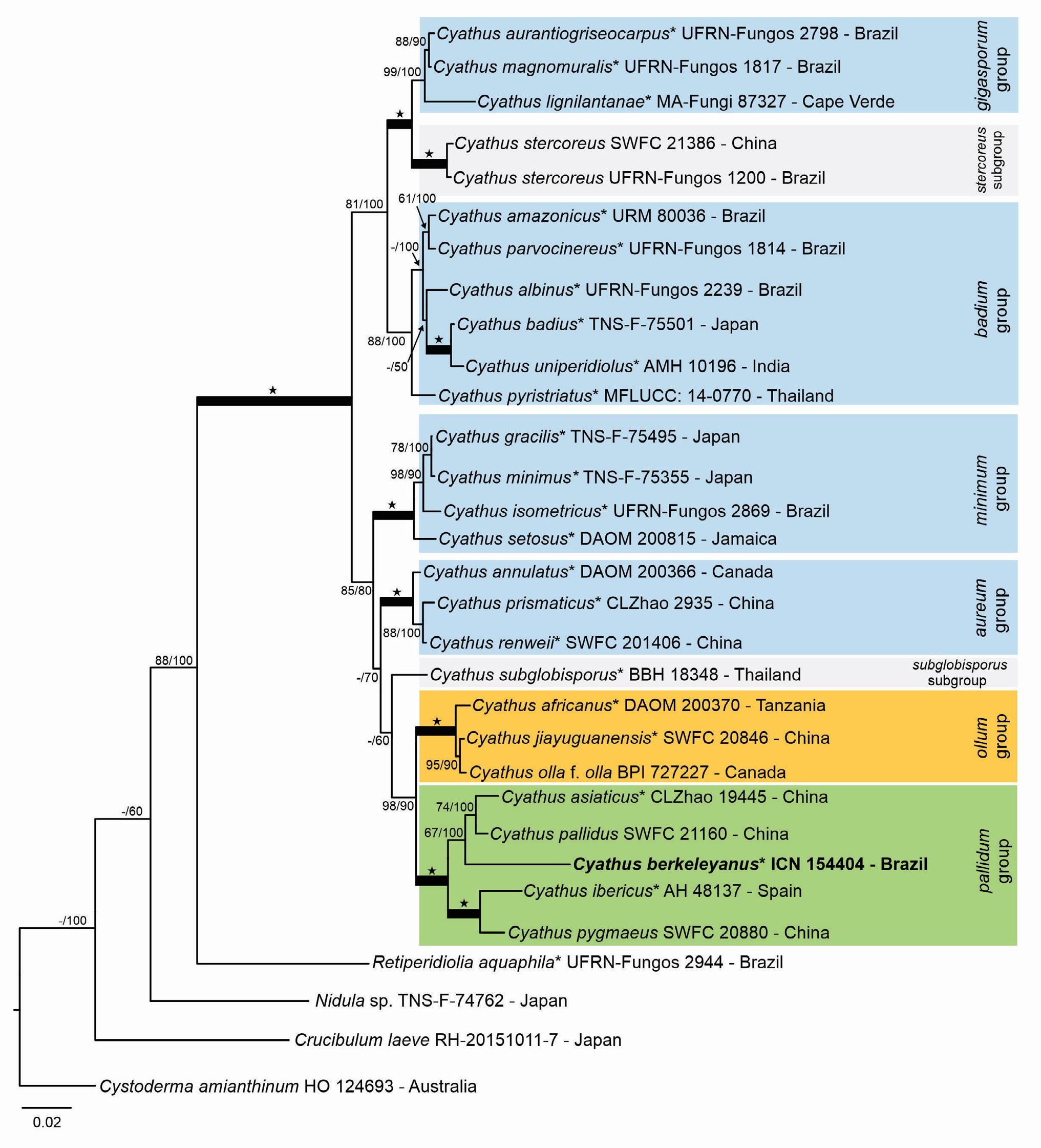
Molecular phylogeny - According to Brodie’s (1975) morphological classification, this species belongs to the group VII - striatus, and based on the proposed groups by Cruz et al. (2023) and Zhao et al. (2007), using morphological and molecular data, this species can belong to the pallidum group. Our study confirms the occurrence of this species in this clade (figures 4 and 5).

Phylogenetic tree of Cyathus obtained through Maximum Likelihood (ML) analysis from ITS sequences. The topologies of the ML and Bayesian analyses were similar. Type species sequences (holotype, lectotype, epitype, or neotype) are marked with an asterisk “*” after the species name. Values less than 50% in MLbs or pp are referenced with “-”. The new sequence is in bold. Accession numbers from the NCBI Genbank nucleotide database are indicated on the tree. The star (★) represents the nodes with PP = 100 and MLbs = 100%. The scale bar indicates the estimated number of nucleotide substitutions per site.

Phylogenetic tree of Cyathus obtained through the Bayesian analysis from the concatenated dataset ITS+LSU. The topologies of the Bayesian and ML analyses were similar. Type species sequences (holotype, lectotype, epitype, or neotype) are marked with an asterisk “*” after the species name. Values less than 50% in MLbs or pp are referenced with “-”. The voucher numbers are indicated on the tree. The star (★) represents the nodes with PP = 100 and MLbs = 100%. The scale bar indicates the estimated number of nucleotide substitutions per site.
Both ML and Bayesian analysis recovered a similar topology for all the datasets. In the ITS tree, the epitype of Cyathus berkeleyanus proposed in this study forms a sister clade with C. pallidus and C. asiaticus Z.Y. Duan & C.L. Zhao (64 MLbs/80 pp). The Chinese sample of C. berkeleyanus (DQ463355) forms a sister clade with C. ibericus J.C. Zamora & Poveda-Molero and C. pygmaeus Lloyd with great support (96 MLbs/100 pp) (Figure 4). As we do not have access to the Chinese sample, we cannot confirm if this taxon is, in fact, C. berkeleyanus; therefore, morphological data from this samples is needed to correctly delimit the species in the near future. Without this taxonomic treatment and based solely on phylogeny, it is possible that this Chinese C. berkeleyanus is, in fact, a distinct species: a cryptic species if the morphology is confirmed to be the same, or another taxon, even a new species, if the morphology is not consistent with the epitype.
The concatenated tree shows that the pallidum group is well supported in both analyses (100 MLbs/100 PP). Cyathus berkeleyanus also forms the same clade with C. pallidus and C. asiaticus (67 MLbs/100 pp), indicating that this is probably the correct position in the phylogeny (Figure 5). The Chinese sample of C. berkeleyanus does not have LSU data; therefore, we could not be able to add it to the concatenated dataset.
Our description of C. berkeleyanus fits the description proposed by Tulasne & Tulasne (1844), Lloyd (1906), and Brodie (1975), even with the inclusion of new morphological features proposed by Cruz (2017) which expanded the description with additional taxonomic features not evaluated before. The nomenclatural update of C. berkeleyanus is essential for future taxonomical studies with the genus, mainly because this species is commonly found in the Atlantic rainforest areas of Brazil and in other tropical and subtropical areas around the world. Studies involving morphological and molecular data are also required for the genus, especially in regions that are subsampled.
Acknowledgements
The authors thank the ICN Herbarium, for the loan of specimens. Jefferson Santos de Góis thanks to the Coordenação de Aperfeiçoamento Pessoal de Nível Superior, for the Master’s scholarship, to the Conselho Nacional de Desenvolvimento Científico e Tecnológico, for the split PhD scholarship (Process 200038/2023-0) and to the Systematic and Evolution Post-Graduation program. Rhudson Henrique Santos Ferreira da Cruz thanks for the support of the Universidade Federal do Oeste da Bahia, through the Public Notice nº 04/2022 for Teaching Qualification at the Post-Doctoral level (Process UFOB 23520.000500/2023-72). The authors also would like to thank to Kyung-OK Nam, for the support in DNA extraction and to the anonymous reviewers, for their useful comments in the manuscript.
Literature cited
- Bas, C. 1969. Morphology and subdivision of Amanita and a monograph on its section Lepidella Persoonia 5: 285-573.
- Baseia, I.G. & Milanez, A.I. 2001. Cyathus (Gasteromycetes) in Areas of the Brazilian Cerrado region, São Paulo state. Mycotaxon 80: 493-502.
- Blenis, P.V. & Chow, P.S. 2005. Evaluating fungi from wood and canola for their ability to decomposed canola stubble. Canadian Journal of Plant Pathology 27(2): 259-267.
- Bottomley, A.M. 1948. Gasteromycetes of South Africa. Bothalia 4: 473-810.
- Bononi, V.L.R., Guzmán, G. & Capelari, M. 1984. Basidiomicetos do Parque Estadual da Ilha do Cardoso V: Gasteromicetos. Rickia 11: 91-97.
- Brodie, H.J. 1967. Cyathus bulleri, a hitherto undescribed fungus of the Nidulariaceae from the West Indies. Bulletin of the Torrey Botanical Club 94(2): 68-71.
- Brodie, H.J. 1975. The Bird’s nest fungi. University of Toronto Press, Toronto, Canada.
- Brodie, H.J. 1984. More bird’s nest fungi (Nidulariaceae). A supplement to “the bird’s nest fungi” (1975). Lejeunia Revue de Botanique. Nouvelle Série 112: 70.
- Brodie, H.J. & Dennis, R.W.G. 1954. The Nidulariaceae of the West Indies. Transactions British Mycological Society 37(2): 151-160.
- Calonge, F.D., Mata, M. & Carranza, J. 2005. Contribución al catálogo de los Gasteromycetes (Basidiomycota, Fungi) de Costa Rica. Annales del Jardín Botânico de Madrid 62(1): 23-45.
- Cortez, V.G., Baseia, I.G. & Silveira, R.M.B. 2014. Gasteroid mycobiota of Rio Grande do Sul, Brazil: Nidulariaceae. Acta Biologica Paraense 43(1-2): 1-21.
- Cruz, R.H.S.F., Assis, N.M., Silva M.A. & Baseia, I.G. 2014. Revision of the genus Cyathus (Basidiomycota) from the herbaria of northeast Brazil Mycosphere 5(4): 531-540.
- Cruz, R.H.S.F. 2017. Revisão Morfológica e Molecular do Gênero Cyathus Haller (Nidulariaceae, Agaricales, Basidiomycota). Tese de Doutorado, Universidade Federal do Rio Grande do Norte, Natal.
- Cruz, R.H.S.F., Góis, J.S., Marinho, P., Baseia I.G. & Hosaka, K. 2023. Rearranging the Bird’s Nest Fungi: molecular review of internal clades in Cyathus (Nidulariaceae, Basidiomycota). IMA Fungus 14(8).
- Darriba, D., Taboada, G.L. & Posada, D. 2012. jModelTest2: more models, new heuristics and parallel computing. Nature methods 9(8): 772.
- De Meijer, A.A.R. 2006. Preliminary list of the macromycetes from the Brazilian State of Paraná. Boletim do Museu Botânico Municipal 68: 1-55.
- Dennis, R.W.G. 1970. Fungus flora of Venezuela and adjacent countries. Kew Bulletin Additional Series III. London.
- Góis, J.S., Cruz, R.H.S.F. & Baseia, I.G. 2021. Taxonomic review and updates of the genus Cyathus (Agaricales, Basidiomycota) from Brazil. Journal of the Torrey Botanical Society 148(3): 155-196.
- Gómez, C.L. & Pérez-Silva, E. 1988. Especies de Nidulariales (Gasteromycetes) comunes en Mexico. Revista Mexicana de Micología 4: 161-183.
- He, M.Q., Zhao, R.L., Hyde, K.D., Begerow, D., Kemler, M., Yurkov, A., McKenzie, H.C.E., Raspé, O., Kakishima, M., Sánchez-Ramírez, S., Vellinga, E.C., Halling, R., Papp, V., Zmitrovich, I.V., Buyck, B., Ertz, D., Wijayawardene, N.N., Cui, B.K., Schoutteten, N., Liu, X.Z., Li, T.H., Yao, Y.J., Zhu, X.Y., Liu, A.Q., Li, G.J., Zhang, M.Z., Ling, Z.L., Cao, B., Antonín, V., Boekhout, T., da Silva, B.D.B., de Crop, E., Decock, C., Dima, B., Dutta, A.K., Fell, J.W., Geml, J., Ghobad-Nejhad, M., Giachini, A.J., Gibertoni, T.B., Gorjón, S.P., Haelewaters, D., Shuang-Hui, H., Hodkinson, B.P., Horak, E., Hoshino, T., Justo, A., Lim, Y.M., Menolli, N., Mešić, A., Moncalvo, J.M., Mueller, G.M., Nagy, L.G., Nilsson, R.H., Noordeloos, M., Nuytinck, J., Orihara, T., Ratchadawan, C., Rajchenberg, M., Silva-Filho, A.G.S., Sulzbacher, M.A., Tkalčec, Z., Valenzuela, R., Verbeken, A., Vizzini, A., Wartchow, F., Wei, T.Z., Weiß, M., Zhao, C.L. & Kirk, P.M. 2019. Notes, outline and divergence times of Basidiomycota. Fungal diversity 99: 105-367.
- Hosaka, K. 2009. Phylogeography of the genus Pisolithus revisited with some additional taxa from New Caledonia and Japan. Bulletin of the National Museum of Nature and Science, Series B (Botany) 35(3): 151-167.
- Hosaka, K. & Castellano, M.A. 2008. Molecular phylogenetics of Geastrales with species emphasis on the position of Sclerogaster Bulletin of the National Museum of Nature and Science, Series B (Botany) 34(4): 161-173.
- Kornerup, A. & Wanscher, J.H. 1978. Methuen Handbook of Colour. 3ª Ed. London, Eyre Methuen.
- Kraisitudomsook, N., Healy, R.A. & Smith, M.E. 2021. Molecular systematics and taxonomic overview of the bird’s nest fungi (Nidulariaceae). Fungal Biology 125(9): 693-703.
- Kraisitudomsook, N., Choeyklin, R., Boonpratuang, T., Pobkwamsuk, M., Anaphon, S. & Smith, M.E. 2022. Hidden in the tropics: Retiperidiolia gen. nov., a new genus of bird’s nest fungi (Nidulariaceae), and a systematic study of the genus Mycocalia Mycological Progress 21: 56.
- Liu, B. 1984. The Gasteromycetes of China. Nova Hedwigia 74: 1-235.
- Lloyd, C.G. 1906. Nidulariaceae. Mycological Writings. Cincinnati. Ohio.
- Miller, M.A., Pfeifer, W. & Schwartz, T. 2010. Creating the CIPRES Science Gateway for Inference of Large Phylogenetic Trees. In: 2010 Gateway Computing Environments Workshio (GCE).
- Ronquist, F., Teslenko, M., van der Mark, P., Ayres, D.L., Darling, A., Höhnas, S., Larget, B., Liu, L., Suchard, M.A. & Huelsenbeck, J.P. 2012. MRBAYES 3.2: Efficient Bayesian Phylogenetic Inference and Model Selection Across a Large Model Space. Systematic Biology 61(3): 539-542.
- Stamatakis, A. 2014. RaxML version 8: a tool for phylogenetic analysis and post-analysis of large phylogenies. Bioinformatics 30(9): 1312-1313.
- Sotão, H.M.P., Gibertoni, T., Maziero, R., Baseia, I.G., Medeiros, O.S., Martins, J.R.A. & Capelari, M. 2009. Fungos macroscópicos da Floresta Nacional de Caxiuanã: Basidiomycota (Agaricomycetes), In: Lisboa, P. L. B. (eds.) Caxiuanã: Desafios para a conservação de uma Floresta Nacional na Amazônia. Pará, Brazil, Museu Paraense Emílio Goeldi, pp. 383-396.
-
Thiers, B. [permanently updated]. Index Herbariorum: a global directory of public herbaria and associated staff. New York Botanical Garden’s Virtual Herbarium. Available at https://sweetgum.nybg.org/ih (access in 10-I-2023).
» https://sweetgum.nybg.org/ih - Tulasne, L.R. & Tulasne, C. 1844. Recherches sur l’organisation et le mode de frutification des champignons de la tribu des Nidulariées, suivies d’un essai monographique. Annales des Sciences Naturalles 3(1): 41-107.
- Turland, N.J., Wiersema, J.H., Barrie, F.R., Greuter, W., Hawksworth, D.L., Herendeen, P.S., Knapp, S., Kusber, W.H., Li, D.Z., Marhold, K., May, T.W., McNeill, J., Monro, A.M., Prado, J., Price, M.J. & Smith, G.F. 2018. International Code of Nomenclature for algae, fungi, and plants (Shenzhen Code): adopted by the Nineteenth International Botanical Congress Shenzhen, China, July 2017. Regnum Vegetabile 159. Glashütten: Koeltz Botanical Books.
- Vilgalys, R. & Hester, M. 1990. Rapid genetic identification and mapping of enzymatically amplified DNA from several Cryptococcus species. Journal of Bacteriology 172: 4238-4246.
- White, T.J., Bruns, T., Lee, S. & Taylor, J. 1990. Amplification and direct sequencing of fungal ribosomal RNA genes for phylogenetics. In: Innis, M.A., Gelfand, D.H., Sninsky, J.J. & White, T.J. (eds.) PCR Protocols: a guide to methods and applications, Academic Press, pp. 315-322.
- White, V.S. 1902. The Nidulariaceae of North America. Bulletin of the Torrey Botanical Club 29(5): 251-280.
- Zhao, R.L., Desjardin, D.E., Soytong, K., & Hyde, K.D. 2008. A new species of bird’s nest fungi: characterisation of Cyathus subglobisporus sp. nov. based on morphological and molecular data. Persoonia 21: 71-76.
- Zhao, R.L., Jeewon, R., Desjardin, D.W., Soytong, K. & Hyde, K.D. 2007. Ribossomal DNA phylogenies in Cyathus: Is the current infrageneric classification appropriate? Mycologia 99(3): 385-395.
Edited by
-
Associate Editor:
Viviana Motato-Vásquez