Abstracts
BACKGROUND: Chagas' disease is an endemic tropical affliction found from southern United States to Argentina. The acute phase of this disease is difficult to study in man because the symptoms are non-specific and most cases require no medical assistance. Experimental models have been developed for sequential studies, and intense parasitism in all organs and tissues, including the pancreas, have been detected in the acute phase. PURPOSE: To evaluate the involvement of the pancreas in acute experimental Chagas' disease in a mouse model by histopathological characterization. CASUISTIC AND METHODS: Ten BALBc mice, about 20 g, injected i.p. with 100 000 forms of the Y strain of Trypanosoma cruzi were used. The animals were sacrificed after 14 days of infection. Fragments of pancreas were processed by conventional paraffin embedding and hematoxylin-eosin staining. RESULTS: Ruptured pseudocysts and release of parasites to the extracellular medium caused by necrosis of acinar and duct cells and foci of fat were the most striking histopathological features of acute Chagasic pancreatitis. CONCLUSION: Parasitism is the main cause of acute pancreatitis in Chagas' disease.
Chagas' disease; Pancreas; Pathology; Mouse model; Trypanosoma cruzi
INTRODUÇÃO: A Doença de Chagas é uma endemia tropical encontrada desde o sul dos Estados Unidos até a Argentina. Os estudos de fase aguda da doença são difíceis de serem realizados em seres humanos porque os sintomas são inespecíficos e a maioria dos casos não requer socorro médico. Em modelos experimentais desenvolvidos a doença aguda aparece com intenso parasitismo em todos os órgãos e tecidos. OBJETIVO: Caracterizar histopatologicamente o envolvimento pancreático na Doença de Chagas aguda experimental. CASUÍSTICA E MÉTODOS: Com esta finalidade utilizamos animais inoculados intraperitonialmente com 100.000 formas de cepa Y de Trypanosoma cruzi. Os animais foram sacrificados após 14 dias de infecção e os fragmentos colhidos foram processados em parafina e corados pela H&E. RESULTADOS: As características histopatológicas mais importantes da pancreatite aguda na Doença de Chagas experimental são: pseudocistos intensamente parasitados, íntegros ou rompidos, parasitas no espaço extracelular, necrose de células acinares e ductais, além de focos de esteatonecrose. CONCLUSÃO: O parasitismo dos tecidos é o principal mecanismo patogenético da pancreatite aguda na Doença de Chagas.
Doença de Chagas; Pâncreas; Patologia; Camundongo; Trypanosoma cruzi
PANCREATIC LESIONS IN ACUTE EXPERIMENTAL CHAGAS' DISEASE
Carlos Eduardo Pereira Corbett, Luciano Henrique Gazoni Scremin, Rafael Arsky Lombardi, Joaquim José Gama-Rodrigues and Masayuki Okumura
RHCFAP/3070
CORBETT CE et al. - Pancreatic Lesions in acute experimental Chagas' Disease. Rev. Hosp. Clín. Fac. Med. S. Paulo 57(2):63-66, 2002.
BACKGROUND: Chagas' disease is an endemic tropical affliction found from southern United States to Argentina. The acute phase of this disease is difficult to study in man because the symptoms are non-specific and most cases require no medical assistance. Experimental models have been developed for sequential studies, and intense parasitism in all organs and tissues, including the pancreas, have been detected in the acute phase.
PURPOSE: To evaluate the involvement of the pancreas in acute experimental Chagas' disease in a mouse model by histopathological characterization.
CASUISTIC AND METHODS: Ten BALBc mice, about 20 g, injected i.p. with 100 000 forms of the Y strain of Trypanosoma cruzi were used. The animals were sacrificed after 14 days of infection. Fragments of pancreas were processed by conventional paraffin embedding and hematoxylin-eosin staining.
RESULTS: Ruptured pseudocysts and release of parasites to the extracellular medium caused by necrosis of acinar and duct cells and foci of fat were the most striking histopathological features of acute Chagasic pancreatitis.
CONCLUSION: Parasitism is the main cause of acute pancreatitis in Chagas' disease.
DESCRIPTORS: Chagas' disease. Pancreas. Pathology. Mouse model. Trypanosoma cruzi.
Chagas' disease, first described by the Brazilian Carlos Chagas at the beginning of the century and formally called American trypanosomiasis, is one of the most important endemic tropical afflictions. It is found from southern United States to Argentina; according to the World Health Organization, there are 16 to 18 million infected people in Central and South America7. This disease is caused by the protozoan Trypanosoma cruzi, which is transmitted to man by hematophagus triatomine insects (Hemiptera: Reduviidae) when infective trypomastigote forms are discharged with the vector's feces during feeding. When the affected person scratches the site of bite, the infection occurs. The trypomastigotes form circulates in the blood, and after penetrating host cells, transform into amastigote form, giving rise to pseudocysts.
Chagas' disease presents an acute course with fever, malaise, and non-specific signs of infection. The clinical course is usually discreet and may be unnoticed. From the pathological point of view, parasitemia and multiple organ injury may be detected. In man, it is difficult to identify the acute phase because the signs and symptoms are those of non-specific infection, and patients normally do not seek medical help. In general, the only symptom of infection is a local inflammatory reaction that occurs at the site of infection in 80% of the cases7. The acute phase in man is poorly characterized because of the ethical impossibility of performing biopsies in multiple organs. To overcome this problem, several experimental models have been developed that permit the study of the acute phase of Chagas' disease and its evolution into the chronic phase. Intracellular parasitism in almost all tissues and organs is the characteristic of acute infection in experimental models4,9,10. Amastigote forms are found within pseudocysts in almost all cell types. Many strains of T. cruzi have been isolated and are used as inoculum in laboratory animals. The Y strain, used in experimental studies since 1953, has fairly uniform behavior and dissemination in the host organism6,9.
Intense loss of body fat during the acute phase of the disease had been described. Pancreatic injury has also been reported, together with a description of parasite dissemination throughout several organs and tissues in experimental models1,4,6,9,10,12. Previous studies have addressed the possible involvement of the pancreas in Chagas' disease5,9,12. However, a systematic description of the course of events in acute Chagasic pancreatitis and the possible underlying pathogenetic mechanism was not the main concern of those authors.
After the acute phase of Chagas' disease when the inflammatory process and symptoms disappear, the parasite assumes an indeterminate form. In this phase, parasites are not easily found in the circulation or in tissues.
In the chronic phase, tissue amastigotes or circulating trypomastigotes are rare. Cellular destruction leads to tissue injury, inflammatory infiltrate, and fibrosis, whether related or not to detectable local parasitism3,11,14.
Descriptions of histopathological changes of the pancreas have not been systematized for Chagas' disease. The objective of this work is a histopathological characterization of the involvement of the pancreas in experimental Chagas' disease.
CASUISTIC AND METHODS
Casuistic
Animals: Ten BALBc mice weighing about 20 g, obtained from the central colony of the University of São Paulo Medical School, were used.
Parasite: T. cruzi, strain Y, maintained through successive inoculations in BALBc mice at the Instituto Adolfo Lutz, São Paulo State Secretary of Health, was used.
Methods
Inoculum: 100 000 blood trypomastigote forms of T. cruzi were injected into mice by the intraperitoneal route (i.p.). The parasites were obtained from blood collected from infected mice by heart puncture. The inoculum was adjusted to the desired concentration by dilution of infected blood with saline.
Protocol: The animals were sacrificed after 14 days of infection, and samples were collected through laparotomy and resection of the pancreas.
Processing of samples: Tissue fragments were fixed in buffered formaldehyde solution 10%, dehydrated, embedded in paraffin, and stained with hematoxylin-eosin.
RESULTS
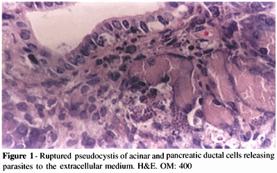
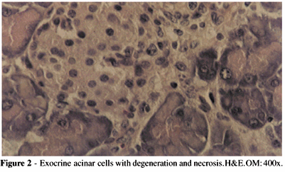
Light microscopy of pancreas fragments showed that T. cruzi was present in all the infected mice; the intensity of parasitism, however, varied between the animals. Parasites were found mainly at the exocrine portion of the organ, with many pseudocysts filled with amastigotes in acinar and epithelial duct cells. The involvement of acinar cells was diffuse all over the organ. Ruptured pseudocysts were seen, with parasites released into the extracellular medium and some penetrating and injuring neighboring cells. In pancreatic duct branches, rupture of pseudocysts and consequent destruction of parasitized cells was observed (Fig. 1). The exocrine acinar cells showed features of degeneration and necrosis, and few were found in an intact state (Fig. 2). Focal areas of fat necrosis were also identified. Debris of cells and tissue were found at injured sites (Fig. 3).
Parasite nests were found at the transition between the exocrine tissue and islets of Langerhans, and also within the endocrine pancreas (Fig. 4).
DISCUSSION
Acute pancreatitis is mainly associated with obstruction of biliary and pancreatic juice drainage caused by biliary calculus pathology. Alcoholism and the action of drugs are also considered causes of acute pancreatitis, even though the underlying mechanism in these cases is not currently fully understood. Changes in pancreatic juice components, such as protein-rich pancreatic fluid secretion that gives rise to protein plugs, may play an important pathogenic role in pancreatitis. The pathology of the disease results from the activation of digestive enzymes trapped within obstructed ducts, thus leading to ductile and acinar necrosis and extravasation of pancreatic juice2..
Features of pancreatic fat necrosis may be found when pancreatic juice extravasation occurs.
This study shows pancreatic lesions caused by a parasitic agent. Fat necrosis occurs in the pancreas when there is obstruction of pancreatic juice drainage or when injured cells rupture and release acids and enzymes to the interstitium, mainly via the action of lipase in its active form. In Chagas' disease, the pathogenic process appears to be related to the presence of parasites in acinar and duct cells and their subsequent rupture, rather than obstruction of drainage of pancreatic juice or changes in its protein components. The presence of amastigotes in most pancreatic acinar and epithelial duct cells, forming pseudocysts, has been previously reported9,12.
The pathogenesis of pancreatic lesions in acute Chagasic pancreatitis seems to be related to local parasitism and consequent necrosis of fat tissue caused by the release of pancreatic enzymes from ruptured parasitized cells.
A neuronal participation in the pathogeneses of pancreatic alterations has been considered in chronic Chagas' disease8 because of the decrease in neuronal populations reported for human Chagasic cases13.
The lesions described herein that relate to the acute phase of Chagas' disease are due to parasite-induced cell destruction. Ganglia cell parasitism in the acute phase may also be the cause of the reduced numbers of these cells observed in the chronic phase.
The occurrence of T. cruzi in acinar and duct cells as well as the interstitium demonstrates the direct involvement of this parasite in acute Chagasic pancreatitis. The necrotic aspect of infected cells and the fat necrosis associated with parasitism suggest that the presence of the parasite is a necessary condition for induction of acute pancreatitis in Chagas' disease.
RESUMO
RHCFAP/3070
CORBETT CE e col. Lesões pancreáticas em Doença de Chagas aguda experimental. Rev. Hosp. Clín. Fac. Med. S. Paulo 57 (2): 63-66, 2002.
INTRODUÇÃO: A Doença de Chagas é uma endemia tropical encontrada desde o sul dos Estados Unidos até a Argentina. Os estudos de fase aguda da doença são difíceis de serem realizados em seres humanos porque os sintomas são inespecíficos e a maioria dos casos não requer socorro médico. Em modelos experimentais desenvolvidos a doença aguda aparece com intenso parasitismo em todos os órgãos e tecidos.
OBJETIVO: Caracterizar histopatologicamente o envolvimento pancreático na Doença de Chagas aguda experimental.
CASUÍSTICA E MÉTODOS: Com esta finalidade utilizamos animais inoculados intraperitonialmente com 100.000 formas de cepa Y de Trypanosoma cruzi. Os animais foram sacrificados após 14 dias de infecção e os fragmentos colhidos foram processados em parafina e corados pela H&E.
RESULTADOS: As características histopatológicas mais importantes da pancreatite aguda na Doença de Chagas experimental são: pseudocistos intensamente parasitados, íntegros ou rompidos, parasitas no espaço extracelular, necrose de células acinares e ductais, além de focos de esteatonecrose.
CONCLUSÃO: O parasitismo dos tecidos é o principal mecanismo patogenético da pancreatite aguda na Doença de Chagas.
DESCRITORES: Doença de Chagas. Pâncreas. Patologia. Camundongo. Trypanosoma cruzi.
Received for publication on May 14, 2001.
From the Departments of Pathology and Gastroenterology, Hospital das Clínicas, Faculty of Medicine, University of São Paulo.
- 1. CALABRESE KS, LAGRANGE PH & COSTA SC - Trypanosoma cruzi: histopathology of endocrine system in immunocompromised mice. Int J Exp Pathol 1994; 75(6): 453-62.
-
2CRAWFORD JM & COTRAN RS - The Exocrine Pancreas. In.: COTRAN RS et al. - Robbins Pathologic Basis of Disease 5th Philadelphia, Saunders, 1995. p. 900.
- 3. CUNHA-NETO E, DURANTI M, GRUBER THE et al. - Autoimmunity in Chagas disease cardiopathy: biological relevance of the cardiac myosin-specific epitope crossreactive to an immunodominant Trypanosoma cruzi antigen. Proc Natl Acad Sci USA 1995; 92(8): 3541-3545.
- 4. HANSON WL & ROBERSON EL - Density of parasites in various organs and the relation to number of trypomastigotes in the blood during acute infection of Trypanosoma cruzi in mice. J Protozool 1974; 21(4): 512-517.
- 5. KÖBERLE F - Patogenia da moléstia de Chagas. Rev Goiana Med 1957; 3:155-180.
- 6. LENZI HL, OLIVEIRA DN & LIMA MT - Trypanosoma cruzi: Paninfectivity of CL strain during murine acute infection. Exp Parasitol 1996; 84(1): 16-27.
-
7LOPES ER, CHAPADEIRO E, TAFURI WL et al. - Chagas Disease. In: Bogliolo l - Bogliolo: Pathology 5th Rio de Janeiro, Guanabara Koogan, 1994. p.1103-1104.
- 8. MOTT CB, HUT DR, SIPAHI AM et al. - Functional evaluation of the pancreas exocrine in carriers of chronic Chagas' disease. Rev Hosp Clin Fac Med S Paulo 1988; 43(6): 279-287.
- 9. OKUMURA M, BRITO T, SILVA LHP et al. - The pathology of experimental Chagas disease in mice: 1. Digestive tract changes, with reference to necrotizing arteritis. Rev Inst Med Trop São Paulo 1960; 2(1): 17-28.
- 10. OKUMURA M, FRANCE LCM & CORRÊA-NETO A - Comentários on the patogenia of the Chagas' disease: Special reference to the experimental infection in camundongos. Rev Hosp Clin Fac Med S. Paulo 1963; 18:151-164.
- 11. OKUMURA M - Pathogenesis of Chagasic myocarditis (an experimental study). Rev Hosp Clin Fac Med S Paulo 1996; 51(5):166-174.
-
12PIZZI T - Localización pancrática predominante y fenómenos de esteatonecrosis en ratones experimentalmente infected con Tripanosoma cruzi. Apud 9.
- 13. OKUMURA M, BRITO T, SILVA LHP et al. - The pathology of experimental Chagas disease in mice: 1. Digestive tract changes, with reference to necrotizing arteritis. Rev Inst Med Trop São Paulo 1960; 2(1): 17-28.
- 14. ROCHA A, de OLIVEIRA LC, ALVES RS et al. - Pancreatic neuronal loss in chronic Chagas' Disease patients. Rev Soc Bras Med Trop 1998; 31(1): 43-49.
- 15. VAGO AR, MACEDO AM, ADAD SJ et al. - PCR detection Trypanosoma cruzi DNA in esophageal tissues of patients with chronic digestive Chagas' disease. Lancet 1996; 348:891-892.
Publication Dates
-
Publication in this collection
18 Oct 2002 -
Date of issue
2002
History
-
Received
14 May 2001