Abstracts
PURPOSE: To compare the hemodynamic effects following a toxic dose of either agent after intravenous injection in swines, as might accidentally occur during regional anesthesia in humans. METHODS: Large White pigs were anesthetized with thiopental, tracheal intubation was performed and mechanical ventilation was instituted. Hemodynamic variables were recorded with invasive pressure monitoring and pulmonary artery catheterization. After a 30-minute resting period, the animals were randomly divided into two groups in a double-blinded fashion and received a bolus intravenous injection of 4 mg.kg-1 of either agent. Hemodynamic results were evaluated at rest and 1, 5, 10, 15, 20 and 30 minutes after intoxication. RESULTS: Hemodynamic repressions of acute intoxication with levobupivacaine were more important and more prolonged than those of ropivacaína. CONCLUSION: In pigs, levobupivacaine was shown to be more toxic than ropivacaine when the same large doses are injected intravenously.
Poisoning; Hemodynamics; Anesthetics, Local; Swine
OBJETIVO: Comparar as repercussões hemodinâmicas após a injeção endovenosa dos dois agentes em suínos simulando a intoxicação que pode ocorrer durante uma anestesia locorregional em humanos. MÉTODOS: Suínos da raça Large-White foram anestesiados com tiopental, realizada intubação traqueal e instituída ventilação controlada mecânica. As variáveis hemodinâmicas foram medidas através de monitorização invasiva da pressão arterial e cateterização de artéria pulmonar. Após período de repouso de 30 minutos os animais foram aleatoriamente e em duplo-cego divididos em dois grupos e receberam por via endovenosa 4 mg.kg-1 de um ou outro agente. Os resultados hemodinâmicos foram avaliados em repouso e 1, 5, 10, 15, 20 e 30 minutos após a intoxicação. RESULTADOS: As repercussões hemodinâmicas da intoxicação aguda com levobupivacaína foram mais importantes e mais prolongadas do que as com ropivacaína. CONCLUSÃO: Em suínos, a levobupivacaína foi mais tóxica do que a ropivacaína quando as mesmas grandes doses são injetadas por via endovenosa.
Envenenamento; Hemodinâmica; Anestésicos Locais; Suínos
9 - ORIGINAL ARTICLE
EFFECTS OF DRUGS
Hemodynamic effects of ropivacaine and levobupivacaine intravenous injection in swines1 1 Research performed at Laboratory of Experimental Anesthesia, State University of Campinas (UNICAMP), Sao Paulo, Brazil.
Efeitos hemodinâmicos da injeção endovenosa de ropivacaína e levobupivacaína em suínos
Artur UdelsmannI; William Adalberto SilvaII; Ana Cristina de MoraesII; Elisabeth DreyerIII
IAssociate Professor, Department of Anesthesiology and Laboratory of Experimental Anesthesia, UNICAMP, Sao Paulo, Brazil
IIBiologist, Center for Experimental Medicine and Surgery, UNICAMP, Sao Paulo, Brazil
IIIRN, MSc, Chief Nurse Intensive Care Unit of the Hospital, UNICAMP, Sao Paulo, Brazil
Correspondence Correspondence: Dr. Artur Udelsmann Av. Prof. Atílio Martini, 213 13083-830 Campinas - SP Brazil Phone / Fax: (55 19)3521-9560 audelsmann@yahoo.com.br
ABSTRACT
PURPOSE: To compare the hemodynamic effects following a toxic dose of either agent after intravenous injection in swines, as might accidentally occur during regional anesthesia in humans.
METHODS: Large White pigs were anesthetized with thiopental, tracheal intubation was performed and mechanical ventilation was instituted. Hemodynamic variables were recorded with invasive pressure monitoring and pulmonary artery catheterization. After a 30-minute resting period, the animals were randomly divided into two groups in a double-blinded fashion and received a bolus intravenous injection of 4 mg.kg-1 of either agent. Hemodynamic results were evaluated at rest and 1, 5, 10, 15, 20 and 30 minutes after intoxication.
RESULTS: Hemodynamic repressions of acute intoxication with levobupivacaine were more important and more prolonged than those of ropivacaína.
CONCLUSION: In pigs, levobupivacaine was shown to be more toxic than ropivacaine when the same large doses are injected intravenously.
Key words: Poisoning. Hemodynamics. Anesthetics, Local. Swine.
RESUMO
OBJETIVO: Comparar as repercussões hemodinâmicas após a injeção endovenosa dos dois agentes em suínos simulando a intoxicação que pode ocorrer durante uma anestesia locorregional em humanos.
MÉTODOS: Suínos da raça Large-White foram anestesiados com tiopental, realizada intubação traqueal e instituída ventilação controlada mecânica. As variáveis hemodinâmicas foram medidas através de monitorização invasiva da pressão arterial e cateterização de artéria pulmonar. Após período de repouso de 30 minutos os animais foram aleatoriamente e em duplo-cego divididos em dois grupos e receberam por via endovenosa 4 mg.kg-1 de um ou outro agente. Os resultados hemodinâmicos foram avaliados em repouso e 1, 5, 10, 15, 20 e 30 minutos após a intoxicação.
RESULTADOS: As repercussões hemodinâmicas da intoxicação aguda com levobupivacaína foram mais importantes e mais prolongadas do que as com ropivacaína.
CONCLUSÃO: Em suínos, a levobupivacaína foi mais tóxica do que a ropivacaína quando as mesmas grandes doses são injetadas por via endovenosa.
Descritores: Envenenamento. Hemodinâmica. Anestésicos Locais. Suínos.
Introduction
Regional anesthesia might require high doses of local anesthetic agents for surgery and there is always the potential risk for toxic reactions. Both the cardiovascular and central nervous systems are the primary target organs of local anesthetic toxicity in case of intravascular injection. Bupivacaine was one of the most widely used local anesthetics, due to its quality of anesthesia and prolonged duration of action1,2. Nevertheless, an Anesthesiology editorial on the severe cardiovascular effects of bupivacaine intoxication was published in 19793 and since then research has focused on discovering new long-acting local anesthetics with lower toxicity. Although bupivacaine is synthesized in the form of its two dextrorotatory R(+) and levorotatory L(-) isomers4, evidences that the levorotatory isomers caused less toxicity was already known since 19725,6. In the last decade the levorotatory isomers ropivacaine and levobupivacaine were developed as safer alternatives. Ropivacaine is the S(-) isomer of the propyl analogue of mepivacaine and bupivacaine, whereas levobupivacaine is the S(-) enantiomer of bupivacaine. Indeed, experimental and clinical data have demonstrated these two agents may be less toxic than bupivacaine7. In some animal models, the lethal dose of levobupivacaine was shown to be up to 1.6 times higher than that of the racemic mixture8! In humans, levobupivacaine would be less potent at producing negative inotropic effects and prolonging the PR and QT intervals of the ECG, typical of racemic mixture intoxication9. Despite having equal analgesic potency to racemic bupivacaína10 the drawback with this new agents would be its less intense motor block11, The aim of this study was to simulate an acute intoxication with either agent in pigs, as might accidentally occur during local and regional anesthesia with high doses of local anesthetics and to evaluate its hemodynamic repercussions.
Methods
After approval from the Ethics Committee for Animal Experiments, 40 healthy Large White pigs (both sexes, body weight 20-27 kg) underwent the following protocol and follow the Council for International Organization of Medical Sciences ethical code for animal experimentation and the principles of the Brazilian College on Animal Experimentation. The pigs were fasted the night before and had free access to water. The morning, the animals were weighed, an auricular vein was cannulated and anesthesia was induced with a 25 mg.kg-1 IV dose of sodium thiopental12. The body surface area of the animal (BSA) was calculated using the formula13: BSA= (9 x weight in grams 2/3) x 10-4 m2, introducing the result into the multiparametric Engstrom AS/3 monitor to calculate body index values. Intubation was performed and the animals were ventilated by a pneumatically driven ventilator using a partial re-breathing system and CO2 absorbent with tidal volume of 15ml.kg-1 and respiratory rate to obtain ETCO2 32 - 34 mmHg. Fresh oxygen flow was 1 l.min-1 and hemoglobin oxygen saturation was measured and kept above 97%. ECG was also monitored. Anesthesia was maintained with an intravenous infusion of sodium thiopental 5mg.kg-1.h. In the medial left hind limb of the animal, dissection and cannulation of the femoral artery was performed for continuous arterial pressure measurement. A Swan-Ganz catheter was inserted into the femoral vein and hemodynamic measurements were made by the Datex Engstron AS/3 multiparametric monitor. Cardiac index (CI) mean arterial pressure (MAP), mean pulmonary artery pressure (MPAP), central venous pressure (CVP), pulmonary capillary wedge pressure (PCWP) stroke index (SI), systemic vascular resistance index (SVRI), pulmonary vascular resistance pulmonary index (PVRI), left ventricular stroke work index (LVSWI), right ventricular stroke work index (RVSWI) were measured. After a resting period of 30 minutes to ensure stabilization, baseline hemodynamic measurements were recorded (T0). The animals were then randomly and in a double-blinded fashion divided in 2 groups: ropivacaine group (R) and levobupivacaine group (L). The animals received intravenously a toxic dose of 4 mg.kg-1 of the local anesthetic14 during 30 seconds and the experimenter was blinded about the group. Additional hemodynamic measurements were recorded at 1, 5, 10, 15, 20 and 30 minutes after intoxication. At the end the animals were sacrificed while still under anesthesia by a 10 ml intravenous injection of 19.1% potassium chloride solution. Then the double-blinded protocol was disclosed and data were statistically treated. For categorical variables the chi-square test was used and to compare the distribution of single numerical variables (measured at a single time point) the Student's t-test. To study changes in numerical variables measured at several time points, repeated-measures analysis of variance was performed (ANOVA). Furthermore, the Duncan test was used to compare the groups at each time point and the profile contrast test was applied to analyze the time course of each group. The significance level was set at 5%, i.e., p < 0.05.
Results
The following Table 1 shows the distribution by sex, mean and standard deviations of weight and body surface areas in both groups. There were no statistic differences.
There were no difference in any of the hemodynamic parameters measured at rest between groups.
Following intoxication, there was a reduction in cardiac index (Figure 1 and Table 2) in both groups but L was lower than R until the end (p<0.001) and did not return to values similar to T0 till the end while R returned to resting values in T10 (p<0.001).
Mean arterial pressure (Figure 2 and Table 3) decreased in both groups but L was lower than R from T1 to T5 and higher from T15 to T30 (p<0.001). In L pressure was lower than T0 till T10 while in R only in T1 (p<0.001).
There was a significant and similar decrease in heart rate (Figure 3 and Table 4) in both groups. L did not reach T0 till the end but R did at T10 (p<0.001). From T15 till T30 L had lower values than R (p<0.001).
Central venous pressure (Figure 4 and Table 5) increased significantly in both groups but more in L which was higher than R till the end (p=0.012). L did not return to T0 similar values but R did since T5 (p<0.001).
Mean pulmonary artery pressure (Figure 5 and Table 6) was higher in L than R from T5 till T30 (p=0.021). There were no differences in times in both groups (p=0.06).
Pulmonary capillary wedge pressure (Figure 6 and Table 7) showed increased values in both groups following intoxication but in L they were more important till T20 (p=0.007). L did not return to resting values but R did at T10 (p=0.042).
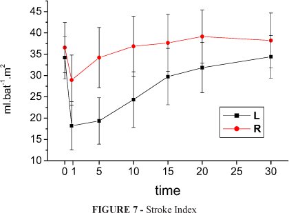
Stroke index (Figure 7 and Table 8) had a significant decline in both groups but in L these fall was more important till T20 (p<0.001). L returned to resting values in T20 while R in T5 (p<0.001).
In L systemic vascular resistance index (Figure 8 and Table 9) had a fall in T1 but from T10 till the end was greater than T0, R had an increase from T1 to T10 (p<0.001). L was greater than R from T10 to T30 (p<0.001).
In L pulmonary vascular resistance index (Figure 9 and Table 10) showed higher values than resting in T1, T5 and T20 (p<0.001) while in R only in T1. L was greater than R from T5 to T30 (p=0.013).
Left ventricular stroke work index (Figure 10 and Table 11) decreased after intoxication in both groups, however the fall was greater in L until T10 (p<0.001). Values returned to similar to resting at T20 in L and T5 in R (p<0.001).
Right ventricular stroke work index (Figure 11 and Table 12) fall in both groups, but this was more important in L until T10 (p=0.03). Values returned to to resting at T20 in L and T5 in R (p<0.001).
Discussion
Cardiac toxicity of local anesthetics is attributed to a blockade of Na+ in the heart leading to a prolonged conduction time with widening of QRS complexes, prolongation of PR interval, AV block and arrythmias15. These drugs cause cardiac toxicity as well by altering mitochondrial metabolism in cardiac cells and thus alter inotropism16. The S(-) isomer may be less cardiotoxic due to its lower affinity for cardiac Na+ channels in comparison to the R(+)isomer as demonstrated in guinea pigs17 and ropivacaine would be less potent than levobupivacaine to block them18. Overall hemodynamic variables events seen with both drugs were similar but quantitative differences were found, with ropivacaine changes were less important and turned over more quickly to the starting values. Our results demonstrated that levobupivacaine had greater hemodynamic repercussions on a swine model of acute intoxication simulating what may occur after accidental intravenous injections of local anesthetics during local and regional anesthesia. Evidence of toxicity with the same doses was shown in the significantly more important decrease in cardiac index mean arterial pressure, heart rate and left ventricular stroke work index. However, further studies must be encouraged since Polley et al.19 found the drugs equipotent, Parpaglioni et al.20 levobupivacaine more potent, Liisanantti et al.21 ropivacaine more potent and different potencies could justify different results with the same doses. Accidental injections of high doses of local anesthetics and toxic reactions during local and regional anesthesia have decreased in the last 30 years, falling from 0.2 to 0.01%. Peripheral nerve blocks still account for the majority of these cases (7.5 per 10.000)22. Such considerations should intensify new efforts to discover drugs and techniques that enable to achieve a significantly low morbidity and mortality rate, protecting patients from the undesirable and unpredictable effects of local and regional anesthesia techniques with high doses of local anesthetics.
Conclusion
In pigs, levobupivacaine was shown to cause more important hemodynamic repercussions than ropivacaine when the same large doses are injected intravenously.
Received: January 20, 2009
Review: March 18, 2009
Accepted: April 15, 2009
Conflict of interest: none
Financial source: none
How to cite this article
Udelsmann A, Silva WA, Moraes AC, Dreyer E. Hemodynamic effects of ropivacaine and levobupivacaine intravenous injection in swines. Acta Cir Bras. [serial on the Internet] 2009 July-Aug;24(4). Available from URL: http://www.scielo.br/acb
- 1. Groban L, Deal DD, Vernon JC, James RL, Butterworth J. Cardiac resuscitation after incremental overdosage with lidocaine, bupivacaine, levobupivacaine and ropivacaine in anesthetized dogs. Anesth Analg. 2001;92:37-43.
- 2. Chang DH-T, Ladd LA, Copeland S, Iglesias MA, Plummer JL, Mather LE. Direct cardiac effects of intracoronary bupivacaine, levobupivaciane and ropivacaine in the sheep. Br J Pharmacol. 2001;132:649-58.
- 3. Albright GA. Cardiac arrest following regional anesthesia with etidocaine or bupivacaine. Anesthesiology. 1979;51:285-7.
- 4. Ohmura S, Kawada M. Ohta T Yamamoto K, Kobayashi T. Systemic toxicity and resuscitation in bupivacaína, levobupivacaína or ropivacaine-infused rats. Anesth Analg. 2001;93:743-8.
- 5. Åberg G. Toxicological and local anesthetic effects of optically active isomers of two local anesthetic compounds. Acta Pharmacol Toxicol. 1972;31:273-86.
- 6. Luduena FP, Bogado EF, Tullar BF. Optical isomers of mepivacaine and bupivacaine. Arch Int Pharmacodyn. 1972;200:359-69.
- 7. Groban L. Central nervous system and cardiac effects from long-acting amide local anesthetic toxicity in the intact animal model. Reg Anesth Pain Med. 2003;28:3-11.
- 8. Foster RH, Markham A. Levobupivacaine. A review of its pharmacology and use as a local anesthetic. Drugs. 2000;59:551-79.
- 9. Bardsley H, Gristwood R, Baker H, Watson N, Nimmo W. A comparison of the cardiovascular effects of levobupivacaine and rac-bupivacaine following intravenous administration to healthy volunteers. Br Clin Pharmacol. 1998;46:245-9.
- 10. Lyons G, Columb M, Wilson RC, Johnson RV. Extradural pain relief in labour:potencies of levobupivacaine and racemic bupivacaine. Br J Anaesth. 1998;81:899-901.
- 11. Héctor JL, Columb MO. The relative motor blocking potencies of bupivacaine and levobupivacaine in labour. Anesth Analg. 2003;97:1509-13.
- 12. Smith AC, Ehler WJ, Swindle MMl. Anesthesia and analgesia in swine. In: Kohn DF, Wixson SK, White WJ, Benson JG. Anesthesia and analgesia in laboratory animals. 1ed. New York: Academic Press; 1997. p.313-6.
- 13. Ettinger SJ. Textbook of veterinary internal medicine. vol.1. 1ed. Philadelphia: WB Saunders Co.; 1975.
- 14. Lefrant JY, Muller L, de La Coussaye JE, Lalourcey L, Ripart J, Peray PA, Mazoit X, Dauzat M, Sassine A, Eledjam JJ. Hemodynamic and cardiac electrophysiologic effects of lidocaine-bupivacaine mixture in anesthetized and ventilated piglets. Anesthesiology. 2003;98:96-103.
- 15. Clarkson CW, Hondeghem LM. Mechanism for bupivacaine depression of cardiac conduction: fast block of sodium channels during the action potential with slow recovery from block during diastole. Anesthesiology. 1985;62:396-405.
- 16. Butherworth JF, Brownlown RC, Leith JP, Prielipp RC, Cole LR. Bupivacaine inhibits cyclic 3.5 adenosine monophosphate production. A possible contribution factor to cardiovascular toxicity. Anesthesiology. 1993;79:88-95.
- 17. Valenzuela C, Snyders DJ, Bennett PB, Tamargo J, Hondeghem LM. Stereoselective block of cardiac sodium channels by bupivacaine in guinea pig ventricular myocytes. Circulation. 1995;92:3014-24.
- 18. Nau C, Strichartz GR. Drug chirality in anesthesia. Anesthesiology. 2002;97:497-502.
- 19. Polley LS, Columb MO, Naughton NN, Wagner DS, van de Ven CJ, Goralski KH. Relative analgesic potencies of levobupivacaine and ropivacaine for epidural analgesia in labor. Anesthesiology. 2003;99:1354-8.
- 20. Parpaglioni R, Frigo MG, Lemma A, Sebastiani M, Barbati G, Celleno D. Minimum local anaesthetic dose (MLAD) of intrathecal levobupivacaine and ropivacaine for caesarean section. Anaesthesia. 2006;61:110-5.
- 21. Liisanantti O, Luukkonen J, Rosenberg PH. High-dose bupivacaine, levobupivacaine and ropivacaine in axillary brachial plexus block. Acta Anaesthesiol Scand. 2004;48:601-6.
- 22. Cox B, Durieux ME, Marcus MA. Toxicity of local anesthetics. Best Pract Res Clin Anaesthesiol. 2003;17:111-36.
Publication Dates
-
Publication in this collection
17 Aug 2009 -
Date of issue
Aug 2009
History
-
Accepted
18 Apr 2009 -
Received
20 Jan 2009 -
Reviewed
18 Mar 2009