Resumos
Tumor benigno de tecido muscular, o piloleiomioma tem origem no músculo eretor do pelo, atingindo ambos os sexos geralmente na terceira década de vida. Apresenta-se como nódulo-pápulas assimétricas nas extremidades, de cor eritêmato-acastanhada e de consistência firme. As lesões, quando múltiplas, podem ser sensíveis ou dolorosas. Sua associação com miomas uterinos, denominada de síndrome de Reed ou leiomiomatose cutis et uteri, é apresentação rara, podendo estar associada a carcinoma de células renais. A abordagem é cirúrgica em casos isolados e medicamentosa se houver sintomas. Relatamos um caso de síndrome de Reed em que se optou por acompanhamento pela ausência de sintomatologia.
Colágeno; Crioterapia; Leiomioma
Piloleiomyoma, a benign smooth-muscle tumor arising from the arrectores pilorum muscles of the skin, affects males and females in the third decade of life. It presents as asymmetrical, reddish-brown nodules or papules with a firm consistency, predominantly located on the limbs. When multiple lesions are present, they may be tender or painful. Their association with uterine fibroids, referred to as Reed syndrome or familial leiomyomatosis cutis et uteri, is rare and may be associated with renal cell carcinoma. The approach consists of surgical excision in cases presenting few lesions and pharmacological treatment if symptomatic. The present paper describes a case of Reed syndrome in which a decision was made to monitor the patient in view of the absence of symptoms.
Collagen; Cryotherapy; Leiomyoma
SÍNDROME EM QUESTÃO
Do you know this syndrome?* * This study was conducted at the Hospital Naval Marcílio Dias, Rio de Janeiro, RJ, Brazil.
Thais Jerez JaimeI; Tatiana Jerez JaimeII; Daniel Fernandes MeloIII; Bianca de Mello GuaraldiIV; César de Souza Bastos JúniorV; Claudio LererVI
IMedical Degree awarded by the Pontifical Catholic University of São Paulo (PUC - SP). Postgraduate student in Dermatology, Hospital Naval Marcílio Dias, Rio de Janeiro, RJ, Brazil
IIProfessor, Head of the Psoriasis Outpatient Clinic, Mogi das Cruzes University, Mogi das Cruzes, SP, Brazil
IIIProfessor, Head of the Alopecia and General Outpatient Clinic, Hospital Naval Marcílio Dias, Rio de Janeiro, RJ, Brazil
IVMedical Degree awarded by the Fundação Educacional Serra dos Órgãos. Postgraduate student in Dermatology, Hospital Naval Marcílio Dias, Rio de Janeiro, RJ, Brazil
VMaster's degree student in Anatomopathology, Federal University of Rio de Janeiro. Assistant at the Anatomopathology Department, Hospital Naval Marcílio Dias. Assistant Professor, Estácio de Sá University, Rio de Janeiro, RJ, Brazil
VIProfessor and Head of the postgraduate medical program in Dermatology of the Carlos Chagas Institute. Professor, Postgraduate Program in Dermatology, Santa Casa de Misericórdia, Rio de Janeiro, RJ, Brazil
Mailing address Mailing address: Thais Jerez Jaime Rua Cesar Zama, s/n, Lins de Vasconcelos 20725-090 Rio de Janeiro, RJ, Brazil E-mail: thaisjerez@yahoo.com.br
ABSTRACT
Piloleiomyoma, a benign smooth-muscle tumor arising from the arrectores pilorum muscles of the skin, affects males and females in the third decade of life. It presents as asymmetrical, reddish-brown nodules or papules with a firm consistency, predominantly located on the limbs. When multiple lesions are present, they may be tender or painful. Their association with uterine fibroids, referred to as Reed syndrome or familial leiomyomatosis cutis et uteri, is rare and may be associated with renal cell carcinoma. The approach consists of surgical excision in cases presenting few lesions and pharmacological treatment if symptomatic. The present paper describes a case of Reed syndrome in which a decision was made to monitor the patient in view of the absence of symptoms.
Keywords: Collagen; Cryotherapy; Leiomyoma
CASE REPORT
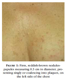
A 52-year old female patient was referred to the dermatology clinic complaining of unsightly lesions on her chest that had been present for the past 15 years. She described the sudden onset of asymptomatic erythematous papules, confined to the left side of her chest. Over the following years, the patient noticed an increase in the number of lesions, which spread to her left arm, now developing a mild burning sensation when exposed to low temperatures. The patient had had a hysterectomy 15 years previously due to uterine fibroids. On dermatological examination, firm, reddish-brown nodules-papules measuring 0.3 cm in diameter, presenting singly or coalescing into plaques, were found on the left side of her chest (Figures 1 and 2). A single papule with the same characteristics was found on her left arm. Biopsy revealed proliferation of bundles of muscle cells with spindle-shaped nuclei in the papillary and reticular dermis (Figure 3). Tomography of the abdomen and pelvis was normal.
DISCUSSION
Leiomyoma, a benign tumor originating in the smooth muscle tissue, is classified into three types according to its site of origin: piloleiomyoma in which the arrector pili muscle is affected; genital and nipple leiomyoma, which develop in these regions, and vascular leiomyoma in which the smooth muscle of the middle layer of the vessels is affected. 1-3,4 Piloleiomyoma is the most common of these, affecting both sexes equally and affecting patients of any age, although it is more common between 10 and 30 years of age 1-6. It presents as a single lesion, more common in males, or as multiple lesions, which are more common in women. It is characterized by reddish-brown papules-nodules of less than 1.5 cm in diameter with a smooth surface and firm consistency, located asymmetrically on the extensor muscles of the limbs, chest and face. The lesions are tender or painful, particularly when presentation is multiple, with patients referring to a burning sensation, tightness or pain that may appear spontaneously or induced by cold temperatures, touch, pressure or excitement. The pain is known to be caused by compression of the cutaneous nerves, by injury or by simple contraction of the muscle fibers of the tumor. 1,3,4,6 The occurrence of multiple cases of leiomyomas in families has been described as autosomal dominant inheritance with incomplete penetrance. The finding of cutaneous leiomyomas with concomitant erythrocytosis has been reported and is explained by an effect of the "erythropoietin-like" behavior of the tumor. 1 The association between piloleiomyoma and uterine leiomyomas, referred to as Reed syndrome or familial leiomyomatosis cutis et uteri, is considered a rare presentation that may be associated with renal cell carcinoma. 1,2,3,5 In this syndrome, the defect occurs in chromosome 1q.42.3-43, which acts as a tumor suppressor2. In these cases, investigation by imaging tests is mandatory. 1,2 As for treatment, surgical excision is recommended when lesions are few; however, there is a recurrence of the lesions in 50% of cases. In cases of numerous and painful piloleiomyomas, a pharmacological approach is recommended; however, effectiveness is limited. Drugs used for this treatment include gabapentin, oral or topical nitroglycerin, lidocaine, nifedipine, verapamil, phenoxybenzamine, phentolamine, hyoscine, painkillers and antidepressants. Cryotherapy and electrocoagulation have been used, but with little success 1-6 In this case, a decision was made to monitor the patient clinically, since her symptoms were mild. 
Received on 30.05.2011.
Approved by the Advisory Board and accepted for publication on 30.06.2011.
Conflito de interesse: Nenhum
Suporte financeiro: Nenhum
- 1. Parreira LML, Sípoli JMS, Mercante AMC, Orfali RL. Caso para diagnóstico. Piloleiomioma múltiplo unilateral. An Bras Dermatol. 2009;84:197-9.
- 2. Kim G. Multiple cutaneous and uterine leiomyomatosis ( Reed's syndrome). Dermatol Online J. 2005;11: 21.
- 3. Alper M, Parlak AH, Kavak A, Aksoy KA. Bilateral multi ple piloleiomyomas on the breast. Breast. 2004;13:146-8.
- 4. Pacheco AP, Ramos AMO, Rolim MLM, Oliveira FM, Lopes JG, Rocha KF. Piloleiomioma múltiplo: relato de caso com diagnóstico diferencial. An Bras Dermatol. 1995;70:43-6.
- 5. Alam M, Rabinowitz AD, Engler DE. Gabapentin treatment of multiple piloleiomyoma-related pain. J Am Acad Dermatol. 2002;46(2 Suppl Case Reports):S27-9.
- 6. Arrua GGA, Cadena GMM, Filippo AA, Azulay DR, Azulay RD. Piloleiomioma múltiplo: relato de três casos tratados com nifedipina. An Bras Dermatol. 1991;66:303-5.
Datas de Publicação
-
Publicação nesta coleção
23 Jan 2012 -
Data do Fascículo
Dez 2011
Histórico
-
Aceito
30 Jun 2011 -
Recebido
30 Maio 2011