Abstracts
Pyodermitis are primary skin infections mainly caused by pyogenic bacteria of the Staphylococcus and Streptococcus genera. They are relatively common diseases that affect adults and children. There have been frequent reports of bacterial resistance to the recommended antibiotics over the last few years; however, new substances are in use or under development, and this represents an evolution in the treatment of pyodermitis. This review aims at describing clinical, diagnostic and therapeutical features of major pyodermitis: impetigo, ecthyma, erysipelas, staphylococcal scalded skin syndrome and folliculitis.
Bacterial infections; Review; Staphylococcus; Streptococcus; Skin infections staphylococcal
As piodermites são infecções cutâneas primárias originadas principalmente por bactérias piogênicas dos gêneros Staphylococcus e Streptococcus. Tratam-se de doenças relativamente comuns, que acometem adultos e crianças. Nos últimos anos há relatos freqüentes de resistência bacteriana aos antibióticos preconizados, no entanto, novas substâncias estão em uso ou mesmo em desenvolvimento, o que representa uma evolução na terapia das piodermites. Esta revisão tem como objetivo descrever aspectos clínicos, diagnósticos e terapêuticos das principais Piodermites: impetigo, ectima, erisipela, síndrome da pele escaldada estafilocócica e foliculites.
Infecções bacterianas; Infecções cutâneas estafilocócicas; Revisão; Staphylococcus; Streptococcus
REVIEW
Pyodermitis* * Work conducted at Universidade Estadual do Oeste do Paraná - UNIOESTE, Cascavel Parana, Brazil.
Piodermites
Júlio César EmpinottiI; Hirofumi UyedaII; Roseli Terezinha RuaroIII; Ana Paula GalhardoIV; Danielle Cristine BonattoIV
IPhD - Professor of Dermatology, Universidade Estadual do Oeste do Paraná - UNIOESTE - Cascavel (PR), Brazil
IIDermatologist, Associate Member of the Brazilian Society of Dermatology - Assistant Professor of Medicine, Universidade Estadual do Oeste do Paraná - UNIOESTE - Cascavel (PR), Brazil
IIIDermatologist, Associate Member of the Brazilian Society of Dermatology - Collaborator Professor of Dermatology, Medical School, Universidade Estadual do Oeste do Paraná - UNIOESTE - Cascavel (PR), Brazil
IVMedical Student, Universidade Estadual do Oeste do Paraná - UNIOESTE - Cascavel (PR), Brazil
Mailing address Mailing address: Júlio César Empinotti Rua São Paulo, 752, Centro Cep: 85801-020 - Cascavel - Paraná, Brazil Fone/Fax: (45) 3225 2011 E-mail: jcempinotti@yahoo.com.br
ABSTRACT
Pyodermitis are primary skin infections mainly caused by pyogenic bacteria of the Staphylococcus and Streptococcus genera. They are relatively common diseases that affect adults and children. There have been frequent reports of bacterial resistance to the recommended antibiotics over the last few years; however, new substances are in use or under development, and this represents an evolution in the treatment of pyodermitis. This review aims at describing clinical, diagnostic and therapeutical features of major pyodermitis: impetigo, ecthyma, erysipelas, staphylococcal scalded skin syndrome and folliculitis.
Keywords: Bacterial infections; Review; Staphylococcus; Streptococcus; Skin infections staphylococcal
RESUMO
As piodermites são infecções cutâneas primárias originadas principalmente por bactérias piogênicas dos gêneros Staphylococcus e Streptococcus. Tratam-se de doenças relativamente comuns, que acometem adultos e crianças. Nos últimos anos há relatos freqüentes de resistência bacteriana aos antibióticos preconizados, no entanto, novas substâncias estão em uso ou mesmo em desenvolvimento, o que representa uma evolução na terapia das piodermites. Esta revisão tem como objetivo descrever aspectos clínicos, diagnósticos e terapêuticos das principais Piodermites: impetigo, ectima, erisipela, síndrome da pele escaldada estafilocócica e foliculites.
Palavras-chave: Infecções bacterianas; Infecções cutâneas estafilocócicas; Revisão; Staphylococcus; Streptococcus
INTRODUCTION
The skin has resident bacteria, which live as commensal bacteria, and transient bacteria, which can occasionally colonize the skin. The resident flora consists mainly of Gram-positive cocci (Staphylococcus epidermidis), diphtheroids (Corynebacterium and Brevibacterium) and anaerobic rods (Propioni bacterium). The organisms of the resident flora contribute to resistance against colonization with pathogenic bacteria by hydrolyzing lipids and producing free fatty acids, which are toxic to many bacteria. 1,2
The transient flora is mainly represented by Staphylococcus aureus (coagulase- positive) and by Streptococcus pyogenes. These bacteria, originating from the environment, show pathogenicity usually in the presence of a disturbance of skin integrity. 1
Pyodermitis are defined as primary skin infections caused mainly by pyogenic bacteria of the Staphylococcus and Streptococcus genera. 3
Among staphylococci, Staphylococcus aureus (S. aureus) is the most important pathogen, causing superficial or deep infections and diseases related to the action of their toxins. The skin is the natural habitat of many species of staphylococci. There is permanent colonization with S. aureus in the anterior nares of 20% of the population. Approximately 60% of healthy individuals have occasional colonization with S. aureus somewhere in the body, especially in the axilla, perineum, pharynx, hands and nares. The main defense against staphylococci is neutrophil phagocytosis. Conditions that predispose to skin colonization due to low resistance to the agent include atopic dermatitis, diabetes mellitus, dialysis, intravenous drug use, liver dysfunction and HIV infection. Direct invasion occurring at small breaks in the mucous membranes, skin and its appendages results in a variety of superficial infections. 1,4,5
Streptococci almost always participate in the microbiota of the oral or gastrointestinal tract, and they are among the most common agents of human diseases. Streptococcus pyogenes (S. pyogenes) is responsible for most infections caused by bacteria of the Streptococcaceae family, being found in the oropharynx of 10% of the general population. Only 1% of individuals have it in normal skin. The most virulent streptococci belong to group A and have the M protein on their surface, which protects against phagocytosis and increases adherence to epithelial tissues. 1 Infections with S. pyogenes are more common in childhood and adolescence. This is due to the fact that successive infections, symptomatic or unapparent, with prevalent serotypes in the community confer lasting and specific immunity against this pathogen. 4
The frequency of pyodermitis varies based on several factors; in general, they affect around 7% of the population. The summer season favors skin infections, as it facilitates the installation and maintenance of the microclimate, heat and moisture necessary for the development of infectious agents. 5
In the pathogenesis of pyodermitis we must consider host factors: presence of resident flora which hinders the colonization with other bacteria and produces unsaturated fatty acids that form a chemical barrier; mechanical barrier; the moisture content of the skin - the higher the moisture, the easier the bacterial multiplication; an alkaline pH that facilitates bacterial colonization, and the immune competence of the individual. With regard to the microorganism, the degree of pathogenicity and virulence is considered, which basically derives from the invasive potential determined by the presence of antiphagocytic elements on the bacterial surface and the ability to produce toxins.
Although the etiology of pyodermitis is basically the same, there are several distinct clinical forms with regard to morphology, evolution, pathogenesis, complications and therapy. 5
Impetigo
Impetigo is a superficial skin infection caused mainly by staphylococci and, less often, streptococci or a combination of both bacteria. The disease is common and more prevalent in children. 1 (Figure 1).
There are two variants of the disease: nonbullous impetigo and bullous impetigo. Staphylococci have been the cause of bullous impetigo and group A streptococci are most frequently the causative organisms in nonbullous impetigo. 3
The nonbullous variant, which accounts for more than 70% of cases, also known as impetigo contagiosa (Tilbury-Fox), begins with a thin-walled vesicle or pustule, hardly noticeable, as it soon ruptures, sometimes on an erythematous base. The purulent exudate dries and forms the classic thick yellowbrown crust (meliceric). When removed, the crust quickly reappears. Although exposed areas of skin are affected more often in adults, in children any site can be affected. 6,7
In staphylococcal bullous impetigo, small breaks in the skin allow the onset of infection with the formation of blisters caused by staphylococcal exfoliative toxins produced on site. The disease affects children, most often in the face and legs; however, it can also involve other areas. 4 Lesions begin as erythematous maculae that progress to superficial blisters with serous content. Nikolsky's sign is not present. 1 These blisters rupture easily, leading to the formation of thin, smooth, honey-colored crusts, similar to a coating film, which, upon scaling, do not leave scars. The lesions are small, multiple and in various stages of development. Staphylococcal bullous impetigo does not jeopardize the general health of the individual, and fever is only present when there are multiple lesions. Locally, there is minor discomfort and itching may occur. 4
In the two variants, after the vesiculobulla ruptures and there is crust formation, central clearing and peripheral extension can lead to a circinate lesion, mimicking superficial fungal infection. Lesions are usually asymptomatic and may sometimes show mild itching or burning. There is rapid development of satellite lesions by contamination of other areas, by scratching, or where there was contact with the exudate. 6
The appearance of lesions of impetigo, which usually follows bruising, abrasions, minor trauma or insect bites, occurs in people whose skin surface was already colonized with group A streptococci, and lack of hygiene is an important predisposing factor. The bacterium can also implant on pre-existing skin lesions caused by eczema or scabies, a condition called impetiginization. 4
The diagnosis is achieved through clinical examination. The smear of the aspirated vesicle fluid subjected to direct microscopy and Gram-stained reveals the presence of Gram-positive cocci, and culture of the exudate beneath the crusts can reveal group A streptococci or staphylococci. 7,8
The differential diagnosis of nonbullous impetigo should include herpes simplex, herpes zoster and scabies. The presence of atopic dermatitis, seborrheic dermatitis, chickenpox and scabies should also be considered. It is important to differentiate bullous impetigo from bullous drug eruption, bullous tinea, chickenpox, herpes simplex, staphylococcal scalded skin syndrome, as well as other less correlated conditions - dermatitis herpetiformis, erythema multiforme, bullous pemphigoid and pemphigus vulgaris. 7
Invasive infection can complicate untreated impetigo caused by S. aureus. It may cause cellulitis, lymphangitis and bacteremia, resulting in osteomyelitis, septic arthritis, pneumonitis and even sepsemia. 6,7
Glomerulonephritis, caused by nephritogenic strains of S. pyogenes, is relatively frequent, and is mediated by immune complex deposition in the subendothelial region of the glomerular capillary wall, which induces an inflammatory process in the basement membrane with loss of barrier of the glomerular capillaries, with manifestation of proteinuria and hematuria. Generally, the patient develops nephritic syndrome 3 to 6 weeks after skin infection. The severity of renal involvement ranges from asymptomatic microscopic hematuria with normal renal function to acute renal failure. The acute phase usually resolves between 6 to 8 weeks. 6, 7, 9
The treatment of impetigo involves cleaning and removing the crusts with warm water and soap, which should be done two to three times daily. An antibiotic ointment or cream should be then applied, preferably one that is exclusively topical and with a low sensitizing power, such as mupirocin or fusidic acid. This approach is sufficient to clear mild to moderate cases, since the infection is often self-limited. 3,7
Recently, retapamulin has been revealed as a new topical antibiotic to treat cases of impetigo. Retapamulin is the first member of a new class of antibiotics called pleuromutilins. It is a semi-synthetic drug, available as 1% ointment for dermatological use only. It was approved by the FDA in April 2007 for the treatment of impetigo, when the disease is caused by S. aureus sensitive to methicillin or S. pyogenes; it is recommended for topical application twice daily for five days or more. It can be used in children nine months old or older. 10,11
When there are widespread or bullous lesions, systemic administration of antibiotics is recommended, such as common or penicillinase-resistant semisynthetic penicillin, e.g. oxacycline, 250 to 500 mg four times daily for 5 to 7 days. Oral azithromycin, 500 mg on the first day followed by 250 mg over the next four days, in adults, has similar efficacy to that of oxacycline in treating skin infections. In patients allergic to penicillin, macrolides, such as erythromycin, are effective at the same dosage as penicillin for 5 to 7 days .3,7
Ecthyma
Ecthyma is a pyogenic infection of the skin caused primarily by Streptococcus; however, Staphylococcus can also be found. The basic lesion consists of an erythematous plaque, often slightly swollen, measuring 2 to 3 cm in diameter. A vesicle or vesiculopustule soon develops and rapidly ruptures, forming a superficial ulcer covered by hard, thick and adherent honey-colored crusts. The border of the ulcer is indurated and violaceous and the granular tissue extends deep into the dermis. As it progresses, it may affect the subcutaneous cell tissue. (Figure 2).3,4,6
This form of the disease manifests mainly in environments with poor hygiene. Factors such as low socio-economic status, malnutrition and immunodeficiency considerably influence its evolution. It may result from impetigo that has not been properly treated. 1 The infection can occur on sites of insect bites, lesions of scabies or pruriginous dermatosis. There are often multiple lesions and they are frequently found in the feet, legs, thighs and buttocks. 3,4 It may be accompanied by fever and satellite adenitis and clearing usually leaves a scar.6
Differential diagnosis should be done with stasis ulcers and cutaneous leishmaniasis.4 Laboratory tests are the same as those to diagnose impetigo.
Several weeks of antibiotic therapy are needed for clearing. Treatment should be administered for 10 days with oral antibiotics such as dicloxacillin or a cephalosporin such as cephalexin. Possible complications are the same as those of impetigo, especially glomerulonephritis. 3
Erysipelas
Erysipelas is an acute inflammatory skin infection characterized by redness, swelling and pain, usually caused by group A β-hemolytic streptococcus, with groups C and G being less common. Staphylococcus aureus can be isolated more rarely. 3,7,12-14 It has an estimated incidence of 10 to 100 cases per 100,000 inhabitants/year. It primarily affects adults between 40 and 60 years of age, predominantly women. 15 It affects the lower limbs in 85% of the cases, but it may also involve the facial region.3,15 The bacteria may enter through a traumatic or surgical injury, but in most cases no entry way is usually found (Figure 3).
The condition is characterized by redness, swelling, heat and pain, accompanied by fever, chills, malaise and oftentimes nausea or vomiting. 16 It is usually characterized by a single elevated lesion, averaging 10 to 15 cm in its largest axis, with a clear border, which advances with the progression of the disease. 3,7,15,17 Blisters may develop, usually flaccid, with a translucent content.3,15
The diagnosis of erysipelas is basically achieved through clinical examination. 3,15 Mild leukocytosis with a nuclear shift to the left at CBC and erythrocyte sedimentation rate slightly increased may be present. 16 It is histologically characterized by edema of the dermis and lymphatic dilation.12 The main differential diagnoses are necrotizing fasciitis, contact dermatitis and deep vein thrombosis. 15
Venous insufficiency, fungal skin infections, lymphedema, diabetes mellitus, obesity, immunosuppression, respiratory tract infection and alcoholism are risk factors for erysipelas. 12,17
After having had erysipelas in a lower extremity, 46% of patients develop persistent edema and 47%, recurrent erysipelas. 17 Patients presenting with a first episode of erysipelas in a lower extremity often show pre-existing signs of lymphatic damage in the clinically unaffected limb, suggesting that subclinical lymphatic dysfunction of both legs may be an important predisposing factor. It is recommended that the treatment of erysipelas act not only on the infection, but also on lymphatic aspects. Long-term therapy for lymphedema is essential to prevent recurrence of erysipelas and aggravation of lymphatic disorder.17
The therapeutic management of erysipelas consists in the introduction of antibiotics together with general measures, such as rest and elevation of the affected limb. 15 The treatment of choice is oral or parenteral penicillin. Patients without signs of local or general severity can be treated orally with amoxicillin, 3-4.5 g/day for 10 to 14 days in an outpatient setting. Severe forms usually require intravenous antibiotics, penicillin G 12-20 million units/day or cefazolin 4g/day. 10 In cases of a favorable outcome to initial intravenous antibiotic therapy, oral treatment for 10-20 days may be administered to complete the therapy. 15
The criteria for assessing the need for hospitalization include age over 60 years, location in the face, signs of local severity such as blisters and necrosis or general severity such as confusion and hypotension, as well as co-morbidity factors such as immunosuppression, heart or kidney failure.15
In a nosocomial environment and when there is strong suspicion of infection with methicillin-resistant Staphylococcus aureus (MRSA), vancomycin and, more recently, linezolid are recommended alternatives. 15 Linezolid is the first of a new class of antibiotics called oxazolidinones. Although relatively new, it shows great promise in treating a variety of gram-positive organisms, including MRSA. 14
Taking into account the low risk of deep vein thrombosis (4.9%), the systematic use of prophylactic anticoagulation drugs is not indicated for the treatment of erysipelas. Only the existence of a clinical context that establishes a moderate to high thromboembolic risk can justify their use. 15
Recurrence is the main complication of erysipelas, which occurs in about 20% of cases. Measures to reduce recurrences include the treatment of predisposing factors.13
Staphylococcal scalded skin syndrome
Staphylococcal scalded skin syndrome (SSSS), also known as Ritter's disease, is caused by exfoliative toxins produced by some strains of S. aureus usually belonging to phage type II, 3,7,12,18 which due to lack of immunity to toxins and kidney immaturity in children leads to poor elimination of toxins. The toxin is antigenic and, when produced, triggers an immune response (Figure 4).
Two antigenically distinct forms of enterotoxins have been identified: toxin A (ET A) and toxin B (ET B). 19 Toxin B is the predominant isoform in SSSS. 20 It usually occurs in newborns or older children and, very rarely, in adults.3 Approximately 62% of children are younger than 2 years and 98% are 6 years old or less. In adults SSSS is associated with underlying diseases related to immunosuppression, altered immunity and renal insufficiency. 19
In general, the infection is not in the skin, but elsewhere, in the form of otitis, conjunctivitis and other infections. A few days after the onset of staphylococcal infection, fever and diffuse erythema appear, over which large flaccid blisters develop and rapidly rupture, resulting in large areas of erosion surrounded by epidermal patches, corresponding to the detached skin. Nikolsky's sign is present. 3 Fluid loss by evaporation of large areas is associated with increased water loss and dehydration.19 There is no mucosal involvement. 12 Clearing occurs within 7 to 10 days. Reepithelization is fast due to increased separation of the epidermis. 19 Childhood mortality is approximately 4%, whereas in adults it is over 60%.21
Histopathology shows mild inflammation and separation in the stratum granulosum of the epidermis near the skin surface. 7,19 There may be some swelling in the dermis and dilation of the superficial vascular plexus. The adjacent skin shows no necrosis and acantholysis is variably present. 12
The main differential diagnosis is made with toxic epidermal necrolysis (TEN), a severe variant of erythema multiforme usually related to drugs. 12 The latter is characterized by dermal-epidermal separation, different from the separation in the granular layer of the epidermis seen in SSSS, as well as by an intense inflammatory infiltrate. 19 It may be useful to conduct cytologic tests which, due to high cleavage in SSSS, will show the presence of epithelial cells without inflammatory cells; in TEN, by virtue of subepidermal cleavage, inflammatory cells are observed. 3
The diagnosis of SSSS is based primarily on clinical criteria; however, isolation and phage typing of S. aureus can be done through material collected in the nares, pharynx or lesions, helping confirm the diagnosis. 7,22 The results of skin culture and blood culture are often negative in children and positive in adults.22
The treatment of SSSS should aim at eradicating S. aureus, which usually requires hospitalization and intravenous antibiotics. 7 Patients with limited disease can be managed at home with oral antibiotics.19 Treatment involves the intravenous administration of semi-synthetic penicillins such as oxacillin, 50-100 mg/kg/day in neonates and 100-200 mg/kg/day in adults. 23 After relevant clinical improvement, the drug can be orally administered, oxacillin 50 mg/kg/day. Some drugs such as linezolid and quinupristin-dalfopristin have shown significant efficacy against resistant gram-positive bacteria. In addition to the aforementioned drugs, daptomycin shows significant evidence of efficacy. Oral or intravenous flucloxacillin is considered first-line therapy. 12
Topical therapy should involve fusidic acid as a first-line treatment, or mupirocin and retapamulin in proven cases of bacterial resistance. General measures are also important, such as proper hydration and proper wound care, including abscess drainage, when indicated. 3
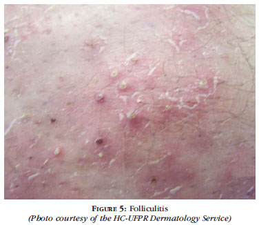
Folliculitis
Folliculitis is inflammation of the hair follicle caused by infection, chemical irritation or physical injury.19 It is histologically defined by the presence of inflammatory cells in the inner wall and ostium of the hair follicles, creating a follicular pustule. 24 The inflammation may be superficial, confined to the upper portion of the hair follicle or extend to the entire hair follicle (Figure 5).19
Infection of the hair follicle is probably the most common form of skin infection, and it affects all ages. 4 Staphylococcal folliculitis, caused by coagulasepositive strains of Staphylococcus (S. aureus), is the most common infectious folliculitis; however, when the host's immune system is weak, the process can be triggered by other microorganisms such as coliform bacilli and plasma coagulase-negative staphylococci.3
Superficial folliculitis, also known as impetigo of Bockhart, is characterized by a small and fragile pustule that occurs in the infundibulum of a hair follicle, usually on the scalp of children and the beard area, axilla, buttocks and extremities of adults. 7 The pustule does not interfere with hair growth.3 Certain conditions make patients more susceptible, including frequent shaving, immunosuppression, pre-existing skin conditions, use of long-term antibiotics, occlusive clothing and/or occlusive dressings, exposure to hot and humid temperatures, diabetes mellitus and obesity. 24 Treatment with topical antibiotics such as mupirocin or fusidic acid 2 times daily for 5-10 days or anti-acne lotion is often sufficient.19
Pseudofolliculitis presents as a papulopustular acneiform eruption on the beard area. It occurs more frequently in men who have curly hair, but it is also observed in women after pubic hair removal.4 This condition is found in 50 to 75% of blacks and 3 to 5% of whites who shave. In general, the problem is more severe in the neck region. 19 It is usually unnecessary to conduct laboratory tests for diagnosis. In exceptional cases, questions may arise regarding differential diagnosis with bacterial or mycotic sycosis. Cytobacteriological and mycological examinations may clarify the diagnosis. 3 A variety of topical and oral drugs that are well tolerated can be used in therapy. Topical retinoids such as tretinoin and adapalene are useful. Benzoyl peroxide and antibiotics such as erythromycin or clindamycin and their combinations are useful as first-line treatments. Tetracycline is a common choice as a systemic antibiotic. Similar to a standard regimen for acne, a dose of 500 mg initially used for 1-3 months is often effective. Doxycycline 50100 mg and minocycline 50-100 mg can be used. Injectable corticoid can be used to reduce inflammation and papules, but due to side effects - skin atrophy and hypopigmentation - it is a temporary treatment. 25 In surgery, laser therapy has revolutionized treatment and allowed individuals who suffer from this condition to be cured. Currently, prevention and early intervention are the mainstays of therapy. 26
Nuchal keloid folliculitis is characterized by deep folliculitis with scarring or perifoliculitis, which occurs in the posterior, inferior occipital and nuchal region of the neck of post-pubescent men. It manifests most commonly in blacks and does not seem to develop in women.4 The characteristic of the process is repair with the formation of isolated or, more commonly, confluent keloid lesions.3 The condition has been recently reported in transplanted Caucasians undergoing cyclosporine therapy. The pathogenesis of the disease is unknown. All identified bacteria probably show a secondary phenomenon. The use of ointments and tight collars on the neck are mentioned as aggravating factors. Hyalinization similar to a true keloid is only an occasional feature. Total loss of sebaceous glands is often observed. 4 Differential diagnosis is done with scalp dissecting folliculitis. Laboratory confirmation is usually unnecessary. 3
Drug therapy includes topical and oral antibiotics, but often with disappointing results. Surgical approaches include excision with primary closure or skin grafting. Another approach is surgical excision with healing by second intention. 27 Studies have shown good efficacy of laser hair removal to treat inflammatory papillae and keloids.28
Folliculitis decalvans (FD) is a form of chronic folliculitis, usually caused by S. aureus, which leads to the destruction of follicles, resulting in scarring alopecia. It occurs predominantly in middle-aged adults. 3,4,6 Lesions develop mainly in the occipital region of the scalp. Clinically, lesions appear as follicular pustules, diffuse and perifollicular erythema, follicular engagement, and often erosive and hemorrhagic crusts. 29 Diagnosis is done based on clinical and histopathological examination and through bacterial culture. Differential diagnoses should include cicatricial alopecia in general, folliculitis abscedens et suffodiens, discoid lupus erythematosus and tinea favosa. 3 Treatment consists in the eradication of S. aureus and use of anti-inflammatory drugs. FD can be very resistant to therapy. Therapeutic options include oral and topical antibiotics, oral and topical corticosteroids, topical antiseptic substances and isotretinoin. 29 Studies have shown good tolerance to dapsone in moderate doses.30 More research studies on the pathogenesis and treatment of this disease are needed for better patient management.29
Sycosis barbae is a deep folliculitis with perifollicular inflammation occurring in the bearded areas of the face and upper lip, caused by infection with S. aureus or dermatophytes. If untreated, the lesions may become chronic. 7 Local inflammation is topically treated with mupirocin. Extensive disease is treated with oral antibiotics such as dicloxacillin or cephalexin for at least two weeks or until all signs of inflammation have disappeared. Relapses are not uncommon and require an additional course of oral antibiotics. 19
Boil is characterized by a painful, hot, red and deep nodule or abscess, which develops from staphylococcal folliculitis (Figure 6). Anthrax is a deeper infection caused by interconnecting abscesses, which usually develop in several adjacent hair follicles. It affects more children, adolescents and young adults, being prevalent in men. The most common etiology is S. aureus. The diagnosis is achieved through clinical symptoms and confirmed by gram stain and culture.
The main risk factor for furunculosis is a positive family history, but cases of anemia, previous anti-biotic therapy, diabetes mellitus, previous hospitalization, multiple injuries, personal hygiene and associated skin diseases need to be considered. In recurrent cases of furunculosis, S. aureus appears as the causative organism in 89% of the cases. Many boils are selflimited and respond well to frequent applications of a hot and humid compress. 30
Treatment includes incision and drainage, often accompanied by systemic antibiotic treatment, which accelerate the regression of the infection in healthy individuals and are essential for any individual at risk of developing bacteremia. The use of the following drugs is recommended: 250 to 500 mg dicloxacillin 4x/day for 10 days; 250 to 500 mg cephalexin 4x/day for 10 days; amoxicillin/clavulanate and macrolides for patients allergic to penicillin. Minocycline, trimethoprim-sulfamethoxazole, ciprofloxacin and vancomycin are used to treat MRSA.19
In recent decades, a growing number of patients with MRSA has been reported. Besides the common problem of MRSA variants in hospitals, recently MRSA acquired in the community (cMRSA) has become a frequent occurrence, even in patients without typical risk factors, causing skin diseases such as refractory furunculosis in young adults. 27 Many drugs such as linezolid and quinupristin-dalfopristin have shown significant efficacy against resistant grampositive bacteria. The use of these drugs should be cautious due to resistance by overuse. Daptomycin has also shown significant evidence of efficacy against resistant bacteria. Novel antibiotics, including oritavancin and dalbavancin, are currently under development. In the future these drugs, with their antibacterial action, may be a relevant option in the fight against serious skin infections. Certain fluoroquinolones such as moxifloxacin and gatifloxacin, which have been available for some years, are FDA-approved for the treatment of infections. 14
Recent works have suggested that 308-nm excimer laser light is a valid therapeutic option for the treatment of resistant forms of folliculitis, especially in areas of difficult therapeutic control. 31
CONCLUSION
Pyodermitis are common diseases and their frequency is related to environmental factors and individual factors, among which are lack of hygiene, and factors related to the degree of virulence and pathogenicity of the microorganism. The main bacteria involved in the etiology of pyodermitis are Staphylococcus aureus and Streptococcus pyogenes.
It is important to identify the more frequent pyodermitis, such as impetigo, boils and erysipelas, and remember that non-cutaneous infections caused by Staphylococcus aureus can cause skin lesions as in staphylococcal scalded skin syndrome.
Pyodermitis can be treated with local care and topical antibiotics in self-limited cases and oral or parenteral antibiotics in extensive cases. We must consider methicillin-resistant Staphylococcus aureus (MRSA) in hospitalized patients who develop symp toms of pyodermitis or even in community patients who do not respond to treatment.

Received on 19.04.2011
Approved by the Advisory Board and accepted for publication on 13.07.2011
Conflict of interest: None
Financial Support: None
- 1. Castro MCR, Ramos-e-Silva, M. Fundamentos de Dermatologia. Rio de Janeiro, RJ: Atheneu; 2009. p. 895-901.
- 2. Rotta O. Guia de Dermatologia: clínica, cirúrgica e cosmiátrica. São Paulo, SP: Manole; 2008. p.109-111.
- 3. Sampaio SAP, Rivitti EA. Dermatologia. 3 ed. São Paulo, SP: Artes Médicas; 2008. p.585-98.
- 4. Veronesi R. Infecções estafilocóccicas e estreptococcias. In: Veronesi R, Focaccia R. Tratado de infectologia. 3 ed. São Paulo, SP: Atheneu; 2005. p.861-96.
- 5. Azulay DR, Azulay RD. Dermatologia 4 ed. Rio de Janeiro, RJ: Guanabara Koogan; 2006. p.287-301.
- 6. Silva MR, Aquino AM, Camilo C. Piodermites na infância: Revisão. Pediatria Atual. 1998;11:27-34.
- 7. Freedberg IM, Eisen AZ, Wolff K, Austen KF, Goldsmith LA, Katz SI, editors. Fitzpatrick`s Dermatology in General Medicine. 7th ed. New York: McGraw-Hill; 2008. p.1689-1723.
- 8. Hirschmann JV. Impetigo: etiology and therapy. Curr Clin Top Infect Dis. 2002;22:42-51.
- 9. Berhman RE, Kliegman RM, Jenson HB. Nelson Tratado de Pediatria. 17a ed. São Paulo, SP: Elsevier; 2005. p.1846-7.
- 10. Hofmann H, Schnopp C. New aspects of bacterial skin infections in children. Ped Dermatol. 2009;60:183-93.
- 11. Lopes HV. Tópicos atuais em Infectologia. Prática Hospitalar. 2007;IX:158-9.
- 12. McKee PH, Calonje E, Granter S. Infectious diseases of the skin. In: McKee PH. Pathology of the Skin with Clinical Correlations. 3rd ed. Philadelphia: Elsevier; 2005. p.872-75.
- 13. Bernard P. Management of common bacterial infections oh the skin. Curr Op Inf Dis. 2008;21:122-8.
- 14. Schweiger SE, Weinberg JM. Novel antibacterial agents for skin and skin struture infections. J Am Acad Dermatol. 2004;50:331-40.
- 15. Caetano M, Amorim I. Erisipela. Acta Med Port. 2005;18:385-394.
- 16. Okajima RMO, Freitas THP, Zaitz C. Estudo clínico de 35 pacientes com diagnóstico de erisipela internados no Hospital Central da Irmandade da Santa Casa de Misericórdia de São Paulo. An Bras Dermatol. 2004;79:295-303.
- 17. Danstra RJ, Van Steensel MAM, Boomsma JHB, Nelemans P, Veraart JC. Erysipelas as a sign of subclinical primary lymphoedema: a prospective quantitative scintigraphic study of 40 patients with unilateral erypelas of the leg. Br J Dermatol. 2008;12:10-5.
- 18. Castellano RL, Teixeira DNS, Antonelli EJ, Rodrigues V Jr, Cavalcanti-Cordeiro MB. Cytokine and nitric oxide production in an adult patient with staphylococcal scalded skin syndrome. Invest Clin. 2008;49:547-552.
- 19. Habif TP. Dermatologia Clínica. 4 ed. São Paulo, SP: Artmed; 2005. p. 281-303.
- 20. Nishifuji K, Sugai M, Amagai M. Staphylococcal exfoliative toxins:``Molecular scissors" of bacteria that attack the cutaneous defense barrier in mammals. J Dermatol Science. 2008;49:21-3.
- 21. Patel GK, Finlay AY. Staphylococcal scalded skin syndrome: diagnosis and management. Am J Clin Dermatol. 2003;4:165-175.
- 22. Porzionato A, Aprile A. Staphylococcal scalded skin syndrome mimicking child abuse by burning. Forensic Sci Int. 2007;168:1-4.
- 23. Johnston GA. Treatment of bullous impetigo and the staphylococcal scalded skin syndrome in infants. Expt Rev Anti-Infec Ther. 2004;2:439-46.
- 24. Satter EK. Folliculitis. Department of dermatology: Naval Medical Center; 2008.
- 25. Scheinfeld NS. Pseudofolliculitis barbae. Skinmed. 2004;3:165-6.
- 26. Perry PK, Cook-Bolden FE, Rahman Z, Jones E, Taylor SC. Defining pseudofolliculitis barbae in 2001: a review of the literature and current trends. J Am Acad Dermatol. 2002;46:S113-9.
- 27. Bajaj V, Langtry JA. Surgical excision of acne keloidalis nuchae with secondary intention healing. Clin Exp Dermatol. 2008;33:53-5.
- 28. Shah GK. Efficacy of diode laser for treating acne keloidalis nuchae. Ind J Dermatol Venereol Leprol. 2005;71:31-4.
- 29. Otberg N, Kang H, Alzolibani AA, Shapiro J. Folliculitis decalvans. Dermatol Ther. 2008;21:238-44.
- 30. El-Gilany AH, Fathy H. Risk factors of recurrent furunculosis. Dermatology Online J. 2009;15:16.
- 31. Nisticò SP, Saraceno R, Carboni I, Chimenti S. Treatment of folliculitis with monochromatic excimer light (308 nm). Dermatology. 2009;218:33-6.
Publication Dates
-
Publication in this collection
03 May 2012 -
Date of issue
Apr 2012
History
-
Received
19 Apr 2011 -
Accepted
13 July 2011