Abstract
Myxosporidiosis is an infectious disease caused by myxozoans of the Phylum Cnidaria, Class Myxosporea, and Order Bivalvulida, considered a common parasite in fresh and saltwater fishes that parasitize many organs, especially gills. In the present study, 49 specimens of fishes belonging to eight genera: Tetragonopterus, Leporinus, Myleus, Pirinampus, Rhapiodon, Pygocentrus, Ageneiosus, and Serrasalmus were collected and blood smears were made, fixed with absolute methanol, and stained with Giemsa 10% to survey hemoparasites. However, myxospores were found in the circulating blood of five (10.20%) fishes belonging to genus Tetragonopterus, Myleus, and Pygocentrus. Two morphological types of Myxobolus spp. were identified in all the five fish specimens analyzed. Usually, investigations on myxozoans in fish are carried out with the search for plasmodia or cysts in the fish organs and observation of the cavity of organs. Nevertheless, this study highlights the importance of also examining the blood of these animals, since these parasites can cause severe pathogenic diseases in fish. Thus, the blood analyses can proportionate preventive sanitary control for commercial fish avoiding economic loss.
Keywords:
Morphology; Myleus; Pygocentrus; Tetragonopterus; occurrence
Resumo
A mixosporidiose é considerada uma doença infecciosa causada por mixozoários pertentences ao Filo Cnidaria, Classe Myxosporea e Ordem Bivalvulida, considerados parasitos comuns de peixes de água doce e salgada, parasitando vários órgãos, principalmente as brânquias. No presente estudo, 49 espécimes de peixes pertencentes a oito gêneros: Tetragonopterus, Leporinus, Myleus, Pirinampus, Rhapiodon, Pygocentrus, Ageneiosus and Serrasalmus foram coletados e extensões sanguíneas foram feitas com a finalidade de encontrar hemoparasitos. Entretanto, mixoesporos foram observados em cinco (10.20%) espécimes de peixes, pertencentes aos gêneros Tetragonopterus, Myleus e Pygocentrus. Dois morfotipos de Myxobolus spp. foram identificados parasitando esses peixes. Normalmente, as investigações sobre mixozoários em peixes é realizada com a procura de plasmódios ou cistos nos órgãos dos peixes e observação da cavidade de órgãos. Porém, esse trabalho ressalta a importância de se examinar também o sangue desses animais, já que esses parasitos podem ocasionar patogenias severas em peixes. Sendo assim, a análise sanguínea pode proporcionar controle sanatório preventivo para peixes comerciais e evitar perdas econômicas.
Palavras-chave:
Morfologia; Myleus; Pygocentrus; Tetragonopterus; ocorrência
1. Introduction
Brazil has a large and dense hydrographic network distributed in eight major basins, with a considerable diversity of fishes and lucrative economy around farmed fish (Graça and Pavanelli, 2007GRAÇA, W. and PAVANELLI, C.S., 2007. Peixes de planície de inundação do alto rio Paraná e áreas adjacentes. Maringá: EDUEM, pp. 241.). However, pathogenic parasites can cause fish diseases resulting in substantial economic losses to aquaculture and fisheries (Okamura et al., 2015OKAMURA, B., GRUHL, A. and BARTHOLOMEW, J., 2015. Introduction. In: B. OKAMURA, A. GRUHL and J.L. BARTHOLOMEW, eds. Myxozoan evolution, ecology and development. USA: Springer Int Publ, pp. 1-20. http://dx.doi.org/10.1007/978-3-319-14753-6_1. ).
Among the diseases reported in fishes, infections with myxozoans are frequently observed (Feist and Longshaw, 2006FEIST, S.W. and LONGSHAW, M., 2006. Phylum Myxozoa. In: P.T.K. WOO, ed. Fish diseases and disorders. Protozoan and metazoan infections. CABI Publishing: Wallingford. pp 230-296 http://dx.doi.org/10.1079/9780851990156.0230.
http://dx.doi.org/10.1079/9780851990156....
). These parasites belong to the Phylum Cnidaria Hatschek, 1888 and Order Bivalvulida Shulman 1959; and until now, more than 2,400 species have been described (Eiras et al., 2014EIRAS, J.C., LIMA, J.T.A.X., CRUZ, C.F. and SARAIVA, A., 2014. A note on infection of Scomberomorus brasiliensis (Osteichthyes, Scombridae) by Kudoa sp. (Myxozoa: multivalvulidae). Brazilian Journal of Biology = Revista Brasileira de Biologia, vol. 74, no. 3, pp. 164-166. http://dx.doi.org/10.1590/1519-6984.23712.
http://dx.doi.org/10.1590/1519-6984.2371...
; Vidal et al., 2017VIDAL, L.G.P., IANNACONE, J., WHIPPS, C.M. and LUQUE, J.L., 2017. Synopsis of the species of Myxozoa Grassé, 1970 (Cnidaria: Myxosporea) in the Americas. Neotropical Helminthology, vol. 11, pp. 413-511.). Regarding the genus Myxobolus Bütschili 1882, about 900 species were described so far, which approximately 60 were described from Brazilian fishes (Lom and Dyková, 2006LOM, J. and DYKOVÁ, L., 2006. Myxozoan genera: definition and notes on taxonomy, life-cycle terminology and pathogenic species. Folia Parasitologica, vol. 53, no. 1, pp. 1-36. http://dx.doi.org/10.14411/fp.2006.001. PMid:16696428.
http://dx.doi.org/10.14411/fp.2006.001...
; Okamura et al., 2015OKAMURA, B., GRUHL, A. and BARTHOLOMEW, J., 2015. Introduction. In: B. OKAMURA, A. GRUHL and J.L. BARTHOLOMEW, eds. Myxozoan evolution, ecology and development. USA: Springer Int Publ, pp. 1-20. http://dx.doi.org/10.1007/978-3-319-14753-6_1. ).
The pathogenesis reported so far, includes reduced respiratory capacity (Adriano et al., 2005ADRIANO, E.A., ARANA, S. and CORDEIRO, N.S., 2005. An ultrastructural and histophatological study of Henneguya pellucida n. sp. (Myxosporea: Myxobolidae) infecting Piaractus mesopotamicus (Characidae) cultivated in Brazil. Parasite (Paris, France), vol. 12, no. 3, pp. 221-227. http://dx.doi.org/10.1051/parasite/2005123221. PMid:16218209.
http://dx.doi.org/10.1051/parasite/20051...
), intestinal necrosis (Alvarez-Pellitero et al., 2008ALVAREZ-PELLITERO, P., PALENZUELA, O. and SITJÀ-BOBADILLA, A., 2008. Histopathology and cellular response in Enteromyxum leei (Myxozoa) infections of Diplodus puntazzo (Teleostei). Parasitology International, vol. 57, no. 2, pp. 110-120. http://dx.doi.org/10.1016/j.parint.2007.09.004. PMid:18373973.
http://dx.doi.org/10.1016/j.parint.2007....
), longitudinal compressions of the body (Longshaw et al., 2003LONGSHAW, M., FREAR, P. and FEIST, S.W., 2003. Myxobolus buckei sp. n.(Myxozoa), a new pathogenic parasite from the spinal column of three cyprinid fishes from the United Kingdom. Folia Parasitologica, vol. 50, no. 4, pp. 251-262. http://dx.doi.org/10.14411/fp.2003.043. PMid:14971593.
http://dx.doi.org/10.14411/fp.2003.043...
), degenerative cardiomyopathy (Yokoyama et al., 2005YOKOYAMA, H., ITOH, N. and TANAKA, S., 2005. Henneguya pagri n. sp. (Myxozoa: Myxosporea) causing cardiac henneguyosis in red sea bream, Pagrus major (Temminck & Schlegel). Journal of Fish Diseases, vol. 28, no. 8, pp. 479-487. http://dx.doi.org/10.1111/j.1365-2761.2005.00655.x.
http://dx.doi.org/10.1111/j.1365-2761.20...
), and significant mortalities in wild and farmed fishes (Naldoni et al., 2009NALDONI, J., ARANA, S., MAIA, A.A.M., CECCARELLI, P.S., TAVARES, L.E.R., BORGES, F.A., POZO, C.F. and ADRIANO, E.A., 2009. Henneguya pseudoplatystoma n. sp. causing reduction in epithelial area of gills in the farmed pintado, a South American catfish: histopathology and ultrastructure. Veterinary Parasitology, vol. 166, no. 1-2, pp. 52-59. http://dx.doi.org/10.1016/j.vetpar.2009.07.034. PMid:19695782.
http://dx.doi.org/10.1016/j.vetpar.2009....
).
During a hemoparasite survey of fishes from Goiás and Mato Grosso States, Brazil, myxospores of myxozoans were observed in the blood of some specimens. This study reported the presence of two different morphotypes of Myxobolus spp. parasitizing Tetragonopterus araguaiensis Silva, Melo, Oliveira, and Benine, 2013, Myleus rubriprinnis (Müller and Troschel, 1844), and Pygocentrus nattereri Kyner, 1858.
2. Material and Methods
In July 2017, blood samples of 49 fishes belonging to eight genera were collected during field surveys in two distinct localities: species of Leporinus, Myleus, Pirinampus, Potamotrygon, Rhapiodon, Pygocentrus, and Tetragonopterus were captured at the Vermelho Red River (14°57'08,4” S and 51°06'30,7” W), municipality of Britânia, Goiás State, Brazil; and two genera, Pygocentrus and Serrasalmus, at the Celeste Lake (14°46’32.27” S and 51°32’50.29” W), municipality of Barra do Garças, Mato Grosso State, Brazil.
All applicable international, national, and/or institutional guidelines for the care and use of animals were followed (Ibama Licences: 60640-1, 52063-3, 17625-4; CEUA-UFU approval: 106/15).
The fishes were captured by fishing rod and were submitted to blood collection with disposable and sterile 1 mL syringes and needles (13 x 4.5 mm), by puncturing the caudal vein. Three blood smears per specimen were made, fixed with methanol, stained with Giemsa Eosin Methylene Blue (Eisen and Schall, 2000EISEN, R.J. and SCHALL, J.J., 2000. Life history of malaria parasite (Plasmodium mexicanum): independent traits and basis for variation. Proceedings. Biological Sciences, vol. 267, no. 1445, pp. 793-799. http://dx.doi.org/10.1098/rspb.2000.1073. PMid:10819149.
http://dx.doi.org/10.1098/rspb.2000.1073...
), and examined using an optical microscope (Leica DMLB 5000, Leica Microsystems, Wetzlar, Germany) at 40x and 100x magnification.
Morphometric measurements of spore length, spore width, polar capsule length, polar capsule width, sporoplasm length, and sporoplasm width from myxospores were taken from digital images at 1000x magnification, under light microscopy with Leica software application suite LAS V3.8 (Leica Microsystems).
3. Results
From 49 specimens screened, myxospores were found in circulating blood of five (10.20%) fishes, being one T. araguaiensis, two M. rubriprinnis, and two P. nattereri (Table 1).
Morphometric characteristics of the Myxobolus spp. (M1 and M2) myxospores reported in the blood of fishes from Goiás State and Mato Grosso State, Brazil.
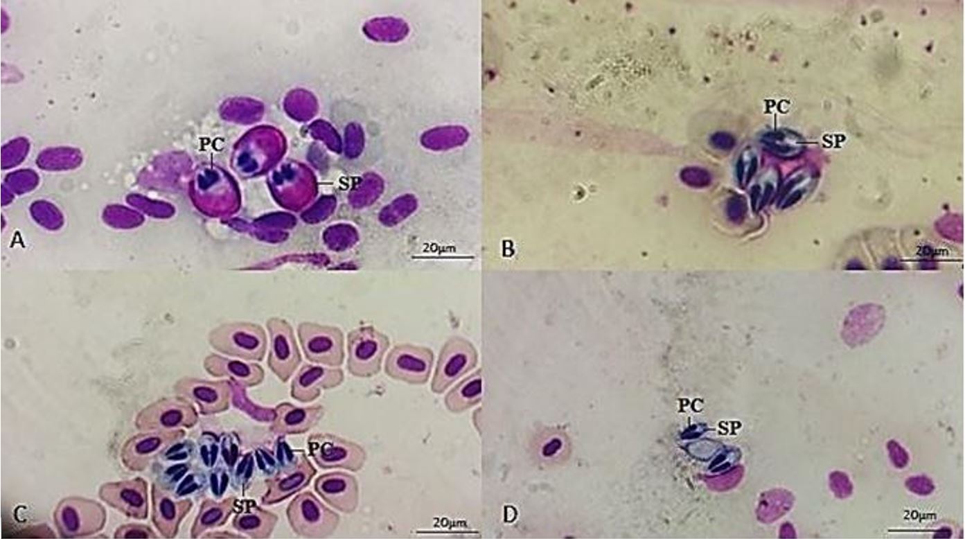
Through the morphological analysis, it is possible to observe myxospores with two polar capsules with similar sizes and without a tail projection. Therefore, these morphological characteristics are compatible with the genus Myxobolus (Figure 1). Moreover, the morphometric analysis revealed two different morphotypes of Myxobolus spp. (Figure 1 and Table 1). The first one (M1), identified in blood smears of T. araguaiensis, revealed larger measures concerning the second morphotype (M2), identified in the blood smears of M. rubriprinnis and P. nattereri (Table 1).

Blood smears of Myxobolus spp. parasites in the circulating blood of fishes from Goiás State and Mato Grosso State, Brazil (A-D): (A) Morphological type 1 (M1) of Myxobolus sp. in the blood of Tetragonopterus araguaiensis; (B-D) Morphological type 2 (M2) of Myxobolus sp. in the blood of Myleus rubriprinnis (b) and Pygocentrus nattereri (c-d). Polar capsule (PC); Sporoplasm (SP).
4. Discussion
In the literature, studies on myxozoans infecting fishes have been developed using morphological, morphometric, and molecular diagnostic tools, with a great number of new species being reported over the years. Regarding the molecular tool, normally, the DNA extraction is made direct from parasite cyst or plasmodia, which contains myxospores (Lom and Arthur, 1989LOM, J. and ARTHUR, J.R., 1989. A guideline for preparation of species descriptions in Myxosporea. Journal of Fish Diseases, vol. 12, no. 2, pp. 151-156. http://dx.doi.org/10.1111/j.1365-2761.1989.tb00287.x.
http://dx.doi.org/10.1111/j.1365-2761.19...
; Úngari et al., 2019ÚNGARI, L.P., VIEIRA, D.H.M.D., DA SILVA, R.J., SANTOS, A.L.Q., DE AZEVEDO, R.K. and O’DWYER, L.H., 2019. A new myxozoan species Henneguya unitaeniata sp. nov. (Cnidaria: Myxosporea) on gills of Hoplerythrinus unitaeniatus from Mato Grosso State, Brazil. Parasitology Research, vol. 118, no. 12, pp. 3327-3336. http://dx.doi.org/10.1007/s00436-019-06533-1. PMid:31728724.
http://dx.doi.org/10.1007/s00436-019-065...
). In this study, few myxospores were found in the circulating blood of fishes, thus the parasite DNA extraction from the blood, and consequently its amplification did not succeed. Therefore, there is a need for a standardized DNA extraction method from the blood for myxozoans.
Regarding the myxobolid group, Myxobolus and Henneguya Thélohan, 1892, are commonly reported in fishes. The main morphological difference between these genera is that Henneguya spp. have spore projections (Lom and Dyková, 2006LOM, J. and DYKOVÁ, L., 2006. Myxozoan genera: definition and notes on taxonomy, life-cycle terminology and pathogenic species. Folia Parasitologica, vol. 53, no. 1, pp. 1-36. http://dx.doi.org/10.14411/fp.2006.001. PMid:16696428.
http://dx.doi.org/10.14411/fp.2006.001...
; Úngari et al., 2019ÚNGARI, L.P., VIEIRA, D.H.M.D., DA SILVA, R.J., SANTOS, A.L.Q., DE AZEVEDO, R.K. and O’DWYER, L.H., 2019. A new myxozoan species Henneguya unitaeniata sp. nov. (Cnidaria: Myxosporea) on gills of Hoplerythrinus unitaeniatus from Mato Grosso State, Brazil. Parasitology Research, vol. 118, no. 12, pp. 3327-3336. http://dx.doi.org/10.1007/s00436-019-06533-1. PMid:31728724.
http://dx.doi.org/10.1007/s00436-019-065...
). Thus, through morphological analysis, it was possible to state the infection with Myxobolus spp. in the circulating blood of the five positive fishes from this study.
There are no reports on myxozoans parasitizing Tetragonopterus spp. and Myxobolus sp. parasitizing P. nattereri, being this study the first one. However, Myxobolus myleus Azevedo, Clemente, Casal, Matos, Alves, Al-Quraishy and Matos, 2012 was described parasitizing the gall bladder from M. rubripinnis (Azevedo et al., 2012AZEVEDO, C., SÃO CLEMENTE, S.C., CASAL, G., MATOS, P., ALVES, A., AL-QURAISHY, S. and MATOS, E., 2012. Myxobolus myleus n. sp. infecting the bile of the Amazonian freshwater fish Myleus rubripinnis (Teleostei: Serrasalmidae): morphology and pathology. Systematic Parasitology, vol. 82, no. 3, pp. 241-247. http://dx.doi.org/10.1007/s11230-012-9360-0. PMid:22711511.
http://dx.doi.org/10.1007/s11230-012-936...
). When compared morphometrically with Myxobolus sp. (M2) found parasitizing the blood of M. rubripinnis in this study, large differences were observed. Myxobolus myleus has a larger body when compared to Myxobolus sp. M2 (19.3 ± 0.5 vs 9.37 ± 1.75). Besides, differences in body width are observed (8.3 ± 0.5 vs 6.43 ± 0.93).
Myxozoans parasitize various organs of fresh and saltwater fishes; can be found in intracellular spaces, inside cells, gills, spleen, liver, kidney, and musculature (Maciel et al., 2011MACIEL, P.O., AFFONSO, E.C., BOIJINK, C.L., TAVARES-DIAS, M. and INOUE, L.A.K.A., 2011. Myxobolus sp. (Myxozoa) in the circulating blood of Colossoma macropomum (Osteichthyes, Characidae). Revista Brasileira de Parasitologia Veterinária, vol. 20, no. 1, pp. 82-84. http://dx.doi.org/10.1590/S1984-29612011000100018. PMid:21439240.
http://dx.doi.org/10.1590/S1984-29612011...
). According to Eiras et al. (2006)EIRAS, J.C., TAKEMOTO, R.M. and PAVANELLI, G.C., 2006. Métodos de estudo e técnicas laboratoriais em parasitologia de peixes. 2. ed. Maringá: Universidade Estadual de Maringá, 199 p., myxozoans can also be found in circulating blood and blood vessels. Maciel et al. (2011)MACIEL, P.O., AFFONSO, E.C., BOIJINK, C.L., TAVARES-DIAS, M. and INOUE, L.A.K.A., 2011. Myxobolus sp. (Myxozoa) in the circulating blood of Colossoma macropomum (Osteichthyes, Characidae). Revista Brasileira de Parasitologia Veterinária, vol. 20, no. 1, pp. 82-84. http://dx.doi.org/10.1590/S1984-29612011000100018. PMid:21439240.
http://dx.doi.org/10.1590/S1984-29612011...
reported myxozoans in blood smears of tambaqui, Colossoma macropomum Cuvier, 1818 (Osteichthyes, Characidae). Although less frequent, the spores present in circulating blood of fish can be explained as a part of the life cycle of some Myxobolus species, reported by Molnár (2002)MOLNÁR, K., 2002. Site preference of fish myxosporeans in the gill. Diseases of Aquatic Organisms, vol. 48, no. 3, pp. 197-207. http://dx.doi.org/10.3354/dao048197. PMid:12033706.
http://dx.doi.org/10.3354/dao048197...
. Another possible explanation is that blood tissue is also considered a form of a dispersion of these pathogens to other organs of the fishes (Maciel et al., 2011MACIEL, P.O., AFFONSO, E.C., BOIJINK, C.L., TAVARES-DIAS, M. and INOUE, L.A.K.A., 2011. Myxobolus sp. (Myxozoa) in the circulating blood of Colossoma macropomum (Osteichthyes, Characidae). Revista Brasileira de Parasitologia Veterinária, vol. 20, no. 1, pp. 82-84. http://dx.doi.org/10.1590/S1984-29612011000100018. PMid:21439240.
http://dx.doi.org/10.1590/S1984-29612011...
).
Lom et al. (1983)LOM, J., DYKOVÁ, I., PAVLÁSKOVÁ, M. and GRUPCHEVA, G., 1983. Sphaerospora molnari sp. nov. (Myxozoa: Myxosporea), an agent of gill, skin and blood sphaerosporosis of common carp in Europe. Parasitology, vol. 86, no. 3, pp. 529-535. http://dx.doi.org/10.1017/S003118200005071X.
http://dx.doi.org/10.1017/S0031182000050...
reported the presence of Sphaerospora molnari Lom, Dyková and Pavlásková, 1983 in the circulating blood of Cyprinus carpio Linnaeus, 1758 from Europa. The blood contained numerous developmental stages, beginning with early pseudoplasmodia containing one sporogonic cell (Lom et al., 1983LOM, J., DYKOVÁ, I., PAVLÁSKOVÁ, M. and GRUPCHEVA, G., 1983. Sphaerospora molnari sp. nov. (Myxozoa: Myxosporea), an agent of gill, skin and blood sphaerosporosis of common carp in Europe. Parasitology, vol. 86, no. 3, pp. 529-535. http://dx.doi.org/10.1017/S003118200005071X.
http://dx.doi.org/10.1017/S0031182000050...
). In this study, only mature myxospores of Myxobolus spp. were observed.
Notwithstanding, it has been suggested that the screening of spores in the fish blood is questionable since the low-intensity parasitosis and few reports in the literature (Eiras et al., 2006EIRAS, J.C., TAKEMOTO, R.M. and PAVANELLI, G.C., 2006. Métodos de estudo e técnicas laboratoriais em parasitologia de peixes. 2. ed. Maringá: Universidade Estadual de Maringá, 199 p.; Maciel et al., 2011MACIEL, P.O., AFFONSO, E.C., BOIJINK, C.L., TAVARES-DIAS, M. and INOUE, L.A.K.A., 2011. Myxobolus sp. (Myxozoa) in the circulating blood of Colossoma macropomum (Osteichthyes, Characidae). Revista Brasileira de Parasitologia Veterinária, vol. 20, no. 1, pp. 82-84. http://dx.doi.org/10.1590/S1984-29612011000100018. PMid:21439240.
http://dx.doi.org/10.1590/S1984-29612011...
). However, the circulating blood and tissue blood should also be investigated for the presence of myxozoans since blood can be part of its life cycle. Also, the blood smear analysis is low cost and can be used as a preventive sanitary control for commercial fish avoiding economic loss.
In conclusion, this study reported 10.20% prevalence of myxobolid mature spores infecting the circulation blood of Brazilian fishes from Goiás and Mato Grosso States, being the first record of myxozoans parasitizing a species of the genus Tetragonopterus and the first record of Myxobolus sp. parasitizing P. nattereri.
Acknowledgements
We thank the team of the Laboratory for Teaching and Research in Wild Animals (LAPAS-UFU) and Non-governmental organization for the preservation of Brazilian wild animals (ONG PAS) for their assistance in the fish blood collection. All applicable international, national, and/or institutional guidelines for the care and use of animals were followed (Ibama Licences: 60640-1, 52063-3, 17625-4; CEUA-UFU approval: 106/15). R.J.S. is supported by CNPq #309125/2017-0. L.P.U. is supported by FAPESP #2018/00754-9 and # 2018q09623-4. L.H.O. is supported by FAPESP #2018/09623-4. D.H.M.D.V. is supported by FAPESP 2019/19060-0.
References
- ADRIANO, E.A., ARANA, S. and CORDEIRO, N.S., 2005. An ultrastructural and histophatological study of Henneguya pellucida n. sp. (Myxosporea: Myxobolidae) infecting Piaractus mesopotamicus (Characidae) cultivated in Brazil. Parasite (Paris, France), vol. 12, no. 3, pp. 221-227. http://dx.doi.org/10.1051/parasite/2005123221 PMid:16218209.
» http://dx.doi.org/10.1051/parasite/2005123221 - ALVAREZ-PELLITERO, P., PALENZUELA, O. and SITJÀ-BOBADILLA, A., 2008. Histopathology and cellular response in Enteromyxum leei (Myxozoa) infections of Diplodus puntazzo (Teleostei). Parasitology International, vol. 57, no. 2, pp. 110-120. http://dx.doi.org/10.1016/j.parint.2007.09.004 PMid:18373973.
» http://dx.doi.org/10.1016/j.parint.2007.09.004 - AZEVEDO, C., SÃO CLEMENTE, S.C., CASAL, G., MATOS, P., ALVES, A., AL-QURAISHY, S. and MATOS, E., 2012. Myxobolus myleus n. sp. infecting the bile of the Amazonian freshwater fish Myleus rubripinnis (Teleostei: Serrasalmidae): morphology and pathology. Systematic Parasitology, vol. 82, no. 3, pp. 241-247. http://dx.doi.org/10.1007/s11230-012-9360-0 PMid:22711511.
» http://dx.doi.org/10.1007/s11230-012-9360-0 - EIRAS, J.C., LIMA, J.T.A.X., CRUZ, C.F. and SARAIVA, A., 2014. A note on infection of Scomberomorus brasiliensis (Osteichthyes, Scombridae) by Kudoa sp. (Myxozoa: multivalvulidae). Brazilian Journal of Biology = Revista Brasileira de Biologia, vol. 74, no. 3, pp. 164-166. http://dx.doi.org/10.1590/1519-6984.23712
» http://dx.doi.org/10.1590/1519-6984.23712 - EIRAS, J.C., TAKEMOTO, R.M. and PAVANELLI, G.C., 2006. Métodos de estudo e técnicas laboratoriais em parasitologia de peixes 2. ed. Maringá: Universidade Estadual de Maringá, 199 p.
- EISEN, R.J. and SCHALL, J.J., 2000. Life history of malaria parasite (Plasmodium mexicanum): independent traits and basis for variation. Proceedings. Biological Sciences, vol. 267, no. 1445, pp. 793-799. http://dx.doi.org/10.1098/rspb.2000.1073 PMid:10819149.
» http://dx.doi.org/10.1098/rspb.2000.1073 - FEIST, S.W. and LONGSHAW, M., 2006. Phylum Myxozoa. In: P.T.K. WOO, ed. Fish diseases and disorders. Protozoan and metazoan infections CABI Publishing: Wallingford. pp 230-296 http://dx.doi.org/10.1079/9780851990156.0230
» http://dx.doi.org/10.1079/9780851990156.0230 - GRAÇA, W. and PAVANELLI, C.S., 2007. Peixes de planície de inundação do alto rio Paraná e áreas adjacentes Maringá: EDUEM, pp. 241.
- LOM, J. and ARTHUR, J.R., 1989. A guideline for preparation of species descriptions in Myxosporea. Journal of Fish Diseases, vol. 12, no. 2, pp. 151-156. http://dx.doi.org/10.1111/j.1365-2761.1989.tb00287.x
» http://dx.doi.org/10.1111/j.1365-2761.1989.tb00287.x - LOM, J. and DYKOVÁ, L., 2006. Myxozoan genera: definition and notes on taxonomy, life-cycle terminology and pathogenic species. Folia Parasitologica, vol. 53, no. 1, pp. 1-36. http://dx.doi.org/10.14411/fp.2006.001 PMid:16696428.
» http://dx.doi.org/10.14411/fp.2006.001 - LOM, J., DYKOVÁ, I., PAVLÁSKOVÁ, M. and GRUPCHEVA, G., 1983. Sphaerospora molnari sp. nov. (Myxozoa: Myxosporea), an agent of gill, skin and blood sphaerosporosis of common carp in Europe. Parasitology, vol. 86, no. 3, pp. 529-535. http://dx.doi.org/10.1017/S003118200005071X
» http://dx.doi.org/10.1017/S003118200005071X - LONGSHAW, M., FREAR, P. and FEIST, S.W., 2003. Myxobolus buckei sp. n.(Myxozoa), a new pathogenic parasite from the spinal column of three cyprinid fishes from the United Kingdom. Folia Parasitologica, vol. 50, no. 4, pp. 251-262. http://dx.doi.org/10.14411/fp.2003.043 PMid:14971593.
» http://dx.doi.org/10.14411/fp.2003.043 - MACIEL, P.O., AFFONSO, E.C., BOIJINK, C.L., TAVARES-DIAS, M. and INOUE, L.A.K.A., 2011. Myxobolus sp. (Myxozoa) in the circulating blood of Colossoma macropomum (Osteichthyes, Characidae). Revista Brasileira de Parasitologia Veterinária, vol. 20, no. 1, pp. 82-84. http://dx.doi.org/10.1590/S1984-29612011000100018 PMid:21439240.
» http://dx.doi.org/10.1590/S1984-29612011000100018 - MOLNÁR, K., 2002. Site preference of fish myxosporeans in the gill. Diseases of Aquatic Organisms, vol. 48, no. 3, pp. 197-207. http://dx.doi.org/10.3354/dao048197 PMid:12033706.
» http://dx.doi.org/10.3354/dao048197 - NALDONI, J., ARANA, S., MAIA, A.A.M., CECCARELLI, P.S., TAVARES, L.E.R., BORGES, F.A., POZO, C.F. and ADRIANO, E.A., 2009. Henneguya pseudoplatystoma n. sp. causing reduction in epithelial area of gills in the farmed pintado, a South American catfish: histopathology and ultrastructure. Veterinary Parasitology, vol. 166, no. 1-2, pp. 52-59. http://dx.doi.org/10.1016/j.vetpar.2009.07.034 PMid:19695782.
» http://dx.doi.org/10.1016/j.vetpar.2009.07.034 - OKAMURA, B., GRUHL, A. and BARTHOLOMEW, J., 2015. Introduction. In: B. OKAMURA, A. GRUHL and J.L. BARTHOLOMEW, eds. Myxozoan evolution, ecology and development USA: Springer Int Publ, pp. 1-20. http://dx.doi.org/10.1007/978-3-319-14753-6_1.
- ÚNGARI, L.P., VIEIRA, D.H.M.D., DA SILVA, R.J., SANTOS, A.L.Q., DE AZEVEDO, R.K. and O’DWYER, L.H., 2019. A new myxozoan species Henneguya unitaeniata sp. nov. (Cnidaria: Myxosporea) on gills of Hoplerythrinus unitaeniatus from Mato Grosso State, Brazil. Parasitology Research, vol. 118, no. 12, pp. 3327-3336. http://dx.doi.org/10.1007/s00436-019-06533-1 PMid:31728724.
» http://dx.doi.org/10.1007/s00436-019-06533-1 - VIDAL, L.G.P., IANNACONE, J., WHIPPS, C.M. and LUQUE, J.L., 2017. Synopsis of the species of Myxozoa Grassé, 1970 (Cnidaria: Myxosporea) in the Americas. Neotropical Helminthology, vol. 11, pp. 413-511.
- YOKOYAMA, H., ITOH, N. and TANAKA, S., 2005. Henneguya pagri n. sp. (Myxozoa: Myxosporea) causing cardiac henneguyosis in red sea bream, Pagrus major (Temminck & Schlegel). Journal of Fish Diseases, vol. 28, no. 8, pp. 479-487. http://dx.doi.org/10.1111/j.1365-2761.2005.00655.x
» http://dx.doi.org/10.1111/j.1365-2761.2005.00655.x
Publication Dates
-
Publication in this collection
11 June 2021 -
Date of issue
2022
History
-
Received
27 Aug 2020 -
Accepted
05 Nov 2020