
Abstract
Laboratory profile of young ovines was studied in order to evaluate and compare their antiserum production from natural and Cobalt-60 irradiated Crotalus durissus terrificus (C.d.t.) venoms. The parameters analyzed included complete blood count, and urea, creatinine, aspartate aminotransferase, total proteins, albumin and globulin serum measurements. Three groups of six animals each were used. Group 1 (G1) received natural C.d.t. venom; Group 2 (G2) received irradiated C.d.t. venom; and Group 3 (G3) was used as control and did not receive venom, only adjuvants, using seven venom inoculations. During the experimental period, animals were fortnightly weighed. According to clinical and weight evaluation, sheep in post-weaning phase showed no changes in their physiological profiles but had excellent weight gain. The parameters analyzed were not statistically different (p<5%) among the groups tested. The hyperimmunization process was successfully accomplished with the production of specific antibodies against Crotalus durissus terrificus venom. Results bring a new possibility of utilizing ovines in the commercial production of anticrotalic serum, which may be used to treat human and animal envenomation. Its production cost may be reduced by subsequent use of hyperimmunized sheep for human consumption.
Crotalus durissus terrificus; hyperimmunization; ovines; antivenom; irradiation
ORIGINAL PAPER
Laboratory evaluation of young ovines inoculated with natural or 60co-irradiated Crotalus durissus terrificus venom during hyperimmunization process
Ferreira Junior R. S.I, II; Nascimento N.III; Couto R.IV; Alves J. B.III; Meira D. A.I; Barraviera B.I, II
IDepartment of Tropical Diseases, Botucatu School of Medicine, São Paulo State University, UNESP, Botucatu, São Paulo, Brazil
IICenter for the Study of Venoms and Venomous Animals, CEVAP, São Paulo State University, UNESP, Botucatu, São Paulo, Brazil
IIIRadiobiology Supervision - Nuclear Energy Research Institute (IPEN/CNEN/SP), São Paulo, Brazil
IVClinical Laboratory of Veterinary, School of Veterinary Medicine and Animal Husbandry, São Paulo State University, UNESP, Botucatu, São Paulo, Brazil
Correspondence to Correspondence to: Rui Seabra Ferreira Júnior Centro de Estudos de Venenos e Animais Peçonhentos CEVAP/UNESP Caixa Postal 577, 18618-000, Botucatu, SP, Brasil Email: rseabra@cevap.org.br
ABSTRACT
Laboratory profile of young ovines was studied in order to evaluate and compare their antiserum production from natural and Cobalt-60 irradiated Crotalus durissus terrificus (C.d.t.) venoms. The parameters analyzed included complete blood count, and urea, creatinine, aspartate aminotransferase, total proteins, albumin and globulin serum measurements. Three groups of six animals each were used. Group 1 (G1) received natural C.d.t. venom; Group 2 (G2) received irradiated C.d.t. venom; and Group 3 (G3) was used as control and did not receive venom, only adjuvants, using seven venom inoculations. During the experimental period, animals were fortnightly weighed. According to clinical and weight evaluation, sheep in post-weaning phase showed no changes in their physiological profiles but had excellent weight gain. The parameters analyzed were not statistically different (p<5%) among the groups tested. The hyperimmunization process was successfully accomplished with the production of specific antibodies against Crotalus durissus terrificus venom. Results bring a new possibility of utilizing ovines in the commercial production of anticrotalic serum, which may be used to treat human and animal envenomation. Its production cost may be reduced by subsequent use of hyperimmunized sheep for human consumption.
Key words:Crotalus durissus terrificus, hyperimmunization, ovines, antivenom, irradiation.
INTRODUCTION
Accidents by the species Crotalus durissus terrificus account for 14% of the ophidic accidents in Brazil with a high mortality rate (12, 28).
Venom from rattlesnakes is extremely toxic although poorly immunogenic (14, 15), which is partially due to the presence of immunosuppressant components (11, 15, 17, 38). Moreover, damage caused to animals after inoculation of crude venom contributes to low antivenom production (31).
Problems observed in patients allergic to equine serum have led to the development of alternative immunization techniques using other animals, which, besides the low cost, have presented excellent results in heterologous serum production (18, 19, 32-37).
Sjostrom et al. (37) produced antivenoms in sheep and compared them with commercial equine serum. Sheep showed tolerance to adjuvants, no local alterations, and fast increase of highly specific antibody titer.
Many researchers have been seeking alternatives to prepare toxoid through venom biological detoxification, which would keep its immunogenicity and minimize the damages to serum producer animals (5, 13, 26, 39).
Gamma radiation has been efficient in attenuating ophidic venoms and in decreasing toxicity without altering immunogenicity, and the addition of substances to the venom was not necessary (1, 2, 8, 10, 16, 20, 21, 32).
Crotalus durissus terrificus venom causes hematological alterations in erythrocytes, leukocytes, platelets, coagulation factors (6) and fibrinogen (3, 40) when inoculated into humans and animals (6).
The aim of the present paper was to evaluate and compare the laboratory profile of young ovines inoculated with natural and 60Co-irradiated Crotalus durissus terrificus venom during the hyperimmunization process. The animals were subjected to hematological exams, biochemical measurements and parasitological tests. These laboratory parameters allowed the evaluation of helminthic infestation, hydration status, and immunological, hepatic and renal functions.
MATERIALS AND METHODS
Crude air-dried venom from a large number of South American rattlesnakes, Crotalus durissus terrificus (C.d.t.), was provided from The Center for the Study of Venoms and Venomous Animals, CEVAP, UNESP, Botucatu, Brazil. Swiss mice (18-22 g) were obtained from the animal facility at the same Institute.
We used 18 male Santa Inês and Ile de France sheep of 60-70 days old, which were kept in the Laboratory for Studies of Reproductive Biotechnology, School of Veterinary Medicine and Animal Husbandry, UNESP, Botucatu, Brazil.
Venom irradiation
Crotalus durissus terificus whole venom was dissolved in saline solution (0.15 M NaCl adjusted to pH 3.0 with concentrated HCl), and its protein concentration adjusted to 2 mg/ml as determined by the Bradford method (9). Samples were irradiated at 5.25 KGy/h with 2000 Gy using gamma rays derived from a 60Co source, Gammacell 220 (Atomic Energy Agency of Canada Ltd), in the presence of O2 at room temperature (30). These experiments were performed at the Institute of Nuclear and Energetic Research, IPEN/CNEM/SP.
Sheep immunization
We used three groups of six animals each. Group 1 (G1) received natural C.d.t. venom; Group 2 (G2) received irradiated C.d.t. venom; and Group 3 (G3) was used as control and did not receive venom, only adjuvants.
Inoculation occurred at six different moments (M): At day one, animals received 500 µg venom diluted with 1 ml saline solution (PBS) homogenized in 1 ml of Freund's Complete Adjuvant (FCA), by intradermal route; at days 14 and 28, they received 1 mg venom diluted with 1 ml PBS homogenized in 1 ml of aluminium hydroxide (AlOH3), by subcutaneous route; at day 42, they received 1.5 mg venom diluted with 2 ml PBS, by subcutaneous route; and at days 56 and 70, animals received 2 mg venom diluted with 2 ml PBS, by subcutaneous route. At day 84, animals were bled and did not receive treatment.
Each animal was injected with 2 ml venom into four different regions of the neck.
Laboratory Evaluation
Complete blood count, biochemical and parasitological exams were performed every 14 days during the experimental period, resulting in seven different moments (M1, M2, M3, M4, M5, M6, M7).
During this period, animals were also weighed.
Total red blood cells and leukocytes count was performed in an automatic cell counter. Hemoglobin was determined by the colorimetric method based on cyanmethemoglobin formation, and globular volume was assessed by the microhematocrit method.
Differential leukocytes count was performed in 100 cells in smears stained according to Rosenfeld method (23).
Plasma total protein concentration was measured by refractometry, and fibrinogen was assessed by the method of heat precipitation (56ºC), as indicated by Kaneko & Harvey (25), and subsequent refractometry.
Samples for cell blood count and plasma total protein and fibrinogen were obtained in EDTA-containing tubes.
Serum samples for the biochemical tests were obtained by centrifugation of total clotted blood. They were stored in 1.5-ml aliquots in polyethylene tubes at -20ºC until use. Biochemical tests were analyzed by spectrophotometry:
All reagents were pro-analysis grade.
Urea: colorimetric enzymatic method.
Creatinine: colorimetric method with kinetic alkaline picrate reaction.
Total serum protein: colorimetric method with biuret reaction.
Albumin: colorimetric method with bromocresol green reaction.
Globulin: difference between total serum protein and albumin concentrations.
Aspartate aminotransferase (AST): optimized kinetic UV method.
Animals received a dose of 1 ml/40 kg weight of vermifuge Levamisol (Ripercol L-150F®), which was repeated after 15 days. The number of eggs per feces [epf] (22) was counted three times: at days 1, 15 and 30, respectively; the first count was before treatment for control.
Statistical analysis
Groups and moments (days) mean values were compared by analysis of repeated measures of the groups mean profiles (or a similar nonparametric procedure) according to Johnson and Wichern (24). Significance level was set at 5% in the F test.
RESULTS AND DISCUSSION
Red blood cells, hemoglobin counts and globular volume showed values within the reference range in the three groups tested with no statistical difference among them. We also noticed a slight increase throughout the experiment.
These results demonstrate that crotalic venom used in the hyperimmunization process did not interfere in sheep red blood cells.
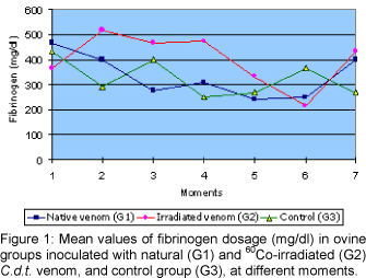
Measurement of plasma protein and fibrinogen showed values within the reference range for the three groups tested with no statistical difference among them.
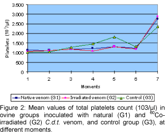
Platelet count values were not within the reference range for the three groups tested but no statistical difference among them was observed. We noticed a significant increase at day 84 (M7) in the three studied groups.
Transitory thrombocytosis may occur due to the epinefrin action during stress, causing splenocontraction and releasing high number of platelets into circulation. Cytokines (IL1, IL3, IL6 and IL11), when produced in inflammatory processes and in reactions that stimulate the immune system, activate megakaryocytes colony factor and can also cause thrombocytosis (27).
As electronic cell count device can count the small ovine erythrocytes as platelets, there may occur technical interference (23), but it would affect all moments of the experiment.
White blood cells count showed that the mean number of leukocytes and neutrophils was within the reference range or limits in the three groups tested, except at day 14 (M2) for the control group, in which an animal had 22,300 leukocytes (/µl) and 13,200 neutrophils (/µl), elevating the group mean values because of an abscess caused by the first inoculation with Freund's Complete Adjuvant. As treatment, we used 5 mg/kg Enrofloxacine (Baytril®) once a day for 5 days. Thereafter, values were within the reference range again. No statistical difference was observed among the groups studied.
Total count of eosinophils and monocytes showed that the mean values found were within the reference range in all the groups without any statistical difference among them.
Serum urea concentration was within the reference range values in the three groups tested, except for Groups 2 and 3 at M1. There was not statistical difference among groups. These animals might have presented values slightly higher than normal due to prerenal causes such as: increased protein ingestion, dehydration, gastrointestinal hemorrhage, heart disease, septic or traumatic shock (7, 27).
Serum creatinine measurement showed values below the reference range in all the groups tested at every moment studied, but no statistical difference was observed among them.
Serum albumin concentration was within the reference range values for the three groups tested, except at days 28 (M3) and 42 (M4), when all groups had values slightly below the ones considered normal for the species. No statistical difference among groups was observed.
According to serum globulin measurement, the values found were within the reference range for all the groups tested at days 01 (M1), 14 (M2), and 70 (M6). There was not statistical difference among groups.
Albumin:globulin relationship might be altered when there is high production of globulins (immunoglobulins). The liver then decreases albumin production to keep the normal relationship. In inflammatory processes, the liver diminishes the albumin yield to produce acute-phase proteins [inflammatory proteins] (29).
Aspartate aminotransferase (AST) measurement showed values within the reference range in the three groups tested. No statistical difference was observed among groups.
The laboratory parameters for the species had been based on Jain NC (23).
All the animals had excellent weight gain throughout the experimental period. The three groups studied did not show any statistical difference.
According to these observations, we can suggest the production of antiserum from young and young-adult sheep, since the hyperimmunization process caused no effects on the animals growth and weight gain; instead, they produced antibodies against Crotalus durissus terrificus venom.
Through parasitological monitoring, we could observe a decrease of endoparasites when counting the number of eggs per gram of feces (epg) at three moments throughout the experiment. The antihelmintic scheme adopted showed a tendency of a decrease of infestation by parasites (4).
Animals from different rural properties showed high parasite infestation rates, which was resolved by the correct use of vermifuge. Animals receiving appropriate parasitological control tend to show better weight gain.
The immunized animals presented an excellent immune response, producing an antiserum of high neutralizing capacity. These results will be shown in a future publication.
Analyzing the results all together, we can conclude that:
In the post-weaning phase, sheep presented no alteration in their physiological profiles and had excellent weight gain, indicating that neither natural nor irradiated venom caused debility or nutritional deficiency.
Since development of the sheep tested was normal, hyperimmunization process was successfully accomplished with the production of specific antibodies against Crotalus durissus terrificus venom.
Utilization of post-weaning ovines as serum producer animals may be an excellent alternative for sheep raisers. According to these results, they could use these animals as food after the hyperimmunization process, and their blood would be another lucrative byproduct available to them.
The present experiment, with confined young animals, will be extended to field animals in order to evaluate the experimental model efficiency. This model will also be tested with venoms from other snakes that commonly cause accidents in Brazil.


Received: September 14, 2006
Accepted: October 31, 2006
Abstract published online: November 6, 2006
Full paper published online: November 30, 2006
- 1 ABIB H., LARABA-DJEBARI F.. Effects of 60Co gamma radiation on toxicity and hemorrhagic, myonecrotic, and edema-forming activities of Cerastes cerastes venom. Can. J. Physiol. Pharmacol., 2003, 81, 1125-30.
- 2 ABIB L., LARABA-DJEBARI F.. Effect of gamma irradiation on toxicity and immunogenity of Androctonus australis hector venom. Can. J. Physiol. Pharmacol., 2003, 81, 1118-24.
- 3 AMARAL CFS., REZENDE NA., PEDROSA TMG., SILVA AO., PEDROSO ERP.. Afibrinogenemia secundária a acidente crotálico (Crotalus durissus terrificus). Rev. Inst. Med. Trop. São Paulo, 1988, 4, 288-92.
- 4 AMARANTE AFT., BAGNOLA JRJ., AMARANTE MRV., BARBOSA MA.. Host specificity of sheep and cattle nematodes in São Paulo State. Brazil Vet. Parasitol., 1997, 73, 89-104.
- 5 ARIARATNAM CA., SJOSTROM L., RAZIEK Z., KULARATNE SAM., THEAKSTON DG., WARRELL DA.. Merit and demerit of polyvalent snake antivenom. Trans. R. S. Trop. Med. Hyg., 2001, 95, 74-80.
- 6 BARRAVIERA B., PERAÇOLI MTS.. Soroterapia heteróloga. In: BARRAVIERA B.. Venenos animais: uma visão integrada. Rio de Janeiro: Editora de Publicações Científicas Ltda, 1994, cap. 28, 411.
- 7 BARSANTI JA., LEES GE., WILLARD MD., GREEN RA.. Urinary disorders. In: WILLARD MD, TVEDTEN H. Small animal clinical diagnosis by laboratory methods. 4 Ed. 2004, 135-164p.
- 8 BONI-MITAKE M., COSTA H., SPENCER PJ., VASSILIEFF VS., ROGERO JR.. Effects of 60Co gamma radiation on crotamine. Braz. J. Med. Biol. Res., 2001, 34, 1531-38.
- 9 BRADFORD MM.. A rapid and sensitive method for the quantitation of microgram quantities of protein utilizing the principle of protein-dye binding. Anal. Biochem., 1976, 72, 248-54.
- 10 CARDI BA., NASCIMENTO N., ANDRADE Jr HF.. Irradiation of Crotalus durissus terrificus crotoxin with 60Co y-rays induces its uptake by macrophages through scavenger receptors. Int. J. Radiat. Biol., 1998, 73, 557-64.
- 11 CARDOSO DF., MOTA I.. Effect of Crotalus venom on the humoral and cellular immune response. Toxicon, 1997, 35, 607-12.
- 12 CARDOSO JLC., FRANÇA FOS., WEN FH., MÁLAQUE CLMS., HADDAD JR. V.. Animais peçonhentos no Brasil: biologia clinica e terapêutica dos acidentes. Ed. Sarvier, 2003, 468p.
- 13 CHIPPAUX JP., GOYFFON M.. Venoms, antivenoms and immunotherapy. Toxicon, 1998, 36, 823-46.
- 14 CHRISTENSEN PA.. The preparation and purification of antivenoms. Mem. Inst. Butantan, 1966, 33, 245-50.
- 15 CLISSA PB., NASCIMENTO N., ROGERO JR.. Toxicity and immunogenicity of Crotalus durissus terrificus venom treated with different doses of gamma rays. Toxicon, 1999, 37, 1131-41.
- 16 DE PAULA RA.. Attainment and evaluation of antisera raised against irradiated whole crotalic venom or crotoxin in 60Co source. J. Venom. Anim. Toxins, 1996, 2, 166.
- 17 DOS SANTOS MC., D'IMPERIO-LIMA R., DIAS DA SILVA W.. Influence of Crotalus venom on the response to sheep red blood cells. Braz. J. Med. Biol. Res., 1986, 19, 636.
- 18 EGEN N., RUSSEL F., CONSROE P., GERRISH K., DART R.. A new ovine Fab antivenom for North American venomous snakes. Vet. Hum. Toxicol., 1994, 36, 362.
- 19 EK N.. Serum levels of the immunoglobulins IgG and IgG(T) in horses. Acta Vet. Scand., 1974, 15, 609-19.
- 20 FERREIRA JUNIOR RS., NASCIMENTO N., MARTINEZ JC., ALVES JB., MEIRA DA., BARRAVIERA B.. Immunization with native and Cobalt 60-irradiated Crotalus durissus terríficus venom in Swiss mice: assessment of the neutralizing potency of antisera. J. Venom. Anim. Toxins. Incl. Trop. Dis., 2005, 11, 300-14.
- 21 FERREIRA JUNIOR RS., NASCIMENTO N., MARTINEZ JC., ALVES JB., MEIRA DA., BARRAVIERA B.. Immunological assessment of mice hyperimmunized with native and Cobalt-60-irradiated Bothrops venoms. J. Venom. Anim. Toxins incl. Trop. Dis., 2005, 11, 447-64.
- 22 GORDON HM., WHITLOCK HUA.. A new technique for counting nematodes eggs in sheep faeces. J. Counc. Sci. Ind. Res., 1939; 12/13, 50-2.
- 23 JAIN NC.. Essentials of veterinary hematology. Philadelphia: Lea & Febiger, 1993, 417p.
- 24 JOHNSON RA., WICHERN DW.. Applied multivariate statistical analysis, 3rd Ed., Prentice-Hall, New Jersey, 1992, 642p.
- 25 KANEKO JJ., HARVEY JW.. Clinical biochemistry of domestic animals. 5ed. New York: Academic Press., 1997, 932p.
- 26 KHAN ZH., LARI FA., ALI Z.. Preparation of toxoids from the venoms of Pakistan species of snakes (Naja naja, Vipera russelii and Echis carinatus). Jpn. J. Med. Sci. Biol, 1977, 30, 19-23.
- 27 MEYER DJ., HARVEY JW.. Evaluation of hemostasis: coagulation and platelet disorders. In: Veterinary laboratory medicine. 1998, 111-137 p.
- 28 MINISTÉRIO DA SAÚDE BRASIL. Manual de diagnóstico e tratamento de acidentes por animais peçonhentos. Brasília: Fundação Nacional de Saúde, 1998, 131 p.
- 29 MOSHAGE HJ., JANSEN JAM., FRANSSEN JH., HAFKENSCHEID JCM.. Study of the molecular mechanism of decreased liver synthesis of albumin in inflammation. J. Clin. Invest., 1987, 79, 1635-41.
- 30 NASCIMENTO N., SEEBART C., FRANCIS B., ROGERO JR., KAISER II.. Influence of ionizing radiation on crotoxin: biochemical and immunological aspects. Toxicon, 1996, 34, 123-31.
- 31 NETTO DP., CHIACCHIO SB., BICUDO PL., ALFIERI AA., NASCIMENTO N.. Hematological changes in sheep inoculated with Crotalus durissus terrificus snake (Laurenti, 1768) venom irradiated with Cobalt60 and in the natural form. J. Venom. Anim. Toxins incl. Trop. Dis., 2004, 10, 34-52.
- 32 NETTO DP., CHIACCHIO SB., BICUDO PL., ALFIERI AA., NASCIMENTO N.. Humoral response and neutralization capacity of sheep serum inoculated with natural and Cobalt 60-irradiated Crotalus durissus terrificus venom (Laurenti, 1768). J. Venom. Anim. Toxins, 2002, 8, 297-314.
- 33 RAWAT S., LAING G., SMITH DC., THEAKSTON D., LANDON J.. A new antivenom to treat eastern coral snake (Micrurus fulvius fulvius) envenoming. Toxicon, 1994, 32, 185-90.
- 34 RUSSEL FE., LAURITZEN L.. Antivenins. Trans. R. Soc. Trop. Med. Hyg., 1966, 60, 797- 801.
- 35 RUSSEL FE., TIMMERMAN WF., MEADOWS PE.. Clinical use of antivenin prepared from goat serum. Toxicon, 1970, 8, 63-5.
- 36 SALAFRANCA ES.. Irradiated cobra (Naja naja philippinensis) venom. Int. J. Appl. Radiat. Isot, 1973, 24, 60.
- 37 SJOSTROM L., AL-ABDULLA IH., RAWAT S., SMITH DC., LANDON J.. A comparison of ovine and equine antivenoms. Toxicon, 1994, 32, 427-33.
- 38 SOUZA E SILVA MCC., GONÇALVES LRC., MARIANO M.. The venom of South American rattlesnake inhibits macrophage function and is endowed with anti-inflammatory properties. Mediat. Inflamm., 1996, 5, 18-23.
- 39 THEAKSTON RDG., WARREL DA., GRIFFITHS E.. Report of a WHO workshop on the standardization and control of antivenoms. Toxicon, 2003, 41, 541-57.
- 40 THOMAZINI IA., IUAN FC., CARVALHO I., HERNANDES DH., AMARAL IF., PEREIRA PCM., BARRAVIERA B.. Evaluation of platelet function and of serum fibrinogen levels in patients bitten by snakes of the genus Crotalus Preliminary report. Rev. Inst. Med. Trop. São Paulo, 1991, 3, 219-20.
Publication Dates
-
Publication in this collection
11 Jan 2007 -
Date of issue
2006
History
-
Received
14 Sept 2006 -
Accepted
31 Dec 2006
