Abstracts
OBJECTIVE: This paper aims to check the proximal tibial valgusing open-wedge osteotomy union with Anthony® plate for the treatment of bowleg with medial osteoarthrosis, final correction of the deformity and clinical improvement. METHODS: Twenty patients (twenty knees) with medial osteoarthrosis of the knee, with mean age of 48.4 years, were evaluated for one year. The patients were submitted to the Lysholm's score, and also to X-ray studies before and after surgery. RESULTS: The osteotomy union occurred after 12 weeks in all cases without complications. The Lysholm's score was regarded as excellent or good in 80% of the cases. The postoperative mechanical alignment was 3.4 ± 3.3 valgus. CONCLUSION: We conclude that the union happened within 3 months with the use of bone grafting and the Anthony® plate to fix the open wedge osteotomy. The open wedge osteotomy is effective in fixing the deformity of the knee providing a significant improvement to patients' lives.
Osteotomy; Osteoarthritis; Knee
OBJETIVO: Este estudo tem por finalidade verificar a consolidação da osteotomia valgizante da tíbia com cunha de abertura fixada com placa tipo calço de Anthony® (OVT), no tratamento da osteoartrose medial do joelho varo, a correção da deformidade e a resposta clínica ao tratamento cirúrgico. MÉTODOS: Vinte pacientes (vinte joelhos) com osteoartrose do compartimento medial do joelho, com idade média de 48,4 ± 9,9, foram avaliados por um período mínimo de um ano. Os pacientes foram submetidos a avaliação radiográfica da consolidação e do eixo mecânico no pré e pós operatório, além da avaliação dos critérios de LYSHOLM. RESULTADOS: A consolidação da osteotomia ocorreu após 12 semanas em 100% dos casos sem complicações. A avaliação do LYSHOLM no pós operatório apresentou 80% de excelentes e bons resultados. A correção final média do eixo mecânico foi de 3,4 ± 3,3 graus de valgo. CONCLUSÃO: Concluímos que a consolidação da osteotomia supra-tuberositária da tíbia com cunha de abertura fixada com placa calço de Anthony® e com enxertia óssea tricortical ocorre num intervalo de três meses. A cirurgia é eficaz para a correção da deformidade em varo do joelho, e propicia melhora clínica significante para o paciente.
Osteotomia; Osteoartrite; Joelho
ORIGINAL ARTICLE
Proximal tibial valgusing open-wedge osteotomy union fixated with Anthony® "support" plate
Cristiano Hossri RibeiroI; Nilson Roberto SeverinoII; Ricardo de Paula Leite CuryIII; Victor Marques de OliveiraIV; Roger Avakian; Tatsuo AyharaIV; Osmar Pedro Arbix de CamargoV
IPost-graduation student, Knee Surgery Group, Department of Orthopaedics and Traumatology, FCMSSP - Medical Sciences School, Santa Casa de São Paulo
IIAssistant Professor, Knee Surgery Group, Department of Orthopaedics and Traumatology, FCMSSP - Medical Sciences School, Santa Casa de São Paulo
IIIAssistant Professor and Head of Knee Surgery Group, Department of Orthopaedics and Traumatology, FCMSSP - Medical Sciences School, Santa Casa de São Paulo
IVAssistant Professor of Knee Surgery Group, Department of Orthopaedics and Traumatology, FCMSSP - Medical Sciences School, Santa Casa de São Paulo
VAssociate Professor and Chairman of the Medicine Course, FCMSSP - Medical Sciences School, Santa Casa de São Paulo
Correspondences to
SUMMARY
OBJECTIVE: This paper aims to check the proximal tibial valgusing open-wedge osteotomy union with Anthony® plate for the treatment of bowleg with medial osteoarthrosis, final correction of the deformity and clinical improvement.
METHODS: Twenty patients (twenty knees) with medial osteoarthrosis of the knee, with mean age of 48.4 years, were evaluated for one year. The patients were submitted to the Lysholm's score, and also to X-ray studies before and after surgery.
RESULTS: The osteotomy union occurred after 12 weeks in all cases without complications. The Lysholm's score was regarded as excellent or good in 80% of the cases. The postoperative mechanical alignment was 3.4 ± 3.3 valgus.
CONCLUSION: We conclude that the union happened within 3 months with the use of bone grafting and the Anthony® plate to fix the open wedge osteotomy. The open wedge osteotomy is effective in fixing the deformity of the knee providing a significant improvement to patients' lives.
Keywords: Osteotomy. Osteoarthritis. Knee.
INTRODUCTION
Osteoarthrosis is the most common form of joint condition. Its prevalence reaches up to 90% of the population above the age of 40, when load joints are radiographically assessed. At least 20 million people in the United States are estimated to have the disease.
Knee is one of the most frequently affected joints, because, additionally of being a load joint, it is usually affected by alignment deformities of the lower limbs, which is recognized as a triggering factor with worse prognosis to osteoarthrosis.
Among the alignment deformities of the knee, the most common one is genuvarus, a change that usually implies in osteoarthrosis at the medial knee compartment, which manifests as pain, deformity, and lost range of motion.
Surgical treatment for osteoarthrosis associated to a misalignment of the limb was first described by Volkman apud Poilvache(1) in Europe, in 1875. That procedure intended, by realigning the limb, to transfer the knee load axis from the affected region to a healthier one, thus increasing a joint's life span. However, osteotomy only became popular in the United States with Coventry(2), in the 1960's.
Since then, many surgical techniques have been suggested and improved, including the tibial valgusing osteotomy (TVO) with open-wedge, fixated with medial support plate, which deserves to be highlighted because it allows for an early mobility due to a stable fixation, because it preserves the bone content of the metaphyseal region, and, finally, because it leads to a lower complication rate.
This technique was developed approximately 15 years ago, and may employ different fixation materials. The present study uses a new fixation method: the Anthony support plate, and gathers information about osteotomy union, the correction achieved by such procedure, and patients' clinical response
METHODS
After the approval granted by the Committee on Ethics in Research of Santa Casa de São Paulo, Department of Orthopaedics and Traumatology, between October 2004 and November 2006, 20 subjects were selected with knee medial osteoarthrosis and deformity in varus. All subjects agreed to participate on the study by signing an informed consent term.
The study dynamics involved the selection of patients from the outpatient facility of the Knee Group, Department of Orthopaedics and Traumatology, clinical evaluation, X-ray studies, and subjective analysis by the Lysholm score, followed by surgical procedure, union evaluation three months later, and clinical and radiographic re-evaluation after one year postoperatively.
The inclusion criteria for the study were the following: presence of idiopathic knee medial unicompartmental osteoarthrosis, genuvarus of up to 20 degrees, preserved range of motion, meaning at least 90 degrees of flexion and less than 15 degrees of contraction at flexion, stable knee and age below 60 years old.
Exclusion criteria were the following: previous knee surgery, ligament instability, deformity in varus above 20 degrees, clinical degree of osteoarthrosis, patellofemoral pain, and rheumatoid arthritis diagnosis.
Osteoarthrosis diagnosis was given according to the clinical and radiographic picture of each patient. It was determined as a clinical criterion the presence of pain at knee medial compartment for over a year. The employed radiographic criteria were the ones described by Ahlbäck(3), who provided a phased osteoarthrosis classification into 5 grades. In our case series, the X-ray evaluation of the patients was bilaterally made at anteroposterior and lateral planes with 30º of orthostatic and axial patellar flexion at 30 degrees, as well as a panoramic plane with bipodal load. These planes enabled the evaluation of the arthrosis grade, of knee mechanical axis, and the measurement of the open wedge.
The mechanical axis was calculated by drawing a line from the center of the femoral head to the center of the knee, and another line from the center of the knee to the center of the ankle. The acute angle formed by the intersection of both lines at the center of the knee comprises the mechanical axis (Figure 1).
The open wedge was calculated by the method described by Dugdale et al(4). This method targets to transfer the load from the lower limb to the lateral plateau at a position corresponding to 62% of tibial joint surface, laterally. For this, tibial plateau length is measured and the desired point is calculated by the rule of three. A line is then drawn from the center of femoral head to the previously determined point on the knee and another line is made from the center of the ankle to the point fixated on the knee. The intersection of both lines will form an angle corresponding to the required tibial opening to achieve, at the end of osteotomy, a final mechanical axis of 5 degrees in valgus (Figure 2).
The subjective evaluation was made with the Lysholm scale(5). In this scale, the patient assigns a score to symptoms of limping, support, knee restraint sensation, instability, presence of joint effusion, difficulty to climb stairs and to squat. According to the score achieved, knee functional performance is rated as excellent (95-100 points), good (84-94 points), fair (65-83 points) and poor (<64 points).
Descriptive variables have been analyzed as mean and standard deviation values. The mechanical axis was regarded as a continuous variable, and pre- and postoperative periods were compared by means of the Student's t test. The Lysholm score was regarded as a categorical and continuous variable. For identifying correlations between study variables, the Pearson's Linear Correlation method was employed. The Kruskall-Wallis' test was used for seeking explicative variables for improvements of Lysholm scores.
RESULTS
Twelve men and eight women participated in the study. The mean age of the subjects was 48.4 years. Eleven right knees and nine left knees were operated. All patients submitted to surgery had arthrosis grade 1 or 2.
Preoperatively, the patients had a mean mechanical axis of 8.1 degrees of varus (-8.1), with standard deviation of 3.1 degrees. The mean correction of the mechanical axis was 11.5 degrees, with standard deviation of 4.6 degrees (Table 1).
The initial clinical evaluation by the Lysholm score showed a mean score of 40.85 points, where 19 patients fit the poor outcome and only one was regarded as fair. Postoperatively, a mean increment of 46.75 points was seen, with a final score of 87.60 points, in average. All patients showed increased scores, and only one was still regarded as poor, three were rated as fair, nine as good, and seven as excellent.
The comparison between pre- and postoperative moments showed that the mechanical axis and the Lysholm score had a significant change (p<0.001).
The mean value obtained from open wedges performed was 10.8 degrees, with a standard deviation of 2.3 degrees.
Correlation analyses showed that the greater the mechanical axis preoperatively, the greater the open wedge employed. All cases showed union within three months postoperatively.
DISCUSSION
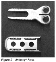
Literature is rich concerning valgusing osteotomies with other synthesis materials in terms of union, deformity correction and clinical improvement of patients. However, our study is one of the first to assess these outcomes with an Anthony® plate (Figure 3).
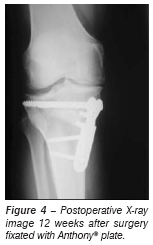
According to literature reports, union occurs within 10 - 16 weeks(6). In our study, TVO union occurred in 100% of the cases within up to 12 weeks (Figure 4). We believe that the use of an Anthony® plate has contributed to this successful outcome, due to fixation stability provided by resistant, long and striated supports. We believe that the use of bone grafting in all cases was another important contributing factor to such result.
The decision of using a three-cortical bone graft was based on studies by Puddu(7), which recommends the use of bone grafting in open wedges above 7.5 degrees. Our smaller wedge had 8 degrees.
The use of bone graft should also have contributed to reduce pseudoarthrosis cases. In our study, we didn't see this complication, but literature data show that pseudoarthrosis may occur in approximately 4% of the cases(8). The open wedge itself - as employed in this technique - is known to be a risk factor for pseudoarthrosis development, because it opens a large space between osteotomy surfaces. There are authors who believe that the presence of a thin proximal fragment also represents a risk for pseudoarthrosis development(9). Therefore, we tried to initiate the osteotomy incision four centimeters below the joint surface, at the upper edge of the anterior tibial tuberosity. Osteotomy should not be performed below tibial anterior tuberosity, because this technique increases the risk of pseudoarthrosis(10).
Concerning deformity correction, literature has demonstrated a good genuvarus correction, i.e., a postoperative mechanical axis between 3 and 6 degrees of valgus with the use of other plates(4,11-13). Our study achieved a mean final mechanical axis of 3.4 degrees of valgus(13). We noticed that the mean correction of the deformity in our study was 11.5 degrees, which is consistent to the study by Hart R, with mean correction of 11.1 degrees. This demonstrates that the use of Anthony plate associated to the calculation of the wedge by Dugdale's method reproduces the satisfactory results found in literature using other plates when we assess deformity correction.
Tibial supra-tuberosity valgusing osteotomy is regarded as an useful therapeutic option for treating knee medial osteoarthrosis, providing pain relief and improved function in approximately 80-90% of the patients in five years, and 50 - 65% in ten years of follow-up(14,15).
In order to assess clinical improvements, we selected the Lysholm score because it is a reproducible, validated and user-friendly instrument allowing for a subjective analysis of the patient.
By following the Lysholm score8, we achieved improvement in 95% of the patients assessed, with 80% of excellent and good results after 12 months postoperatively, but we know that this is a short follow-up period. The patients presented 40.8 points preoperatively (average) and 87.6 points postoperatively, a mean increment of 46.75 points.
The gain by Lysholm subjective analysis was significant: 100% of the patients showed improved scores within one year of follow-up. Of the 20 patients operated, 19 have moved from one classification to a better one, which means that only one subject could not improve rating so as to move to other category. That was the first surgical case of the study.
By making a comparison to the study by Hart(13), where the initial Lysholm score was 55 points, while the final one was 82 (an increment of 27 points after two years of follow-up), we can see that even with a much lower Lysholm score (40.8) we accomplished a very similar final result, but with a greater increment (46.8 against 27 points).
This difference between the increments on Lysholm scores may be a result of a shorter postoperative follow-up time in our study. Perhaps with two years of follow-up patients would present a drop on the Lysholm score, which would reduce this difference. Even so, our results are consistent with literature in what concerns to subjective evaluation of clinical improvement.
CONCLUSION
We conclude that tibial supra-tuberosity osteotomy union with open wedge fixated with Anthony® support plate and three-cortical bone grafting happens within a time span of three months. Surgery is effective in correcting varus deformities of the knee, and provides a significant clinical improvement for the patient.
REFERENCES
- 1. Poilvache P. Osteotomy for the arthritic knee: a Eurepean perspective. In: Insall JN, Scott WN. Surgery of the knee. 3rd ed. Philadelphia: Churchill Livingstone; 2001. p.1465-505.
- 2. Coventry MB. Stepped stable for upper tibialosteotomy. J Bone Joint Surg Am. 1969;51:1011.
- 3. Albäck Sven. Osteoarthrsis of the knee. A radiographic investigation. Acta Radiol Diagn (Stockh). 1968:Suppl 277:7-72.
- 4. Dugdale TW, Noyes FR, Styer D. Preoperative planning for high tibialosteotomy: The effect of lateral tibiofemoral separation and tibiofemoral length. Clin Orthop Relat Res. 1992;(274):248-64.
- 5. Lysholm J, Gillquist J. Evaluation of knee ligament surgery results with special emphasis on use of a scoring scale. Am J Sports Med. 1982;10:150-4.
- 6. Bombaci H, Canbora K, Onur G, Gorgeç M. The effect of open wedge osteotomy on the posterior tibial slope. Acta Orthop Traumatol Turc. 2005;39:404-10.
- 7. Puddu G. Osteotomies about the athetic knee. In: Drez D Jr, DeLee J. editors. Operative techniques in sports medicine. Philadelphia: Saunders; 2000.
- 8. Cameron HU, Park YS. Total Knee replacement after supraconsilar femoral osteotomy. Am J Knee Surg. 1997;10:70-1.
- 9. Insall JN, Joseph DM, Msika C. High tibialosteotomy for varusgonarthrosis. J Bone Joint Surg Am. 1984;66:1040-8.
- 10. Jackson JP, Waugh W.The technique and complications of upper tibialosteotomy: A review of 226 operations. J Bone Joint Surg Br. 1974;56:236-45.
- 11. Marti CB, Gautier E, Wachtl SW, Jakob RP. Accuracy of frontal and sagital plane correction in open wedge high tibialosteotomy. Arthroscopy. 2004;20:366-72.
- 12. Esenkaya I, Elmali N. Proximal tibial medial open-wedge osteotomy using plates with wedges: early results in 58 cases. Knee Surg Sports Traumatol Arthrosc. 2006;14:955-61.
- 13. Hart R, Stipak V, Kucera B, Filian P, deCordeiro J. Precise, computed assisted leg angle correction with open wedge high tibialosteotomy. Orthopade. 2007;36:577-81.
- 14. Billings A, Scott DF, Camargo MP, Hofmann AA. High tibialosteotomy with a calibrated osteotomy guide, rigid internal fixation, and early motion: long term follow-up. J Bone Joint Surg Am. 2000;82:70-9.
- 15. Rinonapoli E, Mancini GB, Corvaglia A, Musiello S. Tibialosteotomy for varusgonarthrosis: a 10 to 21-year follow-up study. Clin Orthop Relat Res. 1998;(353):185-93.
Publication Dates
-
Publication in this collection
02 Dec 2008 -
Date of issue
2008
History
-
Received
01 Mar 2007 -
Accepted
06 May 2007


