Innovative public-health strategies for neurodegenerative disease: leveraging diversified ultraviolet irradiation as a next-generation therapy
Abstract
Neurodegenerative diseases (NDs) such as Alzheimer’s, Parkinson’s, Huntington’s disease, and amyotrophic lateral sclerosis are escalating worldwide, straining healthcare systems and leaving patients with therapies that are largely palliative. Emerging evidence positions diversified ultraviolet (UV) irradiation as a groundbreaking, non-invasive strategy to counter these disorders. Beyond its traditional use in sterilization, specific UV spectra, UV-B (280–320 nm), UV-C (200–280 nm), and far-UV (207–222 nm), are now recognized for modulating oxidative stress, restoring mitochondrial function, correcting apoptotic dysregulation, and enhancing DNA repair. Innovative approaches such as riboflavin-mediated phototherapy and photobiomodulation (PBM) show the capacity to disaggregate toxic protein aggregates like β-amyloid and α-synuclein, boost antioxidant defenses, stimulate neurotrophic factors, and quell neuroinflammation. Preclinical models and early clinical trials reveal preserved cognition, enhanced neurogenesis, and reduced disease biomarkers, suggesting real translational promise. From a public-health perspective, UV-based interventions offer a cost-effective, scalable option for aging populations and resource-limited settings, especially when integrated with community-level health technologies and remote delivery platforms. Continued investigation of optimal dosing, long-term safety, and mechanistic pathways will be pivotal to unlock the full therapeutic and population-wide impact of this novel modality.
Keywords:
neurodegenerative diseases; ultraviolet irradiation; neuroprotection; photobiomodulation; Alzheimer’s Disease; Parkinson’s Disease; amyotrophic lateral sclerosis; Huntington’s disease; oxidative stress modulation; protein aggregation therapy
Resumo
Doenças neurodegenerativas (DNDs), como Alzheimer, Parkinson, doença de Huntington e esclerose lateral amiotrófica, estão aumentando em todo o mundo, sobrecarregando os sistemas de saúde e deixando os pacientes com terapias em grande parte paliativas. Evidências emergentes posicionam a irradiação ultravioleta (UV) diversificada como uma estratégia inovadora e não invasiva para combater essas doenças. Além de seu uso tradicional em esterilização, espectros UV específicos, UV-B (280-320 nm), UV-C (200-280 nm) e UV-distante (207-222 nm), são agora reconhecidos por modular o estresse oxidativo, restaurar a função mitocondrial, corrigir a desregulação apoptótica e aprimorar o reparo do DNA. Abordagens inovadoras, como a fototerapia mediada por riboflavina e a fotobiomodulação (FBM), demonstram a capacidade de desagregar proteínas tóxicas, como β-amiloide e α-sinucleína, aumentar as defesas antioxidantes, estimular fatores neurotróficos e suprimir a neuroinflamação. Modelos pré-clínicos e ensaios clínicos iniciais revelam cognição preservada, neurogênese aprimorada e biomarcadores de doenças reduzidos, sugerindo um potencial translacional real. Do ponto de vista da saúde pública, as intervenções baseadas em UV oferecem uma opção econômica e escalável para populações em envelhecimento e ambientes com recursos limitados, especialmente quando integradas a tecnologias de saúde comunitárias e plataformas de atendimento remoto. A investigação contínua da dosagem ideal, segurança a longo prazo e vias mecanísticas será fundamental para desbloquear o impacto terapêutico e populacional desta nova modalidade.
Palavras-chave:
doenças neurodegenerativas; irradiação ultravioleta; neuroproteção; fotobiomodulação; Doença de Alzheimer; Doença de Parkinson; esclerose lateral amiotrófica; Doença de Huntington; modulação do estresse oxidativo; terapia de agregação de proteínas
1. Introduction
Neurodegenerative diseases such as Alzheimer's, Parkinson’s, Huntington’s, and amyotrophic lateral sclerosis (ALS) represent a mounting global public health challenge. These progressive disorders not only cause substantial disability and reduced quality of life but also impose a significant economic burden on healthcare systems worldwide. In light of limited curative options, the development of scalable, non-invasive, and cost-effective therapies is critical. Ultraviolet (UV) irradiation, historically applied in public health for microbial disinfection, is now emerging as a novel modality for modulating key pathogenic pathways involved in neurodegeneration (Sobti and Bayraktar, 2022; Ruffini et al., 2022).
UV radiation, particularly UV-C (200–280 nm) and UV-B (280–320 nm), can penetrate cells and trigger specific biological responses. In neurodegenerative contexts, controlled UV exposure has been shown to modulate oxidative stress, activate apoptosis in malfunctioning cells, enhance mitochondrial activity, and stimulate DNA repair pathways, mechanisms that collectively may delay or mitigate disease progression (Ruffini et al., 2022). These capabilities are particularly relevant to conditions characterized by oxidative imbalance and aberrant cell survival, such as Alzheimer’s and Parkinson’s disease (Ruffini et al., 2022; Gan et al., 2018).
Modern innovations such as far-UV (207–222 nm) systems, riboflavin-mediated UV treatment, and deep-UV LED technology allow for precise, tissue-selective delivery of UV light with minimal off-target effects, making them suitable for therapeutic use in sensitive neural tissues (Ruffini et al., 2022). Importantly, UV light has shown efficacy in disaggregating toxic protein aggregates like beta-amyloid and alpha-synuclein, hallmark pathologies in Alzheimer’s and Parkinson’s disease, respectively (Gan et al., 2018). From a public health innovation perspective, UV irradiation holds promise as a low-cost, outpatient-compatible intervention that could be deployed even in resource-constrained settings. Its minimal infrastructural requirements and potential to reduce dependency on long-term pharmacological regimens align well with public health objectives aimed at increasing accessibility and reducing the care burden of neurodegenerative disorders (Sobti and Bayraktar, 2022; Wareham et al., 2022).
Moreover, calibrated UV exposure may enhance antioxidant defenses, reduce neuroinflammation through cytokine modulation, and promote neuronal survival by stimulating the production of neurotrophic factors (Wareham et al., 2022; Patel and Patel, 2017). Preclinical studies in animal models have demonstrated cognitive and neuroprotective benefits of UV treatment, while early-stage clinical trials have begun to validate its potential in humans, showing promising outcomes in neurogenesis and biomarker reduction (Wareham et al., 2022). (Li et al., 2020).
Despite ongoing challenges related to selective targeting, dosage optimization, and safety, technological advancements continue to improve the feasibility of UV-based therapies. The rise of photobiomodulation (PBM), which leverages UV-induced activation of mitochondrial chromophores to boost ATP production, synaptic plasticity, and neuronal resilience, offers an additional mechanism of action within a multifaceted treatment strategy (Mohania et al., 2017; Hamblin, 2018). Taken together, these findings underscore the relevance of UV irradiation not only as a scientific innovation but also as a viable, public health-oriented approach to the prevention and management of neurodegenerative diseases.
Neurodegenerative diseases (NDs) represent a heterogeneous group of disorders characterized by the progressive deterioration of neuronal structure and function, culminating in neuronal death within both the central nervous system (CNS) and peripheral nervous system (Sobti and Bayraktar, 2022; Ruffini et al., 2022). Notable examples include Alzheimer’s disease (AD), Parkinson's disease (PD), Huntington’s disease, and Amyotrophic Lateral Sclerosis (ALS) (Ruffini et al., 2022). Patients with these debilitating conditions experience severe impairments in cognitive abilities, motor functions, and overall quality of life, with profound implications for both individuals and their caregivers (Patel and Patel, 2017; Mohania et al., 2017; Batista et al., 2023). Without effective interventions, these disorders frequently result in severe disability, necessitating long-term care by family members or professional healthcare providers, which places a substantial financial burden on individuals and healthcare systems. The costs associated with treatment, rehabilitation, and supportive services are considerable and continue to rise.
The pathophysiological mechanisms driving neurodegeneration remain poorly understood, posing a significant barrier to the development of targeted therapies (Wareham et al., 2022). Existing treatment modalities are primarily palliative, focusing on symptom management rather than addressing the underlying causes of neuronal decline. As a result, current approaches offer limited efficacy in halting disease progression or preventing neuronal loss. Compounding these challenges is the blood-brain barrier (BBB), which significantly impedes the delivery of pharmacological agents to the CNS, reducing the effectiveness of many experimental therapies (Patel and Patel, 2017). Furthermore, the inherent heterogeneity of neurodegenerative disorders, with each condition being associated with distinct pathological processes and clinical presentations, complicates the development of universal treatment strategies.
Among emerging therapeutic modalities, UV irradiation has garnered increasing attention for its multifaceted biological effects. UV light, particularly UVB within germicidal wavelengths (280 to 315 nm), has shown promise in experimental contexts for its ability to influence neuronal health. While chronic UV exposure is associated with adverse effects such as skin damage and oncogenic risks, controlled administration of UV irradiation has demonstrated therapeutic potential in several domains (Mohania et al., 2017). In the context of NDs, UV irradiation has been explored for its ability to modulate neuronal activity, suppress neuroinflammatory pathways, enhance neurogenesis, and promote synaptic plasticity (Batista et al., 2023). These effects are mediated through complex cellular signaling pathways and gene expression regulation, suggesting its potential as a neuroprotective intervention. However, the precise mechanisms through which UV exerts these beneficial effects remain inadequately understood, necessitating further in-depth research to bridge the gap between experimental findings and clinical application.
This study seeks to critically examine recent advances and evolving trends in the use of ultraviolet (UV) irradiation as a therapeutic modality for neurodegenerative diseases. By synthesizing the existing literature, this review aims to elucidate the mechanisms underlying UV-induced neuroprotection, identify challenges in translating these findings into clinical practice, and propose directions for future research. Importantly, it also explores the public health potential of UV-based interventions, emphasizing their scalability, cost-effectiveness, and non-invasiveness, qualities that make them particularly attractive for implementation in aging populations and resource-limited healthcare settings. Ultimately, this work aspires to foster scientific discourse and guide the integration of UV irradiation into broader public health strategies for the prevention, early intervention, and management of neurodegenerative disorders.
2. Methodology
This review employs a in depth structured methodology to systematically explore the role of UV irradiation in the treatment of neurodegenerative diseases (NDs). A comprehensive search strategy was implemented to retrieve peer-reviewed literature from renowned databases, including PubMed, Google Scholar, and Web of Science, covering publications up to the present date. The search encompassed both review articles and primary research studies, with a focus on works that investigate the therapeutic potential, mechanisms of action, and clinical applicability of UV irradiation in the context of NDs.
To ensure relevance and quality, the identified articles were filtered based on specific inclusion criteria, prioritizing studies that offered detailed insights into UV irradiation's biological effects and therapeutic implications for NDs. The data extraction process concentrated on critical aspects of each study, such as objectives, experimental methods, core findings, and conclusions. By synthesizing these elements, the review aimed to provide a nuanced understanding of the research landscape.
A critical evaluation of the selected studies facilitated the identification of emerging trends, existing knowledge gaps, and areas requiring further investigation. This integrative review was conducted in adherence to established systematic review guidelines to ensure transparency, reproducibility, and accuracy. Through this rigorous approach, the review delivers a thorough assessment of the current state of UV irradiation research, underscoring its potential as an innovative therapeutic strategy for NDs (Fowler, 2021).
2.1. Overview of UV irradiation: types and mechanisms of action
UV radiation is classified into three primary categories based on wavelength: UVA (315 to 400 nm), UVB (280 to 315 nm), and UVC (100 to 280 nm) (Farris and Valacchi, 2022). The interaction of these radiation types with biological systems varies significantly, each influencing physiological processes in distinct ways. While UVC is largely absorbed by the Earth's atmosphere and rarely reaches the surface, UVA and UVB penetrate the atmosphere and have direct impacts on biological systems. The exact boundary definitions for these UV types may vary slightly depending on the source.
A critical aspect of UV radiation's biological interaction lies in its ability to affect DNA molecules. UV exposure can induce DNA damage, leading to cellular oxidative stress and inflammatory responses, which may contribute to adverse health outcomes (Kim and He, 2014). However, controlled and moderate exposure to UV radiation offers notable benefits (Hart et al., 2011). For instance, UVB radiation is instrumental in catalyzing the synthesis of vitamin D in the skin, a process vital for bone health, immune regulation, and overall physiological balance (Coussens, 2017).
Moreover, UV radiation influences the central neuroendocrine system via the skin, impacting body homeostasis. These interactions have potential therapeutic implications for managing autoimmune disorders, mood disorders, obesity, and even addiction (Slominski et al., 2018; Mesika and Reichmann., 2019). This dualistic nature of UV radiation highlights its complexity: while excessive exposure poses risks such as DNA damage and inflammation (Ciążyńska et al., 2021), controlled doses can positively impact health. The physiological effects of UV radiation are context-dependent and vary according to wavelength, exposure duration, and biological target (Parrish, 2012). Understanding this balance is essential to harnessing UV radiation's therapeutic potential while minimizing its risks. By delving into these interactions, researchers can optimize UV-based interventions to address a variety of health conditions, including neurodegenerative diseases. Through this nuanced perspective, the review underscores the importance of a balanced and informed approach to UV radiation, emphasizing its potential to act as a catalyst for innovative therapeutic advancements while acknowledging the need for strategies to mitigate associated risks.
2.2. Mechanisms of UV influence in neurodegeneration
UV irradiation primarily interacts with biological tissues through oxidative stress and photobiomodulation (PBM). While excessive UV exposure causes DNA damage via reactive oxygen species (ROS) and direct pyrimidine dimer formation, controlled UV exposure, particularly in the UVA and UVB ranges, has therapeutic effects. UV induces mitochondrial activity through chromophore activation, improving ATP synthesis and cellular resilience (Zhu et al., 2018; Chen et al., 2012; Chen et al., 2021). In neurodegenerative contexts, such activation supports neuronal survival pathways and reduces inflammatory responses, which are critical in conditions like Alzheimer's and Parkinson's diseases. PBM involves applying low-intensity UV or infrared light to modulate cellular processes. Clinical and preclinical studies have shown that targeted UV exposure enhances neurogenesis, reduces β-amyloid deposition, and restores mitochondrial function. PBM promotes expression of neuroprotective factors like BDNF (Brain-Derived Neurotrophic Factor) and heat-shock proteins, enhancing neuronal recovery and synaptic plasticity. Emerging evidence also suggests that PBM attenuates neuroinflammation by modulating glial cells, pivotal in disease pathology (Chae et al., 2004; Gallagher and Lee, 2006; Panich et al., 2016).
2.3. Mechanisms of UV-induced molecular modulation
UV light influences multiple molecular cascades including; PI3K/Akt Pathway which is essential for neuronal survival, it’s activated by UV-induced mitochondrial stimulation. NF-κB Modulation, where UV irradiation reduces inflammatory cytokines via NF-κB inhibition, crucial in mitigating neurodegeneration-associated inflammation; And sirtuin activation, where UV exposure enhances sirtuin (SIRT1) activity, promoting cellular repair and longevity mechanisms implicated in neurodegenerative diseases.
2.4. PI3K/Akt pathway activation
The PI3K/Akt pathway plays a pivotal role in promoting neuronal survival, growth, and resilience, key aspects disrupted in neurodegenerative diseases (NDs). UV irradiation, particularly in the UVA spectrum, stimulates mitochondrial activity by enhancing the electron transport chain's function. This increase in ATP production generates energy reserves crucial for cellular maintenance and stress response. Activation of the PI3K/Akt pathway downstream of UV stimulation enhances anti-apoptotic signals, primarily by upregulating Bcl-2, a protein that maintains mitochondrial integrity and prevents the release of pro-apoptotic factors like cytochrome c. Concurrently, UV-induced Akt activation inhibits pro-apoptotic factors such as Bad and GSK-3β, reducing oxidative stress and neuroinflammation. In neurodegenerative contexts, this mechanism promotes synaptic plasticity and neurogenesis, essential for cognitive function. Recent studies underscore UV photobiomodulation's (PBM) role in increasing Akt phosphorylation, thereby improving neuronal survival and cognitive outcomes in rodent models of Alzheimer’s disease and ischemic stroke. These findings suggest that modulating the PI3K/Akt pathway via UV exposure holds therapeutic potential for treating neurodegenerative conditions by enhancing cellular resilience and reducing apoptotic processes.
2.5. NF-κB modulation and neuroinflammation
Neuroinflammation, mediated by the transcription factor NF-κB, is a hallmark of neurodegenerative diseases such as Alzheimer’s, Parkinson’s, and multiple sclerosis (MS). UV irradiation modulates NF-κB activity, significantly reducing the production of pro-inflammatory cytokines like TNF-α, IL-1β, and IL-6. Mechanistically, UV exposure generates controlled levels of ROS that activate pathways inhibiting IκB kinase (IKK), thereby stabilizing IκB proteins and preventing NF-κB translocation to the nucleus. This inhibition downregulates inflammatory gene expression, resulting in decreased microglial activation and neurotoxicity. In Parkinson’s disease models, reduced NF-κB activity correlates with lower levels of dopaminergic neuronal loss, primarily by limiting α-synuclein-induced neuroinflammation. Similarly, in multiple sclerosis, UV exposure mitigates T-cell activation and reduces autoimmune responses, contributing to lower disease prevalence in regions with higher sunlight exposure. Thus, the controlled modulation of NF-κB via UV irradiation presents a promising strategy to alleviate chronic neuroinflammation, potentially slowing neurodegenerative progression and improving overall neural health (Ryšavá et al., 2021).
2.6. Sirtuin activation (SIRT1)
Sirtuins, particularly SIRT1, are NAD+-dependent deacetylases that regulate cellular stress resistance, metabolism, and longevity. Enhanced SIRT1 activity through UV irradiation plays a crucial role in neuroprotection by orchestrating several mechanisms. First, SIRT1 promotes DNA repair by deacetylating and activating key proteins such as p53, thereby reducing UV-induced genomic damage. Additionally, SIRT1 upregulates PGC-1α, a transcriptional coactivator that drives mitochondrial biogenesis and improves mitochondrial function, essential processes disrupted in neurodegenerative diseases. Autophagy is another critical pathway modulated by SIRT1; UV-induced SIRT1 activation facilitates the clearance of misfolded proteins, including β-amyloid plaques in Alzheimer’s disease and α-synuclein aggregates in Parkinson’s disease, reducing protein toxicity and neuronal death. Furthermore, SIRT1 exerts anti-inflammatory effects by inhibiting NF-κB signaling, creating a dual protective mechanism that enhances neuronal survival while reducing neuroinflammation. Emerging evidence suggests that SIRT1 activation also influences epigenetic modifications, altering gene expression patterns associated with neuronal resilience and longevity. Collectively, these findings underscore the multifaceted role of UV-induced SIRT1 activation in promoting neuroprotection, offering a potential therapeutic approach to mitigate the progression of neurodegenerative diseases. (Hensley et al., 1994) (Il Lee, Chung And Park., 2019)
2.7. The multifaceted impact of UV radiation on the nervous system
UV radiation exerts a complex influence on the nervous system, characterized by both beneficial and detrimental effects (Zhu et al., 2018). Excessive exposure to UV radiation is a well-documented cause of neuronal damage, contributing to neurodegenerative processes through mechanisms such as oxidative stress, DNA damage, and inflammation (Gallagher and Lee, 2006; Panich et al., 2016). Prolonged UV exposure can heighten the risk of neurotoxicity, exacerbating cellular injury and elevating the likelihood of developing neurodegenerative disorders (Slominski et al., 2018).
Conversely, controlled or moderate exposure to UV radiation has demonstrated promising therapeutic potential (Chen et al., 2012). Studies have shown that UV radiation can modulate neuronal activity and promote neurogenesis, the generation of new neurons within the brain (Chen et al., 2021). This process is essential for maintaining neural plasticity, repairing damaged circuits, and enhancing overall brain function. Notably, UV exposure has been found to influence neurotransmitter levels, particularly noradrenaline and serotonin, which are vital for mood regulation, cognitive processing, and emotional stability (Chae et al., 2004). These neurochemical alterations suggest that UV radiation may harbor neuroprotective properties and offer enhancements in cognitive performance under controlled conditions (Gallagher and Lee, 2006).
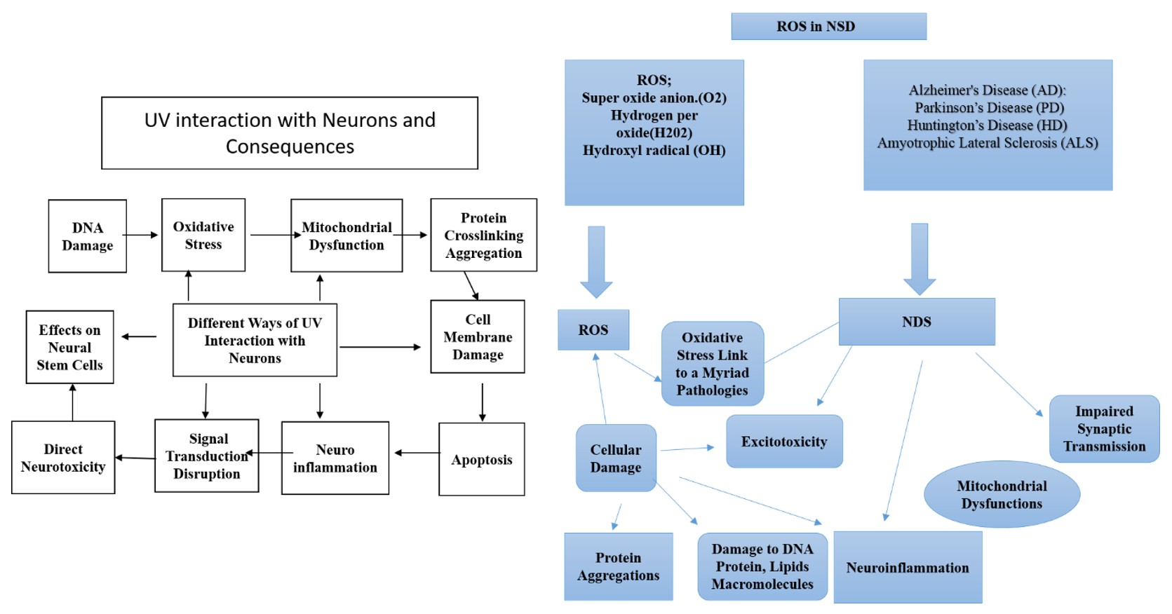
However, leveraging these benefits requires careful consideration of the associated risks. UV-induced DNA damage and oxidative stress can trigger inflammatory responses, undermining neural integrity if exposure thresholds are exceeded (Gallagher and Lee, 2006; Panich et al., 2016). Striking an equilibrium between therapeutic efficacy and safety is critical. By optimizing UV exposure parameters, researchers may harness its neuroprotective potential while minimizing adverse outcomes. Figure 1 illustrates this dynamic interplay, showcasing how UV radiation impacts neural pathways and cellular processes. This nuanced understanding of the relationship between UV radiation and the nervous system could open new avenues for innovative therapeutic strategies targeting neurodegenerative diseases. By balancing the risks and benefits, researchers can explore UV radiation not only as a preventative measure but also as a complementary treatment option, paving the way for advancements in cognitive health and neuroprotection (Li et al., 2017).
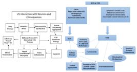
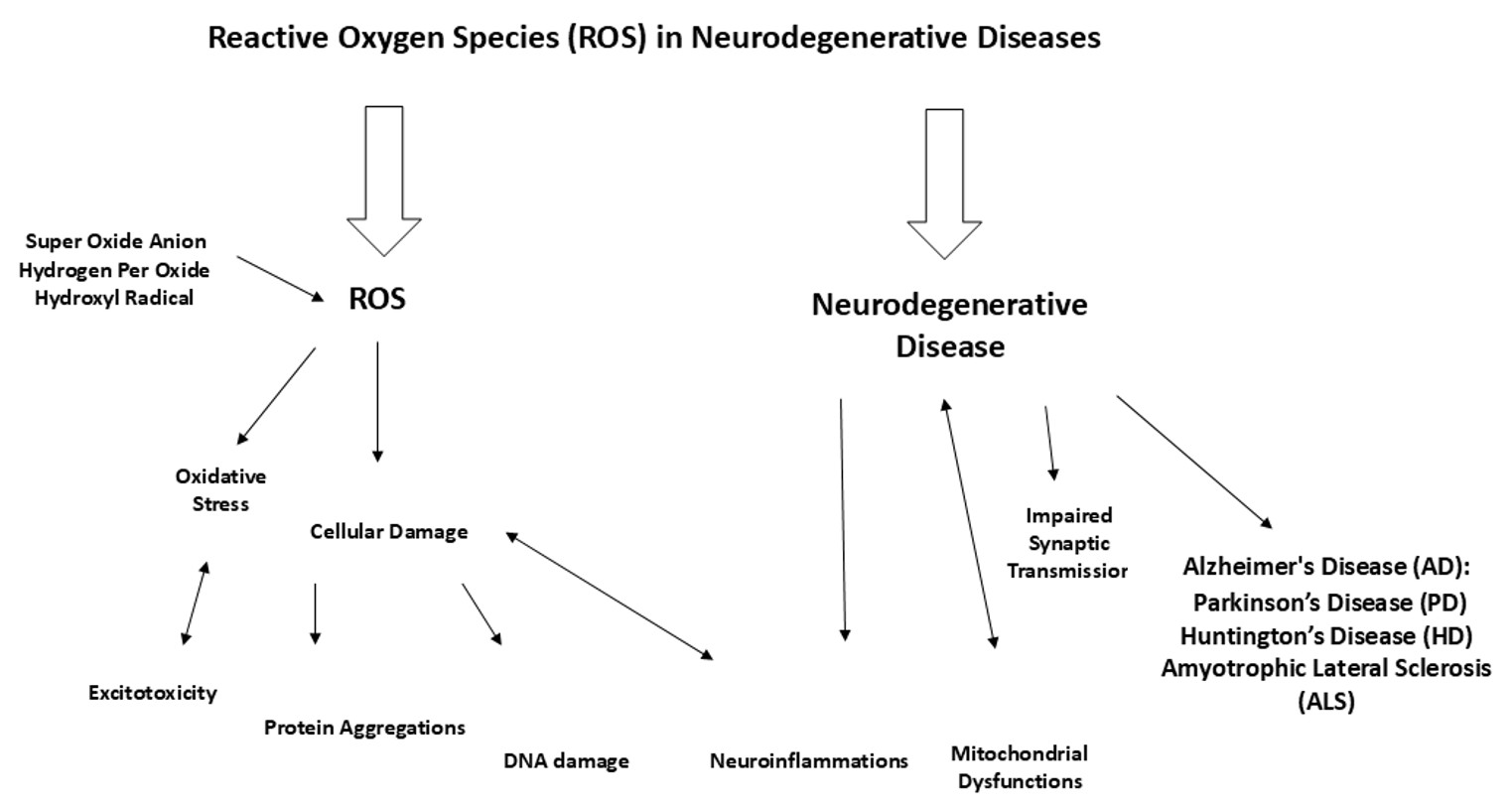
Mechanistic Impact of Ultraviolet Irradiation on Neuronal Networks; And Reactive Oxygen Species (ROS) Pathways in Neurodegenerative Diseases.
The figure illustrates the multifaceted interactions between UV irradiation and neuronal cells, highlighting key pathological pathways. UV irradiation induces oxidative stress, which can lead to mitochondrial dysfunction and subsequent DNA damage. These processes contribute to protein crosslinking and aggregation, as well as cell membrane damage, ultimately triggering neuronal apoptosis. Additionally, UV exposure disrupts neural signal transduction and affects neural stem cell function, contributing to direct neurotoxicity and neuroinflammation. Together, these mechanisms underline the complex role of UV irradiation in neuronal pathology and its potential impact on neurodegenerative disease progression. Furthermore, it depicts the critical role of ROS in the pathogenesis of neurodegenerative diseases. The generation of ROS, including superoxide anions, hydrogen peroxide, and hydroxyl radicals, initiates a cascade of oxidative stress and cellular damage. D These processes lead to excitotoxicity, protein aggregation, and DNA damage, contributing to neuroinflammation and mitochondrial dysfunction. The cumulative impact of these mechanisms disrupts synaptic transmission and accelerates neuronal degeneration, linking ROS activity to the progression of neurodegenerative disorders such as Alzheimer's disease (AD), Parkinson's disease (PD), Huntington's disease (HD), and amyotrophic lateral sclerosis (ALS). This cycle underscores the central role of oxidative stress in neurodegeneration and highlights potential therapeutic targets for mitigating ROS-related damage.
2.8. Alzheimer's Disease (AD): new advances in UV Irradiation for Neurodegenerative Diseases (NDs)
Research on UV irradiation as a potential therapeutic tool for Alzheimer's disease (AD) has largely centered on UVA and UVB radiation, with promising results indicating its neuroprotective and disease-modifying effects (Juzeniene and Moan, 2012). (Akhtar et al., 2017) A significant pathological hallmark of AD is the accumulation of amyloid-beta (Aβ) plaques, which contribute to neuronal dysfunction, synaptic loss, and cognitive decline. UV irradiation has emerged as a potential strategy to mitigate this process by preventing the aggregation of Aβ peptides, a critical step in AD pathogenesis. One of the proposed mechanisms of action is that UV exposure facilitates the proteolysis and disaggregation of amyloid fibrils, potentially reducing the overall burden of Aβ accumulation in the brain (Il Lee et al., 2019).
By inhibiting the aggregation of Aβ, UV radiation could help maintain neuronal structure and function, potentially slowing the cognitive decline and progression characteristic of AD. Among the various UV modalities, photobiomodulation (PBM) therapy, employing controlled UV light exposure—has gained attention for its ability to reduce Aβ plaque load and improve cognitive function in preclinical AD models (Liu et al., 2021). In vitro studies involving AD cell cultures and transgenic animal models have demonstrated that UV irradiation can diminish the extent of Aβ pathology, restore synaptic plasticity, and enhance cognitive performance. These studies suggest that UV therapy might not only provide neuroprotection but also foster recovery of cognitive function in AD patients, offering hope for disease-modifying interventions (Yang et al., 2021).
Despite these promising findings, there is a significant gap in translating UV irradiation therapy from the laboratory to clinical practice. While the therapeutic potential of UV radiation is clear, its safety, optimal dosing, and long-term effects in human populations remain under investigation. Rigorous clinical trials are needed to evaluate the feasibility of UV therapy as an adjunct treatment for AD, and to establish protocols that maximize its efficacy while minimizing adverse outcomes.
2.9. Parkinson’s Disease (PD)
Research on UV irradiation in the context of Parkinson’s disease (PD) has primarily focused on the use of UVA radiation (Jayan et al., 2022). PD is characterized by the progressive loss of dopaminergic neurons in the substantia nigra, a brain region crucial for movement control. This neuronal degeneration is driven largely by oxidative stress, mitochondrial dysfunction, and inflammation, all of which contribute to the clinical manifestations of PD, including motor dysfunction, tremors, and rigidity.
UV radiation has been hypothesized to mitigate these pathological processes by enhancing the activity of endogenous antioxidant enzymes, such as superoxide dismutase (SOD) and catalase, which play key roles in neutralizing oxidative stress (Steenvoorden and Van Henegouwen, 1997). By bolstering these antioxidant defenses, UV exposure may protect dopaminergic neurons from oxidative damage, thereby preserving neuronal function and mitigating the effects of neurodegeneration. In preclinical PD models, UVA irradiation has been shown to preserve dopaminergic function, reduce oxidative damage, and slow the progression of neurodegeneration (Kim et al., 2020).
These findings support the potential of UV therapy as a neuroprotective treatment for PD. However, additional research is required to validate these results in clinical settings. Longitudinal studies are needed to assess the long-term efficacy and safety of UV therapy, including its effects on motor function and overall disease progression. Further investigation into optimal dosing regimens and the most effective UV wavelengths for PD treatment is also warranted (Iacopetta, 2019; Christine et al., 2022).
2.10. Huntington’s Disease (HD)
Although research on UV therapy for Huntington’s disease (HD) is still in its nascent stages, early studies have provided intriguing insights into its potential benefits. HD is a genetic neurodegenerative disorder characterized by the progressive loss of motor control, cognitive decline, and psychiatric symptoms. The disease is caused by an expansion of CAG repeats in the huntingtin gene, leading to the production of a toxic protein that disrupts neuronal survival and function.
UV radiation appears to influence several key intracellular signaling pathways involved in neuronal survival and apoptosis, both of which are disrupted in HD (Magalhaes et al., 2020). Preliminary evidence suggests that UVA exposure may exert neuroprotective effects by enhancing neuronal resilience and counteracting cellular damage associated with HD pathogenesis (Borrell-Pages et al., 2006). Early preclinical studies have demonstrated that UV irradiation can improve motor function, support neuronal survival, and promote the survival of striatal neurons, which are particularly vulnerable in HD models (Andres et al., 2008).
While these findings are promising, further research is necessary to fully understand the efficacy and safety of UV irradiation for HD. The mechanisms by which UV radiation might modulate huntingtin protein aggregation, neuroinflammation, and cellular stress responses require deeper investigation. Additionally, clinical trials in HD patients will be essential to establish whether UV therapy could offer tangible benefits in terms of motor function, cognitive preservation, and overall disease progression. (Muralidharan and Mandrekar, 2013).
2.11. Amyotrophic Lateral Sclerosis (ALS)
Amyotrophic lateral sclerosis (ALS) is a progressive neurodegenerative disorder that primarily affects motor neurons, leading to muscle weakness, atrophy, and eventual paralysis. While UV therapy has long been used in dermatology for conditions like psoriasis and eczema, its application in ALS remains relatively underexplored. However, growing interest is emerging around the potential of UV irradiation to modulate the inflammatory processes and neuroprotective pathways that are central to ALS pathophysiology (Myers et al., 2021)
One of the hypothesized mechanisms through which UV radiation may provide neuroprotection in ALS is by reducing inflammation within the nervous system, particularly within motor neurons. In ALS, inflammation plays a key role in disease progression, exacerbating neuronal damage and accelerating motor neuron degeneration. UV exposure has been shown to modulate immune responses, reducing the activation of microglia and astrocytes, which are often implicated in neuroinflammation (Pandya et al., 2013). By modulating these inflammatory pathways, UV radiation could reduce motor neuron degeneration, slow disease progression, and potentially improve functional outcomes.
Initial preclinical studies in ALS models have demonstrated that UV irradiation can enhance motor function, extend survival, and reduce the burden of neuroinflammation. These studies suggest that UV therapy may have the potential to modify the course of ALS, improving both quality of life and disease prognosis (Nakamura and Kawasaki, 2017). However, as with other neurodegenerative disorders, additional research is needed to determine the optimal parameters for UV exposure, assess long-term safety, and confirm the clinical benefits in ALS patients.
2.12. Concrete findings and areas needing further research
Recent preclinical studies have provided compelling evidence that UV irradiation could serve as a novel therapeutic approach for neurodegenerative diseases (NDs) (Ashrafizadeh et al., 2020). While these findings are promising, critical gaps in our understanding remain, particularly regarding the precise mechanisms through which UV-induced neuroprotection occurs and how therapeutic efficacy can be optimized. It is essential to conduct further research to delineate the specific wavelengths, doses, and exposure durations that offer maximum benefit while minimizing potential harm. The heterogeneity of NDs, combined with variations in individual responses to UV radiation, underscores the need for personalized UV therapy protocols tailored to each disease and patient.
Future studies should focus on identifying the molecular signaling pathways activated by UV irradiation that contribute to neuroprotection. These pathways may include the modulation of mitochondrial function, autophagy, and apoptosis, which are integral to maintaining neuronal health in NDs (Zhou et al., 2015). Additionally, exploring the synergistic effects of UV therapy in combination with conventional pharmacological treatments or stem cell-based therapies may open new avenues for enhancing therapeutic outcomes. The role of UV light in modulating epigenetic mechanisms, such as DNA methylation and histone modification, presents an exciting area for exploration that could reveal new molecular targets for intervention.
Moreover, translating these promising findings from preclinical studies into clinical practice is a crucial next step. Long-term, multicenter clinical trials are necessary to evaluate the safety, efficacy, and tolerability of UV therapy in human patients. Special attention must be given to the potential systemic effects of UV exposure, including its impact on the immune system, skin health, and ocular function. Furthermore, a deeper understanding of the neuroplastic effects of UV radiation, particularly in the context of neurogenesis and cognitive enhancement, is needed to unlock its full potential as a therapeutic tool for NDs.
2.13. Prospects of UV irradiation for NDs
UV irradiation offers significant promise as a non-invasive therapy for NDs, reducing the need for invasive procedures and associated complications. The ability of UV radiation to penetrate tissues and target different cellular processes provides a unique advantage in treating neurological conditions. Specifically, UV light's effects on neuronal metabolism, synaptic plasticity, and oxidative stress could offer a multi-targeted approach to neuroprotection.
UV therapy, including forms like Low-Level Light Therapy (LLLT), has demonstrated potential in treating a wide array of nervous system disorders, such as Alzheimer’s, Parkinson’s, and psychiatric conditions. By stimulating mitochondrial activity and improving cellular bioenergetics, LLLT offers a non-toxic alternative to more traditional therapies, especially in conditions characterized by mitochondrial dysfunction and energy depletion, which are hallmark features of many NDs (Rojas and Gonzalez-Lima, 2011). This is particularly important given the growing evidence that mitochondrial dysfunction is a central pathogenic mechanism in diseases like Parkinson’s and Alzheimer's, where neurons are highly energy-dependent.
Furthermore, UV therapy may have a broader therapeutic role beyond neuroprotection by addressing key pathological processes common across many NDs. For example, UV light has been shown to modulate neuroinflammation, a central feature of most neurodegenerative conditions, by regulating the activation of microglia and the production of pro-inflammatory cytokines. Similarly, UV irradiation can induce cellular repair mechanisms, such as DNA repair pathways and protein misfolding responses, which could help prevent the accumulation of toxic protein aggregates like amyloid-beta or alpha-synuclein, commonly implicated in AD and PD, respectively (Bickers and Athar, 2006). As such, UV therapy’s ability to modulate multiple molecular pathways makes it a potentially transformative strategy for tackling the multifaceted pathophysiology of NDs.
2.14. Challenges and limitations (adverse effects, protocol optimization)
Despite its potential, the use of UV therapy for NDs is not without its challenges. The adverse effects of prolonged UV exposure, including DNA damage, oxidative stress, and inflammation, must be carefully managed to avoid exacerbating neurological damage or contributing to other health conditions, such as skin cancer or cataracts (Davis et al., 2024; Algorri et al., 2023). In order to mitigate these risks, precise UV dosing protocols must be established. This includes determining the optimal wavelength for targeting specific neurodegenerative processes, as well as defining safe exposure durations and frequencies.
One of the most important considerations in optimizing UV therapy for NDs is balancing the therapeutic effects with the need for safety. Notably, UV exposure at different wavelengths interacts with tissues at varying depths, necessitating careful selection of wavelengths depending on the anatomical site and the therapeutic goal. For instance, UVA light, which penetrates deeper into tissues, may be more suitable for targeting the central nervous system, while UVB and UVC could be more effective for surface-level applications. Another consideration is the use of adjunctive therapies, such as antioxidants or pharmacological agents, which may help mitigate the pro-inflammatory effects of UV exposure and enhance its neuroprotective properties.
To further refine UV therapy protocols, personalized medicine approaches will be crucial. Variations in patient characteristics, including age, genetic predisposition, and the stage of the disease, may influence how individuals respond to UV treatment. Biomarker identification, such as the presence of specific neuroinflammatory cytokines or protein aggregation profiles, could be used to tailor UV therapy to the individual’s specific needs. Additionally, UV therapy’s potential to improve outcomes in patients with co-morbid conditions, such as depression or anxiety, which are common in individuals with NDs, should also be explored. By understanding how UV light affects mood regulation and the broader neuroendocrine system, clinicians may be able to offer more comprehensive therapeutic interventions.
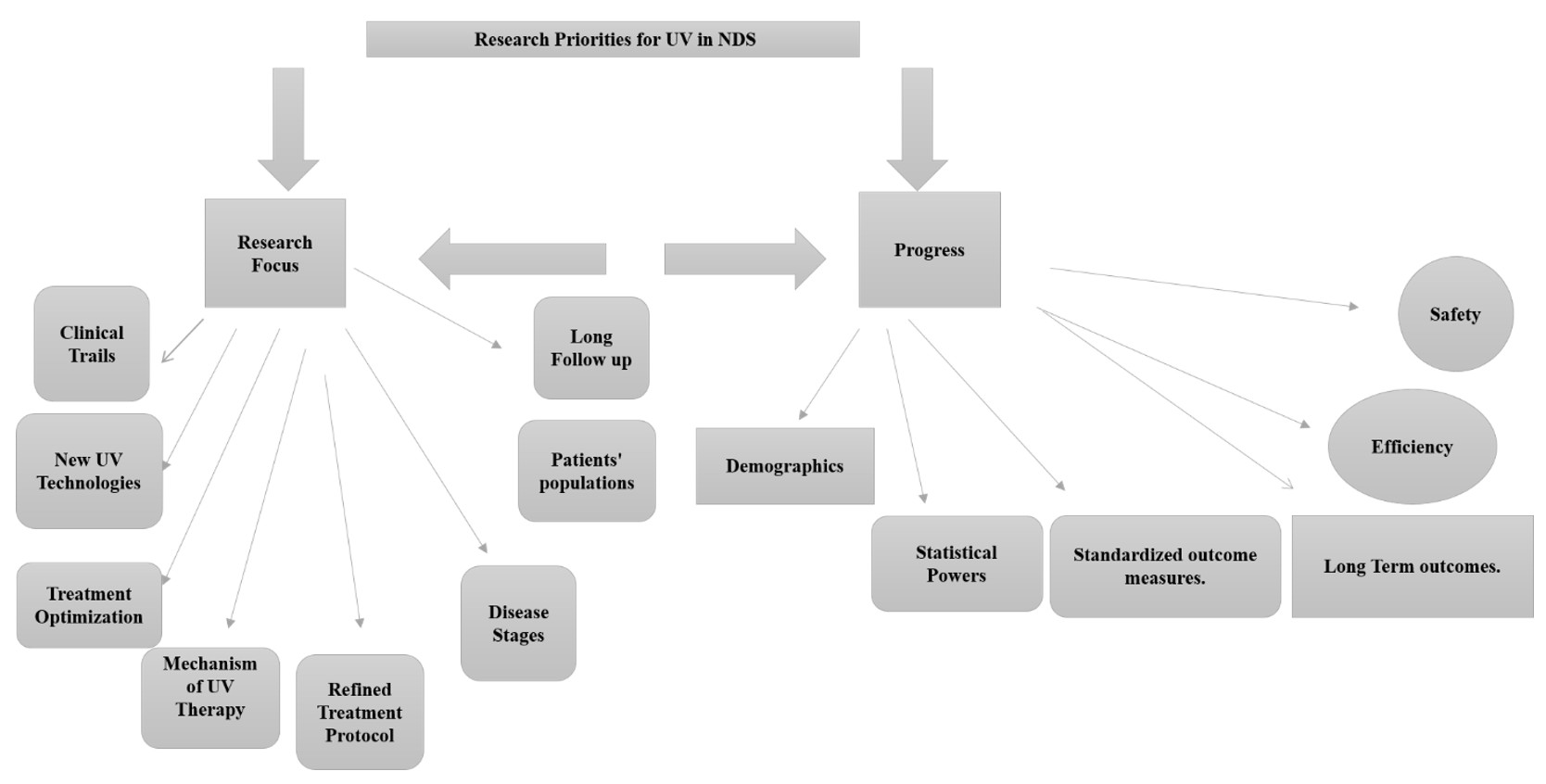
The future of UV therapy in treating NDs depends largely on ongoing efforts to optimize treatment protocols and overcome the existing barriers to clinical implementation. The development of advanced UV delivery systems, such as portable devices or wearable technologies, could make UV therapy more accessible and practical for long-term use, ensuring patient compliance and comfort. Furthermore, the integration of UV therapy with other therapeutic modalities, including gene therapy, stem cell therapy, and immunotherapy, may provide synergistic effects that enhance neuroprotection and promote neuroregeneration. While UV irradiation offers a promising avenue for the treatment of NDs, substantial research efforts are needed to address its challenges and unlock its full clinical potential. By combining cutting-edge technologies, multidisciplinary approaches, and a deep understanding of the molecular mechanisms involved, UV therapy has the potential to revolutionize the way we approach the treatment of neurodegenerative diseases. Figure 2 represents priority research areas for ultraviolet therapy in neurodegenerative diseases.
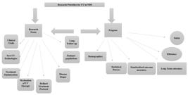
The figure delineates the essential research directions and benchmarks required to establish UV therapy as a viable treatment for neurodegenerative diseases. The "Research Focus" section highlights key areas for exploration, including optimizing treatment protocols, developing innovative UV technologies, elucidating the precise mechanisms underlying UV therapy, and conducting robust clinical trials. Emphasis is also placed on refining patient selection criteria, addressing disease stage-specific responses, and implementing extended follow-up periods to assess long-term effects. On the "Progress" side, critical milestones such as ensuring treatment safety, improving therapeutic efficiency, enhancing statistical robustness, and standardizing outcome measures are outlined. This comprehensive framework underscores the interconnected efforts necessary to translate UV therapy research into clinical practice, ensuring both efficacy and reliability in neurodegenerative disease management.
2.15. Synergistic neuroprotection: exploring the combined effects of ultraviolet irradiation and ursolic acid in neurodegenerative disease management
Ursolic acid, a pentacyclic triterpenoid found in a variety of plants, has recently gained significant attention for its potential neuroprotective properties, particularly in the context of neurodegenerative diseases such as Alzheimer’s disease (AD) and Parkinson's disease (PD) (Habtemariam, 2019). This naturally occurring compound offers a multifaceted mechanism of action that positions it as a promising therapeutic agent. Its effectiveness is largely attributed to its potent antioxidant, anti-inflammatory, and neurogenic properties, all of which are vital in the treatment of neurodegenerative conditions.
One of the primary mechanisms by which ursolic acid exerts its neuroprotective effects is through its robust antioxidant activity. Oxidative stress, driven by an imbalance between ROS and the body's ability to neutralize them, is a hallmark of many neurodegenerative diseases, contributing to neuronal damage and cell death. Ursolic acid’s ability to scavenge free radicals mitigates the damaging effects of oxidative stress, thereby preventing the onset of neurodegeneration and promoting cellular longevity (Habtemariam, 2019). Moreover, by modulating the activity of antioxidant enzymes, ursolic acid enhances the brain's intrinsic defense systems, thereby fostering a healthier neuronal environment.
In addition to its antioxidant properties, chronic inflammation in the brain is another critical factor in the progression of neurodegenerative diseases. Ursolic acid has shown considerable promise in modulating the inflammatory pathways involved in these conditions. By inhibiting pro-inflammatory cytokines and reducing the activation of microglial cells—immune cells in the brain that exacerbate inflammation—ursolic acid may help alleviate the neuroinflammatory response that accelerates neuronal loss (Habtemariam, 2019). This anti-inflammatory effect is crucial in the management of AD and PD, where neuroinflammation plays a central role in disease progression.
In Alzheimer's disease, the accumulation of amyloid-beta plaques in the brain is a key pathological feature that disrupts neuronal function. Ursolic acid has been identified as an inhibitor of amyloid-beta peptide aggregation. Through its interference with amyloid-beta binding and fibril formation, ursolic acid helps in reducing plaque deposition, potentially slowing the progression of Alzheimer's disease (Maggio et al., 1992). Furthermore, by enhancing neuronal signaling pathways, ursolic acid promotes neuroplasticity and neuronal survival, which are essential for combating the degenerative processes inherent in AD (Huang et al., 2017).
The therapeutic potential of ursolic acid extends beyond its antioxidant and anti-inflammatory actions. Emerging evidence suggests that it may also promote neurogenesis, the process by which new neurons are formed in the brain. This could be particularly beneficial for neurodegenerative diseases that impair the brain's ability to regenerate lost cells, such as PD and AD. Ursolic acid’s modulation of signaling pathways involved in neuronal survival and differentiation, such as the PI3K-Akt pathway, underpins its ability to promote neuronal health and restore lost function (Huang et al., 2017).
Integrating the neuroprotective effects of ursolic acid with UV irradiation opens a compelling avenue for exploring synergistic strategies in the management of neurodegenerative disorders like AD and PD. Both approaches exhibit mechanisms that target oxidative stress, inflammation, and cellular repair processes, offering complementary pathways for therapeutic interventions. Ultraviolet irradiation, particularly low-dose UVB light, has been investigated for its potential neuroprotective effects mediated through systemic and localized pathways. UV exposure stimulates the production of vitamin D, a neuroprotective molecule that plays a significant role in brain health by regulating calcium homeostasis, reducing inflammation, and promoting synaptic plasticity. Beyond vitamin D synthesis, UV light can induce the release of other neuroactive molecules, including nitric oxide (NO), which has been implicated in vascular and neuronal health, and beta-endorphins, which modulate stress responses and neuronal survival.
Key to UV irradiation's role in neuroprotection is its ability to modulate oxidative stress and inflammation, hallmarks of neurodegenerative diseases. UV exposure enhances the activity of antioxidant enzymes, including catalase and glutathione peroxidase, bolstering the brain’s defense against ROS. This mechanism aligns with ursolic acid's potent ROS-scavenging capabilities, amplifying the reduction of oxidative damage in neuronal tissues. UV-induced immune modulation reduces systemic inflammation by downregulating pro-inflammatory cytokines, including IL-1β, IL-6, and TNF-α. These pathways are mirrored by ursolic acid’s ability to inhibit microglial activation and suppress neuroinflammation, providing a dual approach to mitigating chronic inflammation in conditions like AD and PD.
Both UV exposure and ursolic acid combat oxidative stress but via complementary pathways. UV-triggered enzymatic antioxidant defenses could act in concert with ursolic acid’s direct free radical scavenging, resulting in a more comprehensive reduction of oxidative damage. This synergistic effect is critical in preventing neuronal apoptosis and ensuring cellular longevity in neurodegenerative conditions. Ursolic acid and UV irradiation share the ability to attenuate neuroinflammation through the inhibition of pro-inflammatory pathways. While UV light primarily downregulates systemic inflammation through regulatory T cells and cytokine modulation, ursolic acid directly inhibits microglial activation within the brain. Together, they form a multifaceted anti-inflammatory strategy capable of slowing disease progression in neurodegeneration.
Emerging evidence suggests UV exposure can influence neurogenesis indirectly through the release of neurotrophic factors, such as brain-derived neurotrophic factor (BDNF). Ursolic acid enhances neurogenesis by modulating intracellular pathways like PI3K-Akt, which govern neuronal differentiation and survival. When combined, UV-stimulated BDNF release could enhance ursolic acid's neurogenic effects, offering a powerful approach to regenerate neuronal networks compromised in AD and PD. Ursolic acid's ability to inhibit amyloid-beta aggregation may benefit from UV-induced systemic changes, such as enhanced clearance of beta-amyloid via improved glymphatic flow, mediated by UV-stimulated nitric oxide signaling. UV's effects on synaptic plasticity, through vitamin D and BDNF pathways, could complement ursolic acid’s promotion of neuroplasticity and neuronal survival, addressing cognitive deficits in AD. UV's antioxidant effects, coupled with ursolic acid's ROS scavenging, may reduce the oxidative damage that leads to dopaminergic neuron loss. Additionally, the anti-inflammatory actions of both agents could mitigate neuroinflammation in the substantia nigra, a critical site of damage in PD. Both UV exposure and ursolic acid have been implicated in improving mitochondrial function, crucial for energy metabolism in neurons. Their combined effects could enhance cellular energy production and prevent mitochondrial-mediated neuronal apoptosis.
The intersection of UV irradiation and ursolic acid presents an innovative therapeutic paradigm for neurodegenerative diseases. Determining safe and effective UV irradiation doses that synergize with ursolic acid supplementation to enhance neuroprotection. Elucidating the interplay between UV-induced vitamin D and NO pathways with ursolic acid’s modulation of signaling cascades like PI3K-Akt. Investigating combined UV and ursolic acid therapies in animal models and clinical populations to assess efficacy in slowing neurodegeneration and improving cognitive and motor functions. By leveraging their complementary mechanisms, UV irradiation and ursolic acid offer a multifactorial strategy to counteract the complex pathophysiology of neurodegenerative diseases, paving the way for integrative approaches in neuro-medicine.
2.16. Mechanism of ultraviolet irradiation signaling from skin receptors to the brain
The mechanism by which UV irradiation influences the brain via the skin is an intricate process that involves several complex physiological pathways. When UV light strikes the skin, it is detected by specialized photoreceptors embedded in the skin, known as cutaneous sensory receptors. These receptors respond to the specific wavelengths of UV radiation and initiate a series of biochemical reactions known as phototransduction, converting UV light into electrical signals that can be processed by the nervous system (Jenkins, 2014; Fell et al., 2014).
These electrical signals travel through the peripheral nervous system to reach the central nervous system (CNS), where they are processed in various brain regions responsible for sensory integration. Once in the brain, these signals can trigger a range of physiological responses, including the release of neurotransmitters and hormones that regulate mood, circadian rhythms, and other biological processes (Luo et al., 2008). This complex communication between the skin and the brain suggests that UV light, through its influence on sensory receptors, plays a crucial role in modulating brain function and may offer therapeutic benefits for mood disorders, sleep regulation, and even neurodegenerative diseases. (Katiyar et al., 2001).
One of the more intriguing aspects of UV irradiation is its ability to influence neuroendocrine function. UV exposure has been shown to impact the production of serotonin and dopamine, neurotransmitters integral to mood regulation, cognitive function, and reward systems (Bellono and Oancea, 2013). This effect is particularly relevant in the context of neurodegenerative diseases, where neurotransmitter imbalances are a hallmark of conditions such as Parkinson’s and Alzheimer’s. By enhancing neurotransmitter release and promoting neuroplasticity, UV irradiation may offer a non-invasive adjunct therapy for these debilitating disorders.
2.17. Infrared therapy for neurodegenerative diseases
Infrared therapy, which utilizes light in the infrared spectrum (typically 700 nm to 1 mm), is another promising therapeutic modality with potential applications for neurodegenerative diseases. Preliminary research indicates that infrared therapy may benefit patients with neurodegenerative conditions such as Parkinson's disease (PD) and Alzheimer's disease (AD) by improving mitochondrial function, enhancing blood circulation, and reducing neuroinflammation—key factors that contribute to neurodegeneration (Eells et al., 2011).
Infrared therapy improves mitochondrial bioenergetics, which is essential for maintaining ATP production in neurons. Mitochondrial dysfunction is a hallmark of several neurodegenerative diseases, including PD and AD, and is closely linked to neuronal death. By stimulating mitochondrial activity, infrared therapy has the potential to counteract energy deficits in the brain, improving cognitive function and potentially slowing disease progression (Tsai and Hamblin, 2017).
Moreover, infrared radiation has potent anti-inflammatory effects, which can help alleviate the chronic brain inflammation that is characteristic of many neurodegenerative diseases. Inflammation in the brain not only damages neurons but also contributes to disease progression by promoting oxidative stress. By reducing inflammation, infrared therapy could protect neuronal health, slow disease progression, and potentially improve quality of life for individuals with neurodegenerative disorders (Johnstone et al., 2015).
In addition to its anti-inflammatory properties, infrared therapy also supports neuroplasticity—the brain’s ability to reorganize and form new neural connections. This capability is particularly beneficial in neurodegenerative diseases, where neural circuits are often impaired or lost. By promoting neuroplasticity, infrared therapy may help the brain compensate for lost function, enabling better outcomes for patients with cognitive and motor impairments associated with PD and AD (Rajan, 2025).
Another significant advantage of infrared therapy is its antioxidant properties. Oxidative stress is a major contributor to neuronal damage and degeneration, and infrared therapy has been shown to reduce ROS levels in the brain, protecting neurons from oxidative damage and apoptosis. This antioxidant effect could be pivotal in mitigating the progression of neurodegenerative diseases and restoring some degree of neuronal function (Salehpour et al., 2018).
As research continues to unfold, both ursolic acid and light-based therapies, such as UV and infrared therapy, hold immense promise for treating neurodegenerative diseases. These non-invasive, multi-targeted approaches offer significant advantages over traditional pharmacological treatments, particularly in terms of safety and ease of administration. However, more research is needed to optimize treatment protocols, understand the precise mechanisms at play, and translate these therapies into clinical practice. Future studies should focus on refining dosage regimens, identifying synergistic combinations of therapies, and exploring the long-term effects of these treatments on disease progression. By advancing our understanding of these innovative therapies, we may open the door to new, more effective treatments for conditions that have long eluded traditional interventions.
2.18. Concept of visible light in neurodegenerative disease intervention
Photobiomodulation (PBM) therapy, leveraging red or near-infrared (NIR) light within the 600–1100 nm wavelength range, has emerged as a revolutionary tool in addressing neurodegenerative diseases. This approach primarily targets intracellular chromophores, such as cytochrome c oxidase in the mitochondrial respiratory chain. By stimulating this key enzyme, PBM catalyzes a cascade of beneficial cellular processes. It enhances ATP production, thereby restoring energy homeostasis in neurons compromised by neurodegeneration. Additionally, it reduces oxidative stress by mitigating the overproduction of ROS and boosting antioxidant enzyme activity, protecting lipids, proteins, and DNA from oxidative damage. PBM also exerts anti-inflammatory effects, modulating microglial activity to favor the anti-inflammatory M2 phenotype over the pro-inflammatory M1 phenotype. This shift helps attenuate neuroinflammation, a central feature in diseases such as Alzheimer’s and Parkinson’s. Furthermore, PBM promotes neurogenesis and synaptic plasticity by upregulating brain-derived neurotrophic factor (BDNF) and other growth factors, thereby supporting synaptic repair, neuronal survival, and regeneration (Loving et al., 2005).
In clinical applications, PBM has demonstrated promising results across a spectrum of neurodegenerative conditions. For Alzheimer’s disease (AD), PBM reduces amyloid-beta aggregation, restores synaptic function, and enhances cognitive performance, with transcranial PBM emerging as a non-invasive method for targeting affected brain regions. In Parkinson’s disease (PD), PBM improves mitochondrial bioenergetics and mitigates oxidative stress in dopaminergic neurons, potentially slowing disease progression and alleviating motor symptoms. For amyotrophic lateral sclerosis (ALS), preclinical studies suggest that PBM delays motor neuron degeneration, enhances muscle strength, and extends survival. Moreover, PBM shows potential in addressing stroke and traumatic brain injury (TBI) by promoting neurovascular repair and reducing secondary neuronal damage, highlighting its versatility in neurorehabilitation (Salehpour et al., 2018).
Expanding the landscape of light-based therapies, photo-excited small molecules are gaining recognition as innovative tools for neurodegenerative disease management. These molecules are engineered to absorb specific light wavelengths, triggering localized therapeutic effects. Photodynamic therapy (PDT), traditionally used in oncology, is now being explored for neurodegeneration. Activated photosensitizers produce singlet oxygen and reactive species, selectively targeting pathogenic cells or amyloid plaques. Similarly, photopharmacology employs light to activate drug molecules, enabling spatially and temporally precise interventions. Photo-activated compounds can target misfolded proteins, offering novel treatments for conditions like Alzheimer’s and Huntington’s disease. Optogenetics, although not strictly radiation-based, complements this approach by using light-sensitive proteins to modulate neuronal activity, offering unparalleled precision in restoring disrupted neuronal circuitry (Deisseroth, 2015).
Despite these advances, challenges remain in fully integrating PBM and radiation-based therapies into clinical practice. Optimization of dosimetry, including the ideal wavelength, energy density, and treatment duration, is critical for maximizing efficacy while ensuring safety. Delivering light effectively to deep brain regions poses another obstacle, with innovative solutions like intranasal probes and implantable light sources under development. Furthermore, the heterogeneity of neurodegenerative diseases necessitates individualized therapeutic approaches tailored to specific pathophysiological profiles.
Photobiomodulation and radiation-based therapies, including the use of photo-excited small molecules, represent cutting-edge approaches to neurodegenerative disease management. PBM’s ability to enhance mitochondrial function, reduce oxidative stress, and modulate inflammatory responses provides a robust foundation for neuroprotection. Meanwhile, emerging technologies like photodynamic therapy and optogenetics offer targeted and precise solutions to address complex neurodegenerative pathologies. With continued advancements in light-based interventions and their integration into personalized medicine frameworks, these therapies hold transformative potential in altering the trajectory of neurodegenerative disorders.
One of the primary mechanisms by which visible light therapy exerts its beneficial effects is through the enhancement of mitochondrial function. Mitochondria are the powerhouses of the cell, responsible for generating ATP, which fuels various cellular processes. In neurodegenerative diseases, mitochondrial dysfunction is often a contributing factor, leading to energy deficits, oxidative stress, and neuronal death. Visible light therapy has been shown to improve mitochondrial bioenergetics, thereby enhancing ATP production and restoring cellular vitality. By repairing and optimizing mitochondrial function, this therapy could potentially mitigate the cellular injuries that occur in neurodegenerative disorders, thereby slowing disease progression (Salehpour et al., 2018).
Beyond its effects on mitochondrial function, visible light therapy also has significant implications for neuroplasticity—the brain's ability to reorganize itself by forming new neural connections. This ability to adapt and repair itself is crucial in the context of neurodegenerative diseases, where neuronal loss and synaptic dysfunction impede cognitive function and memory. Research suggests that visible light therapy may promote neuroplasticity by stimulating neuronal grsowth and facilitating the formation of new synaptic connections. This process could help restore cognitive function and slow the cognitive decline typically seen in Alzheimer's disease (AD), Parkinson's disease (PD), and other neurodegenerative conditions (Amaroli et al., 2019).
Furthermore, visible light therapy has been shown to have a direct impact on the hallmark pathological features of Alzheimer’s disease, specifically beta-amyloid plaques. The accumulation of these plaques disrupts neuronal communication and is considered one of the central causes of neurodegeneration in AD. Studies suggest that visible light therapy can help reduce the formation of amyloid-beta plaques, potentially slowing the progression of Alzheimer’s disease. By modulating the production and aggregation of amyloid-beta peptides, visible light therapy offers a novel avenue for combating the disease at a cellular level (Grillo et al., 2013; Choi et al., 2012).
Another significant benefit of visible light therapy is its potential to enhance cerebral blood flow. Proper blood circulation is vital for maintaining brain health, as it ensures that oxygen, nutrients, and waste products are effectively transported throughout the brain. Reduced blood flow is often observed in individuals with neurodegenerative diseases, leading to cognitive impairment and further neuronal damage. Visible light therapy has been shown to promote vasodilation and improve blood circulation in the brain, ensuring that neural tissue receives the necessary oxygen and nutrients to maintain optimal function. This effect is especially important in diseases like Alzheimer's and Parkinson's, where ischemia and poor circulation exacerbate disease progression (Naeser et al., 2016).
Moreover, visible light therapy has notable anti-inflammatory properties. Chronic neuroinflammation is a hallmark of many neurodegenerative diseases, contributing to neuronal injury, synaptic dysfunction, and disease progression. By reducing the activation of microglia and astrocytes, the immune cells in the brain, visible light therapy helps modulate the inflammatory response, protecting neurons from the harmful effects of excessive inflammation. This anti-inflammatory action can alleviate symptoms and improve brain function in patients suffering from neurodegenerative diseases (Johnstone et al., 2015).
2.19. Photo-excited small molecules in neurodegenerative disease therapy
The innovative utilization of photo-excited small molecules is revolutionizing therapeutic strategies for neurodegenerative diseases, providing unparalleled precision in addressing these conditions' multifaceted pathologies (Zhang et al., 2021). These small molecules, designed to respond to specific wavelengths of light, undergo functional or structural changes that enable highly localized and temporally controlled therapeutic effects. This precise mechanism targets cellular and molecular abnormalities while minimizing systemic side effects, a critical advantage in managing complex neurodegenerative disorders.
Photo-excited small molecules function through highly sophisticated mechanisms that enable targeted therapeutic effects. They initiate photochemical reactions, such as electron transfer, singlet oxygen generation, and radical formation, which selectively modify pathological targets like protein aggregates and damaged DNA. Additionally, these molecules undergo light-induced conformational changes that enhance molecular binding affinities, facilitating precise interactions with specific biological targets. Acting as prodrugs, they remain inert until activated by light, ensuring therapeutic effects are confined to diseased tissues, thereby minimizing systemic side effects. Furthermore, some photo-excited molecules transfer absorbed light energy to adjacent targets, amplifying their therapeutic impact and enhancing efficacy in addressing complex pathological conditions.
The therapeutic applications of photo-excited small molecules are highly relevant in the treatment of various neurodegenerative conditions. They effectively disrupt toxic aggregates, such as amyloid-beta plaques and tau tangles, which are central to Alzheimer’s and related diseases, with molecules like porphyrins and flavins showing significant efficacy in this regard. Photodynamic therapy (PDT), initially developed for oncology, is adapted for neurodegenerative diseases, utilizing these molecules to generate cytotoxic species that neutralize pathogenic cells and reduce neuroinflammation while sparing healthy tissue. These molecules also play a key role in synaptic restoration by modulating neurotransmitter dynamics and receptor activity, thus addressing cognitive decline. In addition, light-activated antioxidants, including flavonoids and polyphenols, mitigate oxidative stress by reducing reactive oxygen species and enhancing the body’s endogenous antioxidant defenses. Furthermore, photo-excited molecules that mimic neurotrophic factors, such as BDNF and NGF, promote neuronal repair and survival, offering vital therapeutic benefits for conditions characterized by significant neuronal loss.
Technological advancements have significantly enhanced the efficacy and broadened the scope of photo-excited small molecules. Photochromic compounds, which function as light-activated switches, allow for the modulation of receptor-ligand interactions and synaptic functions, enabling dynamic control over biological processes. Photoswitchable drugs, such as azobenzenes, toggle between active and inactive states when exposed to light, providing precise control over biochemical pathways. Nanoparticle-assisted delivery systems, using functionalized nanoparticles like gold and carbon dots, improve light penetration and enable effective targeting of deeper brain regions, overcoming previous limitations in tissue depth. Additionally, photosensitizing dyes, such as methylene blue, offer dual benefits by simultaneously reducing protein aggregation and enhancing mitochondrial function, addressing multiple pathological features. Light-sensitive ion channels further contribute by regulating neuronal excitability, providing potential solutions to excitotoxicity and the neurodegenerative symptoms associated with it. These technological innovations represent significant strides in optimizing the therapeutic potential of photo-excited small molecules (Zhou and Kihara, 2023; Liu et al, 2024)
The application of photo-excited small molecules offers numerous advantages in the treatment of neurodegenerative diseases. Their localized effects minimize collateral damage to healthy tissues, ensuring targeted therapy with reduced systemic side effects. Temporal precision allows interventions to be tailored to specific stages of disease progression, offering a more personalized treatment approach. Additionally, many light-based delivery methods are non-invasive, which enhances patient safety and compliance. These molecules are also capable of addressing multiple pathological mechanisms simultaneously, providing a comprehensive therapeutic effect that targets various aspects of neurodegeneration. However, certain challenges remain. One significant limitation is light penetration, particularly when trying to reach deeper brain regions affected by subcortical pathologies. Emerging solutions, such as infrared light technologies, upconversion nanoparticles, and implantable LEDs, are being developed to address these barriers. Another challenge is phototoxicity, as prolonged light exposure may damage healthy tissues. To mitigate this, precision dosimetry and safer activation protocols are currently under development to ensure optimal therapeutic outcomes while minimizing risks (Salehpour et al., 2018).
Photo-excited small molecules represent a transformative leap in neurodegenerative disease therapy, offering a precision-driven, multifaceted approach. Their capacity to degrade toxic aggregates, restore synaptic function, alleviate oxidative stress, and stimulate neuronal repair underscores their potential to revolutionize treatment paradigms. As photopharmacology and related technologies continue to evolve, these molecules are poised to become central to personalized neurotherapeutic strategies, addressing the intricate challenges of neurodegeneration with unparalleled accuracy and efficacy.
2.20. Technological advances and clinical translation of UV therapy for neurodegenerative diseases
The future of UV therapy for neurodegenerative diseases (NDs) depends heavily on the continuous advancement of technology and the rigorous execution of clinical trials. Emerging innovations in UV-emitting devices, such as LEDs and lasers, offer enhanced precision, safety, and efficacy for UV therapy applications in NDs. These devices allow for targeted delivery of UV radiation, optimizing treatment outcomes while minimizing potential side effects. Moreover, the ability to fine-tune the wavelength, intensity, and duration of exposure enhances the therapeutic potential of UV therapy, making it a more viable option for clinical use (Slominski et al., 2018).
One of the most promising advancements in UV therapy is the integration of photobiomodulation (PBM) with near-infrared (NIR) light. NIR light, which penetrates deeper into the tissues, has shown potential in regulating neurodegenerative processes and reducing inflammation in the brain. The combined use of UV and NIR light could offer synergistic benefits, targeting both superficial and deeper layers of tissue to maximize therapeutic effects. As these technologies evolve, their increased specificity and precision offer the possibility of personalized treatment protocols that cater to individual patient needs, enhancing both safety and efficacy ([Q4: Q4]]; Heinig et al., 2020).
Incorporating cutting-edge imaging techniques, such as fluorescence microscopy and optical coherence tomography, into UV therapy protocols allows for real-time monitoring and adjustment of treatment strategies. These advanced imaging tools provide valuable insights into the cellular and tissue-level effects of UV therapy, enabling clinicians to optimize treatment parameters and evaluate the therapeutic response more effectively. Real-time monitoring ensures that UV therapy can be applied with maximum precision, improving its clinical outcomes and minimizing risks associated with overexposure (Engelbrecht at al., 2022).
However, to transition UV therapy from experimental studies to routine clinical practice, large-scale clinical trials are essential. These trials must assess not only the safety and efficacy of UV therapy but also its long-term effects on disease progression and patient outcomes. Randomized controlled trials with diverse patient populations, including various disease subtypes and stages, are crucial for gathering robust data on the therapy’s effectiveness. Longitudinal studies with extended follow-ups are particularly important for evaluating potential adverse effects that may emerge over time. Collaboration among academic institutions, industry leaders, and regulatory agencies will be essential in designing and implementing these trials to ensure that UV therapy reaches its full potential as a treatment for neurodegenerative diseases (Zhang et al., 2021).
2.21. Reactive Oxygen Species (ROS) and their role in neurodegenerative diseases
ROS, which include molecules such as superoxide anions (O2−), hydrogen peroxide (H2O2), and hydroxyl radicals (•OH), are highly reactive molecules derived from oxygen. They are produced as byproducts of normal cellular metabolism, particularly during oxidative phosphorylation in the mitochondria. While ROS play essential roles in cellular signaling, immune defense, and regulation of metabolism, their excessive accumulation can lead to oxidative stress—an imbalance between ROS production and the antioxidant defenses of the cell (Cui et al., 2022; Baev et al., 2022). Oxidative stress is a critical pathological factor in the development and progression of neurodegenerative diseases (NDs), including Alzheimer's disease (AD), Parkinson's disease (PD), Huntington's disease (HD), and amyotrophic lateral sclerosis (ALS) (Angelova and Abramov, 2018).
In neurodegenerative diseases, oxidative stress exacerbates neuronal damage by promoting inflammation, impairing cellular function, and inducing apoptosis (programmed cell death). ROS can damage cellular components such as proteins, lipids, and DNA, leading to cellular dysfunction and death. In the context of neurodegeneration, oxidative stress accelerates neuronal loss, exacerbates disease symptoms, and impairs the brain's ability to repair itself. Therefore, mitigating oxidative stress through antioxidant therapies, such as visible light therapy or pharmacological interventions, represents a promising strategy for treating NDs (Galli et al., 2005; Krumova and Cosa, 2016)
Emerging research suggests that visible light therapy may reduce ROS production and enhance the brain's antioxidant defenses. By stimulating mitochondrial function and promoting the production of antioxidant enzymes, visible light therapy helps restore the delicate balance between ROS and antioxidants, protecting neurons from oxidative damage. This effect could slow the progression of neurodegenerative diseases and improve long-term outcomes for patients suffering from conditions like Alzheimer's and Parkinson's disease (Salehpour et al., 2018). The integration of visible light therapy, UV irradiation, and infrared therapy represents a promising frontier in the treatment of neurodegenerative diseases. By targeting mitochondrial dysfunction, promoting neuroplasticity, reducing inflammation, and modulating ROS levels, these light-based therapies offer a multifaceted approach to managing diseases like Alzheimer's, Parkinson's, and amyotrophic lateral sclerosis. With technological advancements in light-emitting devices and improved clinical trial designs, these therapies have the potential to revolutionize the treatment landscape for neurodegenerative disorders, offering safer, non-invasive options for patients.
2.22. Reactive Oxygen Species (ROS) and their role in neurodegenerative diseases
ROS are highly reactive molecules derived from oxygen, including superoxide anions (O2−), hydrogen peroxide (H2O2), and hydroxyl radicals (•OH) (Cui et al., 2022). These molecules are essential byproducts of normal cellular processes, predominantly produced during mitochondrial oxidative phosphorylation, as well as through inflammatory responses and various stress reactions. While ROS are crucial for cellular signaling, immune response modulation, and pathogen defense, an imbalance between their production and neutralization leads to oxidative stress, which has profound consequences on cellular integrity and function (Bhattacharya, 2015; Zhou et al., 2022).
ROS are typically generated in response to environmental stimuli or during metabolic processes such as respiration and immune activation. In the mitochondria, ROS are produced as side products of the electron transport chain, where electrons are transferred to oxygen molecules. Under normal conditions, ROS production is tightly regulated by cellular antioxidants, including enzymes like superoxide dismutase (SOD), catalase, and glutathione peroxidase. However, when ROS production exceeds the antioxidant defense mechanisms, a cascade of damaging events occurs, resulting in oxidative stress. Oxidative stress is recognized as a fundamental pathological factor in the progression of many chronic diseases, including neurodegenerative disorders (NDs) such as Alzheimer's disease (AD), Parkinson's disease (PD), Huntington's disease (HD), and amyotrophic lateral sclerosis (ALS) (Angelova and Abramov, 2018; Kim et al., 2020).
2.23. Impact of oxidative stress on neuronal health
Neurons, with their high metabolic activity and elevated oxygen consumption, are particularly vulnerable to the deleterious effects of ROS. Unlike other cell types, neurons have a limited capacity to regenerate and repair themselves, which makes them more susceptible to oxidative damage. The high rate of oxygen utilization in neurons, coupled with their relatively low levels of endogenous antioxidants, renders them especially sensitive to ROS-induced damage. When ROS accumulate beyond the capacity of antioxidant systems, they can inflict widespread damage to cellular components such as lipids, proteins, and DNA, disrupting normal cellular processes and ultimately leading to neuronal death (Liguori et al., 2018; Branca et al., 2020).
2.24. Cellular targets of oxidative damage
Lipids: Oxidative stress causes lipid peroxidation, a process in which ROS attack lipids in cell membranes, particularly polyunsaturated fatty acids. This reaction leads to the formation of reactive aldehydes, such as malondialdehyde, which further damage cellular structures. Lipid peroxidation destabilizes cellular membranes, impairing membrane fluidity and integrity, and contributing to synaptic dysfunction, which is a hallmark of neurodegenerative diseases (Radak et al., 2013).
Proteins: Oxidative damage to proteins is one of the most significant contributors to cellular dysfunction in neurodegenerative diseases. ROS induce the oxidation of amino acid side chains, leading to protein misfolding, aggregation, and loss of function. These oxidized proteins can form toxic aggregates, disrupting normal cellular processes. For example, in Alzheimer's disease, oxidative damage to tau proteins promotes the formation of neurofibrillary tangles, while in Parkinson's disease, oxidative modifications of alpha-synuclein contribute to the formation of Lewy bodies. Protein misfolding and aggregation are central to the pathological features of many neurodegenerative conditions (Uttara et al., 2009).
DNA: Oxidative stress also induces DNA damage, including base modifications, strand breaks, and cross-linking. This genomic instability can lead to mutations and chromosomal aberrations, further exacerbating cellular dysfunction. In neurons, this DNA damage is particularly detrimental, as these cells are post-mitotic and cannot easily repair or replace damaged genetic material. Accumulated mutations can impair neuronal function and accelerate neurodegeneration. In addition, oxidative damage to mitochondria can impair their function, contributing to further ROS production in a vicious cycle of damage and dysfunction (Butterfield and Halliwell, 2019).
2.25. Mitochondrial dysfunction and excitotoxicity
Mitochondria are central to the pathophysiology of neurodegenerative diseases, and their dysfunction plays a pivotal role in ROS generation. In healthy neurons, mitochondria efficiently generate ATP through oxidative phosphorylation. However, in neurodegenerative conditions, mitochondrial dysfunction leads to the overproduction of ROS and a subsequent energy deficit. This disruption in mitochondrial function is a critical factor in the initiation and progression of NDs. In addition to increased ROS generation, mitochondrial dysfunction impairs the regulation of calcium homeostasis, leading to excitotoxicity—an overstimulation of neurons that causes further cellular injury and death.
Excitotoxicity, driven by an excessive release of glutamate and subsequent overstimulation of N-methyl-D-aspartate (NMDA) receptors, is another key mechanism by which oxidative stress contributes to neurodegeneration. The resulting calcium influx activates a cascade of intracellular events, including the activation of proteases, phospholipases, and nitric oxide synthase, leading to further mitochondrial dysfunction, oxidative damage, and ultimately neuronal apoptosis (Dong et al., 2009).
2.26. Oxidative stress, inflammation, and neurodegenerative disease progression
Oxidative stress is tightly linked with neuroinflammation, a prominent feature of neurodegenerative diseases. The accumulation of ROS triggers the activation of microglia, the resident immune cells of the central nervous system. When activated, microglia release pro-inflammatory cytokines, chemokines, and additional ROS, which further exacerbate neuronal injury and disrupt synaptic function. Chronic inflammation perpetuates the cycle of oxidative stress, accelerating neuronal loss and disease progression (Bhat et al., 2015; Kennedy et al., 2012).
In diseases like Alzheimer's and Parkinson's, neuroinflammation is considered both a response to injury and a driver of the disease. Microglial activation and the subsequent release of inflammatory mediators contribute to the pathogenesis of these disorders by fostering an environment of sustained oxidative stress. This chronic inflammatory state accelerates cognitive decline, motor dysfunction, and other debilitating symptoms commonly observed in NDs (Block and Hong, 2007).
2.27. Chronic oxidative stress: a catalyst for disease progression
The continuous accumulation of ROS and oxidative damage over time is a key contributor to the aging process, and when coupled with neurodegenerative diseases, it accelerates cellular aging and disease progression. In the context of Alzheimer's disease, for example, oxidative stress not only accelerates amyloid-beta plaque formation but also promotes tau phosphorylation and the formation of neurofibrillary tangles, which together contribute to the dysfunction and death of neurons. Similarly, in Parkinson’s disease, oxidative damage to dopaminergic neurons in the substantia nigra leads to the characteristic motor symptoms, including tremors, rigidity, and bradykinesia.
Chronic oxidative stress also impairs neurogenesis, the brain’s ability to generate new neurons, which is essential for cognitive function and repair. As neurons lose their regenerative capacity, cognitive decline and motor dysfunction become more pronounced. Additionally, oxidative stress disrupts synaptic plasticity, which is necessary for learning and memory processes. As synaptic connections weaken, cognitive abilities deteriorate, further exacerbating the symptoms of neurodegenerative diseases (Schieber and Chandel, 2014).
2.28. Future directions: targeting ROS in neurodegenerative disease therapy
Given the central role of ROS in the pathophysiology of neurodegenerative diseases, targeting oxidative stress presents an attractive therapeutic strategy. Antioxidant therapies, such as the use of synthetic antioxidants (e.g., N-acetylcysteine) or natural compounds (e.g., curcumin, resveratrol), hold promise in counteracting oxidative damage. Additionally, strategies that enhance the brain's endogenous antioxidant defense systems, such as the activation of nuclear factor erythroid 2-related factor 2 (Nrf2) pathways, are being actively explored. These interventions aim to restore the balance between ROS production and antioxidant defense, protecting neurons from further damage and slowing disease progression (Ma, 2013).
In addition to pharmacological approaches, light-based therapies, including photobiomodulation (PBM), offer novel ways to mitigate oxidative stress. By enhancing mitochondrial function and reducing ROS production, PBM has demonstrated neuroprotective effects in animal models of neurodegenerative diseases. As research into these therapies advances, their integration into clinical practice may offer new hope for patients suffering from conditions like Alzheimer's, Parkinson's, and Huntington's diseases.
ROS are a double-edged sword, vital for normal cellular function but capable of causing extensive damage when not properly regulated. In neurodegenerative diseases, oxidative stress plays a central role in the initiation and progression of neuronal damage. Through the impairment of mitochondrial function, protein misfolding, DNA damage, and activation of inflammatory pathways, ROS contribute to the pathophysiology of Alzheimer's, Parkinson's, Huntington's, and other neurodegenerative disorders. Understanding the precise mechanisms by which ROS induce neurodegeneration is critical for developing effective therapeutic strategies aimed at mitigating oxidative stress. Advances in antioxidant therapies, light-based interventions, and mitochondrial-targeted treatments hold promise in offering new avenues for the treatment and management of neurodegenerative diseases, as shown in Figure 1 and Figure 2 respectively.
2.29. Impact of ROS accumulation on enzyme function and cellular homeostasis
Oxidative stress, driven by the excessive accumulation of ROS, is a central pathological mechanism in neurodegenerative diseases (NDs), as shown in Figure 3. This phenomenon is intricately linked to the disruption of cellular homeostasis. ROS, which include highly reactive molecules such as superoxide anions (O2•−), hydroxyl radicals (•OH), and non-radical species like hydrogen peroxide (H2O2) (Zhao et al., 2021; Zhou and Kihara, 2023), are byproducts of cellular metabolism, primarily produced during aerobic respiration in the mitochondria. While ROS are crucial for regulating normal cellular processes, including cell signaling, immune defense, and apoptosis, an overproduction of these species can overwhelm the body's antioxidant defenses, leading to oxidative stress and widespread cellular damage (Kim et al., 2020).
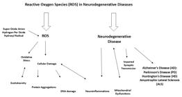
In neurodegenerative diseases, the accumulation of ROS is particularly detrimental to neuronal health. Neurons, which consume high levels of oxygen due to their metabolic activity and are rich in lipid content, are highly vulnerable to oxidative damage. Moreover, the central nervous system (CNS) has relatively low levels of antioxidant enzymes and limited regenerative capacity, making it especially prone to ROS-induced injury. Prolonged exposure to oxidative stress leads to mitochondrial dysfunction, DNA damage, protein oxidation, and inflammation, hallmarks of various neurodegenerative conditions such as Alzheimer's disease (AD), Parkinson's disease (PD), Huntington's disease (HD), and amyotrophic lateral sclerosis (ALS) (Angelova and Abramov, 2018; Butterfield and Halliwell, 2019; Islam, 2017). The underlying cellular damage caused by ROS accumulation is deeply tied to enzyme function and the maintenance of cellular homeostasis, two critical components for neuronal survival.
2.30. Enzymatic dysfunction and cellular homeostasis in the context of ROS
Enzymes are critical catalysts in maintaining cellular homeostasis. They facilitate numerous biochemical reactions, including those involved in energy production, DNA repair, protein synthesis, and the regulation of antioxidant defense systems. However, when ROS levels exceed the cellular antioxidant capacity, these enzymes become highly susceptible to oxidative modifications. This can lead to the inactivation of enzymes essential for cellular metabolism and neuroprotection, contributing to disease progression in NDs.
2.31. Enzyme inactivation through oxidative modification
ROS, particularly superoxide anions and hydroxyl radicals, can directly modify key amino acid residues in the enzyme active sites, resulting in functional impairment. Enzyme function can be disrupted through oxidation of cysteine, methionine, and histidine residues, which are common targets for ROS due to their nucleophilic nature. For instance, the oxidation of cysteine thiol groups can lead to the formation of disulfide bonds or sulfonic acid derivatives, which may alter the enzyme's structure, rendering it inactive or less efficient. Such oxidative modifications prevent enzymes from catalyzing critical reactions necessary for maintaining cellular functions (Liguori et al., 2018).
2.32. Disruption of antioxidant enzyme function
Enzymes responsible for maintaining cellular antioxidant defenses, such as superoxide dismutase (SOD), catalase, and glutathione peroxidase, are particularly vulnerable to ROS-induced damage. These antioxidant enzymes play pivotal roles in neutralizing ROS by converting superoxide anions into less harmful molecules, such as hydrogen peroxide, which is subsequently broken down into water and oxygen. When these enzymes are damaged by oxidative stress, their ability to neutralize ROS is compromised, leading to a vicious cycle of ROS accumulation and cellular damage. This breakdown in cellular defense mechanisms accelerates the pathogenesis of neurodegenerative diseases and further exacerbates neuronal injury (Ma, 2013).
2.33. Disruption of mitochondrial function
Mitochondria are both a major source of ROS and highly sensitive to oxidative damage. In neurons, mitochondrial dysfunction caused by ROS accumulation leads to impaired ATP production, increased ROS generation, and a breakdown in cellular energy homeostasis. Mitochondrial enzymes involved in oxidative phosphorylation, such as complex I and complex IV, are particularly prone to oxidative modifications. The impairment of these enzymes results in decreased energy production, which is critical for maintaining cellular processes such as neurotransmitter release, synaptic plasticity, and neuronal communication. Furthermore, mitochondrial dysfunction can trigger apoptotic pathways, leading to cell death and contributing to the loss of neurons in conditions like Parkinson’s and Alzheimer’s diseases (Kim et al., 2020; Ma., 2013).
2.34. Impaired neurotransmitter regulation and synaptic function
ROS-induced enzyme dysfunction also affects the synthesis, release, and reuptake of neurotransmitters, which are vital for synaptic communication and plasticity. Enzymes involved in neurotransmitter synthesis, such as tyrosine hydroxylase (for dopamine production) and choline acetyltransferase (for acetylcholine production), are susceptible to oxidative damage. In Parkinson’s disease, for example, oxidative stress impairs dopaminergic neurons by damaging these enzymes, resulting in reduced dopamine production and contributing to motor dysfunction. In Alzheimer's disease, the oxidation of enzymes involved in acetylcholine synthesis leads to decreased cholinergic transmission, a hallmark of cognitive decline. The disruption of neurotransmitter signaling also affects synaptic plasticity, the process by which synapses strengthen or weaken over time in response to activity, which is essential for learning and memory. Synaptic dysfunction is a key feature of neurodegeneration and is exacerbated by oxidative stress (Uttara et al., 2009).
2.35. Pathways of ROS-induced cellular dysfunction: a cascade of damage
The detrimental effects of ROS accumulation on enzyme function initiate a cascade of cellular dysfunction that accelerates neurodegeneration. The accumulation of oxidative damage can impair key signaling pathways within the cell, leading to mitochondrial dysfunction, altered calcium homeostasis, and activation of pro-apoptotic pathways. For example, excessive ROS can activate c-Jun N-terminal kinases (JNK) and p38 mitogen-activated protein kinases (MAPKs), which in turn trigger the release of pro-apoptotic factors such as cytochrome c from mitochondria. This cascade leads to the activation of caspases, the enzymes responsible for initiating cell death. Additionally, oxidative stress can impair the activity of protein chaperones and proteases that facilitate protein folding and degradation, leading to the accumulation of misfolded proteins that contribute to the formation of toxic protein aggregates, a hallmark of neurodegenerative diseases (Butterfield and Halliwell, 2019).
2.36. ROS, neuroinflammation, and cellular imbalance
As ROS accumulate in the brain, they also trigger neuroinflammation, which exacerbates the damage to neurons and their surrounding environment. Microglia, the resident immune cells of the CNS, are activated by oxidative stress and release inflammatory cytokines such as TNF-α and IL-1β. This inflammatory response further increases ROS production, creating a feed-forward loop of oxidative damage and inflammation. Chronic neuroinflammation leads to synaptic dysfunction, neuronal death, and the progression of neurodegenerative diseases (Block and Hong, 2007).
Moreover, oxidative stress-induced enzyme dysfunction and disrupted cellular homeostasis impair the ability of neurons to repair and regenerate. Cellular repair mechanisms, including DNA repair enzymes and protein degradation pathways, are often overwhelmed by the excessive accumulation of ROS, leading to further genomic instability and protein aggregation. This imbalance between cellular damage and repair is a critical factor in the accelerated progression of neurodegenerative diseases (Ma., 2013).
2.37. Targeting ROS and Enzyme Dysfunction in Neurodegenerative Diseases
The accumulation of ROS and their impact on enzyme function represent a pivotal mechanism in the pathophysiology of neurodegenerative diseases. The excessive production of ROS leads to the inactivation of critical enzymes, disrupting cellular homeostasis and initiating a cascade of events that drive neuronal dysfunction and death. From mitochondrial dysfunction to impaired neurotransmitter regulation and synaptic plasticity, the effects of ROS on enzyme function are far-reaching and contribute to the progression of diseases such as Alzheimer's, Parkinson's, Huntington's, and ALS.
Targeting ROS accumulation and restoring enzyme function represent promising therapeutic strategies for neurodegenerative diseases. Approaches such as antioxidant therapies, mitochondrial-targeted treatments, and interventions aimed at modulating neuroinflammatory responses may offer new opportunities for slowing disease progression and improving patient outcomes. Furthermore, enhancing cellular repair mechanisms and protecting enzyme activity through pharmacological or genetic approaches could help preserve neuronal health and functionality, providing hope for patients affected by these debilitating disorders.
2.38. The crucial role of ROS-sensitive enzymes, such as ATPases, in Neuronal Health
Enzymes that are sensitive to ROS play an essential role in preserving neuronal health, with ATPases being among the most pivotal. ATPases are a diverse class of enzymes that hydrolyze adenosine triphosphate (ATP) into adenosine diphosphate (ADP) and inorganic phosphate (P), releasing the chemical energy necessary for various cellular processes. Proper ATPase function is vital for neuronal survival, synaptic signaling, and maintaining cellular homeostasis. These enzymes are responsible for crucial functions such as neurotransmitter release, axonal transport, and regulating membrane potential. As highly active cells, neurons are particularly reliant on ATPases to sustain these functions. (Ozougwu., 2016).
2.39. Energy regulation in neurons
Neurons, due to their continuous activity and complex signaling networks, require substantial energy. ATPases are critical for maintaining cellular energy levels and ensuring the proper functioning of neurons. These enzymes facilitate the active transport of ions across the neuronal membrane, which is necessary for generating the electrochemical gradients that allow signal transmission. The most prominent example of this is the Na+/K+-ATPase, which pumps sodium ions out of the cell and potassium ions in, helping to maintain the resting membrane potential and facilitating the repolarization of the cell following an action potential. This electrochemical gradient is fundamental for electrical impulse conduction, and any disturbance in its maintenance can impair neurotransmission, ultimately disrupting neuronal function (Wang et al., 2020; Zhang et al., 2021).
In addition to the Na+/K+-ATPase, calcium-ATPases are also critical for regulating calcium levels within neurons. Calcium ions are central to processes such as synaptic transmission, neuronal excitability, and plasticity, all of which are crucial for cognitive functions like learning and memory. These ATPases, located on both the plasma membrane and the endoplasmic reticulum, help transport calcium out of the cytosol to prevent excessive calcium buildup, thus avoiding excitotoxicity—a condition that leads to neuronal death and dysfunction (Zhu et al., 2022).
2.40. ROS impact on atpase function
Neurons are particularly vulnerable to oxidative stress due to their high metabolic demands and oxygen consumption, as well as the lipid-rich composition of their membranes. ROS—such as superoxide anions (O2•−), hydroxyl radicals (•OH), and hydrogen peroxide (H2O2)—are produced during cellular metabolism, particularly within the mitochondria during oxidative phosphorylation. While ROS are essential for several physiological functions, including immune responses and cellular signaling, an accumulation of ROS can lead to oxidative stress, resulting in neuronal damage and contributing to neurodegenerative diseases (Kim et al., 2020; Angelova and Abramov, 2018).
ROS can directly affect ATPases by causing oxidative modifications to key amino acids in their active sites, such as cysteine, methionine, and histidine. These modifications can impair the enzymatic activity of ATPases, which disrupts cellular energy production and homeostasis. For example, oxidative modifications of Na+/K+-ATPase can disturb ion gradients, impair neurotransmission, and increase neuronal vulnerability to further oxidative damage. This leads to a vicious cycle in which ROS-induced damage reduces ATPase function, exacerbating energy deficits and promoting further ROS production (Butterfield and Halliwell, 2019; Ma, 2013).
ROS accumulation in neurons is not limited to ATPases. It also impacts other critical enzymes involved in cellular maintenance and antioxidant defense, such as superoxide dismutase (SOD), catalase, and glutathione peroxidase. When ROS levels exceed the neutralizing capacity of these enzymes, neuronal defenses are compromised, further accelerating the neurodegenerative process (Liguori et al., 2018; Jaitovich and Bertorello, 2006). As such, maintaining optimal ATPase function and protecting these enzymes from ROS-induced damage represents a key strategy for preserving neuronal health and mitigating neurodegeneration.
2.41. ROS-mediated enzyme inhibition and neurodegeneration
The inhibition of enzymes by ROS is a central mechanism driving neurodegeneration. ROS-induced modifications can lead to enzyme malfunction, destabilizing cellular systems and disrupting neuronal functions. For example, ROS can impair ATPases and other critical enzymes that regulate mitochondrial function, excitotoxicity, and neuronal apoptosis, all of which are hallmarks of neurodegenerative diseases like Alzheimer's disease (AD), Parkinson's disease (PD), Huntington's disease (HD), and amyotrophic lateral sclerosis (ALS) (Kim et al., 2020; Schieber and Chandel, 2014).
In Alzheimer's disease, ROS-induced damage to enzymes involved in protein degradation and energy metabolism can contribute to the accumulation of beta-amyloid plaques. Similarly, in Parkinson's disease, oxidative stress can impair enzymes involved in mitochondrial health and energy production, promoting the aggregation of alpha-synuclein and exacerbating neuronal dysfunction. These examples highlight the crucial role of ROS in enzyme inhibition and the pathophysiology of neurodegenerative diseases (Butterfield and Halliwell, 2019; Ma, 2013).
2.42. Photochemical interactions with UV irradiation
Interestingly, UV irradiation has garnered attention as a potential therapeutic modality for counteracting the harmful effects of ROS and enzyme inhibition in neurodegenerative diseases. Photobiomodulation therapy (PBM), which uses specific wavelengths of light, has shown promise in modulating ROS levels, enhancing mitochondrial function, and promoting cellular repair mechanisms. UV light exposure has been found to stimulate antioxidant defenses and improve the function of enzymes such as ATPases, potentially reducing oxidative stress and protecting neurons from damage.
In particular, near-infrared (NIR) light therapy has been studied for its potential to reduce oxidative stress and improve mitochondrial bioenergetics, thus offering a neuroprotective strategy for conditions such as Alzheimer's and Parkinson's diseases (Hamblin, 2019; Nawashiro et al., 2012). PBM therapy is thought to promote cellular signaling pathways that enhance ATP production and ion transport, which in turn supports neuronal survival and function.
ROS-sensitive enzymes like ATPases are fundamental to neuronal health, and their dysfunction plays a critical role in the progression of neurodegenerative diseases. The damage caused by ROS can impair these enzymes, leading to disruptions in energy metabolism, ion regulation, and cellular homeostasis. Protecting ATPases and other critical enzymes from ROS-induced inhibition represents a promising strategy for mitigating neurodegeneration. Moreover, emerging therapies like photobiomodulation therapy offer potential solutions to restore enzyme function and reduce oxidative stress in neurons, presenting a novel avenue for future treatment strategies. By further understanding the mechanisms through which ROS affect enzyme activity and neuronal function, we can develop more effective therapeutic approaches aimed at preserving neuronal health and preventing neurodegenerative diseases.
2.43. Role of UV irradiation in modulating ROS levels and enzyme function
UV irradiation plays a complex role in the modulation of ROS levels and enzyme functionality, significantly impacting neurodegenerative processes. While high-dose UV exposure can lead to the overproduction of ROS, causing oxidative stress, controlled and low-dose UV exposure has been found to activate the body's antioxidant defense mechanisms, thereby reducing oxidative damage and promoting cellular resilience. Understanding the dual role of UV radiation in both inducing and counteracting oxidative stress is essential for exploring its therapeutic potential, particularly in neurodegenerative diseases. (Sena and Chandel, 2012).
2.44. High-Dose UV irradiation and ROS generation
High-intensity UV radiation, particularly UV-B and UV-C, can directly induce the formation of ROS within cells. These ROS, including superoxide anions (O2•−), hydrogen peroxide (H2O2), and hydroxyl radicals (•OH), result from the photochemical interactions between UV light and cellular components like lipids, proteins, and DNA. This uncontrolled ROS production can overwhelm the cell's antioxidant defenses, leading to oxidative stress. In neurons, this oxidative burden can exacerbate neuronal damage, impair mitochondrial function, and disrupt the integrity of cellular structures—events commonly associated with neurodegenerative diseases such as Alzheimer's disease (AD) and Parkinson's disease (PD).
However, UV-A radiation, typically considered less harmful than UV-B, can also influence cellular processes differently. At low doses, UV-A radiation has been shown to activate hormetic responses, where mild stress triggers adaptive mechanisms that enhance cellular resistance to subsequent oxidative stress. This response includes the upregulation of antioxidant enzymes and the activation of cellular repair mechanisms, which may offer neuroprotective effects in certain contexts. As a result, UV-A exposure in controlled doses may provide a therapeutic strategy to combat oxidative stress, a major contributor to neurodegeneration.
2.45. Modulation of antioxidant enzyme activity by UV radiation
UV irradiation can also directly stimulate the production and activity of antioxidant enzymes such as superoxide dismutase (SOD) and catalase. These enzymes play a crucial role in mitigating oxidative damage by neutralizing harmful ROS. Superoxide dismutase converts superoxide radicals into hydrogen peroxide, while catalase breaks down hydrogen peroxide into water and oxygen, further reducing ROS levels and protecting the cell from oxidative damage. When UV radiation is administered at specific wavelengths and doses, it can enhance the activity of these enzymes, bolstering the cell’s innate antioxidant defenses. (Gilgun-Sherki et al., 2002).
Moreover, UV exposure activates redox-sensitive transcription factors, such as Nrf2, which upregulate the expression of genes involved in antioxidant defense and detoxification pathways. Nrf2 activation induces the transcription of numerous protective genes, including those that encode for enzymes like glutathione peroxidase and NAD(P)H dehydrogenase, thereby enhancing the cell’s ability to combat ROS-induced damage. This process is critical for maintaining cellular homeostasis, particularly in neurons, which are highly susceptible to oxidative stress. By promoting the upregulation of antioxidant pathways, UV irradiation may offer a novel approach for neuroprotection in the context of neurodegenerative diseases (Baird and Yamamoto, 2020; Zhang et al., 2022).
2.46. Low-Dose UV radiation and hormetic responses
Research indicates that low doses of UV radiation, particularly in the UVA spectrum, can induce hormesis—a phenomenon where low levels of stressors, such as UV exposure, elicit adaptive cellular responses that improve the cell's capacity to resist further damage. These responses include the activation of molecular pathways involved in oxidative stress resistance, DNA repair, and protein homeostasis. In neuronal cells, the hormetic effects of low-dose UV radiation may enhance mitochondrial function, improve synaptic plasticity, and reduce neuronal apoptosis, offering neuroprotective benefits in the face of chronic oxidative stress (Narasimhamurthy et al., 2022).
Low-dose UV exposure can also trigger a mild inflammatory response, which activates reparative processes in neuronal tissue. This adaptive response may mitigate the neuroinflammatory processes seen in neurodegenerative diseases, where chronic inflammation exacerbates neuronal injury and accelerates disease progression.
2.47. Mechanisms of ROS-induced enzyme inhibition
Despite the potential benefits of controlled UV exposure, ROS-induced enzyme inhibition remains a significant concern in the context of neurodegenerative diseases. ROS can impair enzyme function through several mechanisms, which contribute to neuronal dysfunction and degeneration.
2.48. Direct oxidation of enzyme active sites:
ROS can interact with the active sites of enzymes, leading to structural changes that impair their catalytic activity. This modification can involve the oxidation of critical amino acid residues, such as cysteine, methionine, and histidine, which are integral to the enzyme's function (Davies, 2016).
2.49. Disruption of metal cofactors
Many enzymes, including ATPases, require metal cations (such as zinc, magnesium, or copper) as cofactors for their enzymatic activity. ROS can oxidize these metal ions, disrupting their coordination with the enzyme and impairing its function. For instance, the Na+/K+-ATPase, which relies on magnesium ions, may experience reduced activity when exposed to oxidative stress, leading to disrupted ion gradients and impaired neurotransmission (Aguilera et al., 2018; Christianson and Cox, 1999; De Lores ARNAIZ and Ordieres, 2014; Dunn and Grider, 2020).
2.50. Post-translational modifications
ROS can induce post-translational modifications (PTMs) on enzymes, such as phosphorylation, nitrosylation, or carbonylation, which alter the enzyme's activity or stability. These PTMs can either inhibit enzyme function directly or change the enzyme's conformation, making it more susceptible to degradation or misfolding (Forman and Zhang, 2021).
2.51. Protein misfolding and aggregation
Excessive ROS can accelerate protein misfolding, which promotes the formation of insoluble aggregates. These aggregates can sequester enzymes, including key ones involved in mitochondrial function and antioxidant defense, leading to a loss of function and contributing to the pathogenesis of diseases like Alzheimer's and Parkinson's disease. These misfolded proteins further perpetuate neurodegeneration through mechanisms like apoptosis and excitotoxicity (Butterfield and Halliwell, 2019).
2.52. Oxidative overload of antioxidant systems
When ROS levels exceed the capacity of cellular antioxidant systems, the protective mechanisms become overwhelmed, leading to oxidative damage to enzymes and other cellular components. This exacerbates the cellular dysfunction and accelerates the pathological processes associated with neurodegeneration (Kim et al., 2020; Hasanuzzaman et al., 2012; He et al., 2017)
UV irradiation plays a dual role in modulating ROS levels and enzyme functionality within neurons. While high-dose UV exposure can lead to excessive ROS generation and oxidative stress, controlled, low-dose UV irradiation has shown promise in activating cellular defense mechanisms, enhancing antioxidant enzyme activity, and promoting hormetic responses. These responses may provide a neuroprotective effect, reducing oxidative damage and mitigating enzyme dysfunction that contributes to neurodegenerative processes. Understanding the precise dosages and wavelengths of UV radiation that can activate protective mechanisms, without inducing harmful oxidative stress, is critical for developing UV-based therapeutic strategies for neurodegenerative diseases. (Lushchak., 2014).
The sensitivity responses of enzymes toward ROS and their consequent roles in brain health are shown in Table 1.
2.53. Therapeutic implications and future directions
UV therapy has emerged as a promising strategy for modulating ROS levels, enhancing antioxidant defenses, and promoting cellular resilience in neurodegenerative diseases. Given the central role of oxidative stress in the pathophysiology of disorders like Alzheimer’s disease (AD), Parkinson’s disease (PD), and other neurodegenerative conditions, UV therapy holds significant potential in mitigating the cellular damage associated with these diseases. While preclinical studies have demonstrated the therapeutic promise of UV radiation, translating these findings into clinical applications requires comprehensive exploration and optimization of UV treatment protocols. To fully harness UV's potential, it is essential to assess its safety, long-term effects, and mechanisms of action in humans (Hamblin, 2019; Kumar et al., 2022).
2.54. Optimizing UV therapy: challenges and opportunities
Despite the exciting prospects, UV therapy remains an area of active research, with several challenges to overcome. One critical aspect is determining the optimal UV dosage that can provide therapeutic benefits without inducing harmful oxidative stress. The balance between sufficient UV exposure to activate protective antioxidant responses and preventing the detrimental effects of excess UV-induced ROS generation is crucial. While preclinical studies in animal models have shown promise in reducing ROS levels and alleviating symptoms of neurodegeneration, human trials are still needed to validate these results (Hamblin and Salehpour, 2021).
Understanding the mechanisms of UV action is another fundamental step in optimizing therapy. UV radiation’s effects on antioxidant enzyme expression, mitochondrial function, and neuroinflammatory pathways need to be further elucidated to design effective treatment regimens. Specifically, UV-induced activation of redox-sensitive transcription factors like Nrf2 and NF-κB could provide a foundation for therapeutic strategies that stimulate endogenous antioxidant enzyme systems in patients (Zhang et al., 2022). Moreover, long-term studies are essential to determine whether repeated UV exposure could lead to cumulative beneficial effects or increase the risk of harmful outcomes, such as genotoxicity, skin aging, or increased cancer risk, which must be carefully studied (Rünger et al., 2022; Baird and Yamamoto, 2020).
Future research should also focus on determining the most effective wavelengths of UV radiation, as different types of UV (UV-A, UV-B, UV-C) have varying impacts on cellular processes. While UV-A radiation is typically less harmful compared to UV-B and UV-C, it still possesses the capacity to modulate cellular activity and promote the desired hormetic responses that may enhance cellular resilience and prevent oxidative damage. By refining the specific parameters of UV exposure, such as intensity, duration, and frequency, future therapies could be better tailored to meet the needs of neurodegenerative disease patients (Baird and Yamamoto, 2020).
2.55. Long-term impacts of UV exposure on neuronal health
One of the most important goals in advancing UV therapy is understanding its long-term effects on neuronal health and functionality. In neurodegenerative diseases, oxidative stress leads to the dysfunction and death of neurons, thus exacerbating disease progression. Preliminary findings suggest that UV therapy may improve mitochondrial function, reduce neuroinflammation, and enhance synaptic plasticity, all of which are crucial for maintaining neuronal health. However, the long-term consequences of repeated UV exposure—especially in terms of potential side effects such as genotoxicity, skin damage, or photodamage to the ocular tissues—must be carefully studied (Hamblin, 2019; Talbot et al., 2012).
It is also important to consider individual patient variability in response to UV therapy. Genetic factors, baseline antioxidant levels, and pre-existing conditions such as skin cancer risk must all be considered when designing personalized treatment protocols. Patient stratification based on these factors could help mitigate risks and optimize the therapeutic efficacy of UV radiation (Zhang et al., 2022).
2.56. UV therapy in neurodegenerative disease prevention and treatment
Recent evidence suggests that UV irradiation could play a dual role in both preventing and treating neurodegenerative diseases. In Alzheimer’s disease, for example, UV radiation has been shown to inhibit the aggregation of amyloid-beta (Aβ) peptides, which are central to the disease’s progression. This inhibition could potentially prevent the formation of toxic amyloid fibrils, slowing disease progression and improving patient outcomes. Similarly, in Parkinson's disease, where oxidative stress significantly contributes to neuronal damage, UV radiation may offer neuroprotective effects by activating endogenous antioxidant enzymes like superoxide dismutase (SOD) and catalase, which neutralize ROS and maintain cellular homeostasis (Hamblin, 2019).
Furthermore, neuroprotection via UV radiation could help modulate neuroinflammation—a hallmark of both Alzheimer’s and Parkinson’s diseases. Studies have indicated that UV exposure can reduce microglial activation and prevent the release of pro-inflammatory cytokines, processes that play a crucial role in neurodegeneration. This anti-inflammatory effect could be beneficial in halting the progression of diseases characterized by chronic inflammation, including Alzheimer's, Parkinson's, and amyotrophic lateral sclerosis (ALS) (Hamblin and Salehpour, 2021).
2.57. Safety considerations and risk management
While the therapeutic potential of UV therapy is significant, it is not without risks. UV exposure can induce skin damage, DNA mutations, and photodamage to the ocular tissues, particularly when UV-B or UV-C radiation is used in high doses. To mitigate these risks, protocols should be designed to limit exposure to safe levels, ensure adequate protection for the skin and eyes, and minimize the potential for cumulative damage over time. Rigorous clinical trials are needed to define the safety profile of UV radiation in the context of neurodegenerative diseases, particularly for patients who may already have compromised health due to aging or pre-existing conditions (Hamblin, 2019; Reglodi et al., 2011; Tenkate, 1999).
The therapeutic use of UV radiation must also account for individualized dosing to avoid unintended consequences. Personalized treatment regimens, considering factors such as age, genetic predispositions, and baseline oxidative stress levels, will be critical in developing safe and effective UV-based therapies (Zhang et al., 2022).
2.58. New horizons in neurodegenerative disease treatment
UV therapy represents an innovative approach to treating neurodegenerative diseases like Alzheimer's, Parkinson’s, and others, offering potential benefits in modulating ROS levels, enhancing antioxidant defenses, and improving overall cellular resilience. While preclinical studies and early-stage research suggest a promising future, further investigations are essential to optimize dosing protocols, evaluate long-term safety, and better understand the mechanisms behind UV-induced neuroprotection. Comprehensive clinical trials will be necessary to validate UV therapy as a viable therapeutic strategy for neurodegenerative conditions.
UV therapy offers a unique opportunity to intervene in the complex pathological processes of neurodegeneration, particularly by modulating oxidative stress, promoting neuronal survival, and enhancing brain function. With careful research and development, UV therapy could become a valuable component in the treatment of neurodegenerative diseases, offering a new avenue for improving patient outcomes and quality of life. Future research will help refine this approach, potentially providing a groundbreaking tool in the battle against these debilitating condition (Hamblin and Salehpour, 2021; Zhang et al., 2022).
2.59. Risks of UV irradiation in neurodegenerative disease management
In the context of harnessing ultraviolet irradiation in neurodegenerative disease management, it is crucial to address the risks associated with UV irradiation, particularly the potential for DNA damage and inflammation, which can complicate therapeutic interventions in sensitive neural tissues. UV irradiation, while promising as a tool for photo-excited small molecules, also carries inherent risks that must be carefully managed to ensure therapeutic efficacy without adverse effects.
UV radiation, particularly UV-B (280–315 nm) and UV-C (100–280 nm), is well-known for its ability to induce DNA damage, primarily through the formation of pyrimidine dimers. These dimers disrupt the normal structure of the DNA helix, leading to mutations that may initiate carcinogenesis or impair normal cellular functions. Neuronal cells, due to their long lifespan and limited regenerative capacity, are particularly vulnerable to DNA damage, which may exacerbate neurodegeneration or accelerate cell death.
UV irradiation can activate inflammatory pathways through the release of pro-inflammatory cytokines, chemokines, and ROS. In the brain, chronic inflammation is a significant contributor to neurodegenerative diseases, including Alzheimer’s, Parkinson’s, and Huntington's diseases. UV exposure can also lead to microglial activation, which can result in neuroinflammation. This inflammatory response, when exacerbated, could worsen neuronal damage and contribute to the progression of neurodegenerative diseases.
The UV-induced formation of ROS can overwhelm the body’s antioxidant defense mechanisms, leading to oxidative stress—a condition linked to cellular injury. In neural tissue, oxidative stress is a central player in the pathology of neurodegeneration, contributing to protein misfolding, mitochondrial dysfunction, and impaired cellular signaling.
2.60. Strategies to mitigate risks of UV irradiation in therapeutic contexts
To minimize the potential risks associated with UV irradiation while maximizing its therapeutic benefits in the treatment of neurodegenerative diseases, several strategies can be employed. UV radiation spans a broad range of wavelengths, with differing effects on biological tissues. UV-B and UV-C wavelengths are more likely to induce DNA damage and inflammation, while UV-A (315–400 nm) has less potential for inducing direct DNA damage and is often considered a safer option for phototherapeutic applications. Employing precise control over the wavelength and ensuring the use of non-harmful UV spectra can help reduce the potential for DNA damage while still allowing for the therapeutic activation of photo-excited molecules.
One of the most effective ways to mitigate the risk of UV-related harm is by localizing the irradiation to the area of interest, thereby reducing exposure to surrounding healthy tissues. This can be achieved through targeted light-delivery systems, such as fiber-optic or micro-LED devices, that focus UV light only on the pathological regions in the brain, minimizing the impact on non-target tissues.
Additionally, spatial and temporal control over UV light application can allow for more precise delivery, reducing overexposure and ensuring that the therapy only affects the disease site while sparing healthy tissue.
Incorporating photoprotective agents into the therapeutic approach can significantly mitigate the risks of UV irradiation. For instance, antioxidants and photoprotective molecules that can absorb UV radiation and prevent DNA damage might be co-administered with photo-excited molecules. Such agents, including flavonoids, polyphenols, and carotenoids, help to neutralize the excess ROS generated by UV exposure, thereby reducing oxidative stress and minimizing inflammation.
Photo-stabilizing molecules can also be used to protect DNA from UV-induced damage. These molecules absorb harmful UV energy, dissipating it in a non-damaging manner or shielding the DNA from direct interaction with UV radiation. To prevent inadvertent or prolonged activation of photo-excited molecules that could result in harmful UV exposure, the use of prodrugs is an important strategy. Prodrugs remain inactive until exposed to light, ensuring that therapeutic activation is restricted to the target area, thus minimizing the possibility of systemic UV exposure and collateral tissue damage.
Proper dosimetry is essential in mitigating the risks of UV-induced damage. The use of light dosimeters allows precise measurement of the light dose delivered to tissues, ensuring that the UV exposure is within safe limits. Employing real-time monitoring systems can also help control the light exposure and avoid excess irradiation that may lead to phototoxicity or inflammation.
Given the potential for UV exposure to activate inflammatory pathways, it is important to combine UV-based therapies with anti-inflammatory agents. Corticosteroids, non-steroidal anti-inflammatory drugs (NSAIDs), or specific inhibitors of microglial activation can be used alongside UV treatment to counteract any potential neuroinflammatory responses. By suppressing the activation of inflammatory cascades, these agents can reduce the likelihood of inflammation-induced neuronal damage during the treatment process (Butterfield and Halliwell, 2019; Kim et al., 2020; Aguilera et al., 2018; Rünger et al., 2022; Baird and Yamamoto, 2020).
Advancements in light-delivery technologies, such as the use of implantable LEDs, light-emitting nanoparticles, and upconversion nanoparticles, hold promise for reducing UV exposure risks. These technologies provide deeper penetration of light into brain tissues while controlling light wavelengths and intensities, minimizing the potential for damage to surrounding healthy tissues.
Continuous monitoring of the patient after UV therapy is crucial to identify any emerging side effects, such as increased oxidative stress or inflammation. Supportive therapies, such as antioxidant supplementation and anti-inflammatory medications, can be employed post-treatment to mitigate any delayed adverse effects. Additionally, neuroprotective strategies can be introduced to support neuronal health and enhance the repair processes post-therapy.
While UV irradiation holds great potential for neurodegenerative disease management, it is critical to understand and mitigate its risks, particularly DNA damage and inflammation, which could exacerbate the progression of neurodegenerative disorders. By adopting a multifaceted approach that incorporates targeted irradiation, precise dosimetry, prodrug systems, antioxidant and anti-inflammatory agents, and advanced light-delivery technologies, it is possible to harness the therapeutic potential of UV irradiation while minimizing its associated risks. This integrated strategy will ensure the safe and effective application of photo-excited molecules in the treatment of neurodegenerative diseases, unlocking new possibilities for precision medicine (Hamblin, 2019) Forman and Zhang, 2021; Butterfield and Halliwell, 2019).
3. Conclusion
Neurodegenerative diseases (NDs) such as Alzheimer’s, Parkinson’s, Huntington’s, and amyotrophic lateral sclerosis (ALS) are rapidly becoming one of the most pressing public health challenges of the 21st century. With aging populations worldwide and limited curative treatments, these disorders contribute substantially to long-term disability, caregiver burden, healthcare expenditure, and reduced quality of life. The growing prevalence of NDs underscores the urgent need for innovative, accessible, and cost-effective therapeutic strategies that can be integrated into diverse healthcare systems, particularly in low- and middle-income countries where advanced neurotherapeutics remain out of reach. In this context, ultraviolet (UV) irradiation emerges as a promising public health-oriented modality, offering the potential for non-invasive, low-cost interventions that can be tailored to aging populations and decentralized care models. This study comprehensively explores the role of reactive oxygen species (ROS) in the onset and progression of NDs, highlighting oxidative stress as a central pathological mechanism. ROS, although necessary for various physiological processes, become detrimental when their production overwhelms the body’s antioxidant defenses. This imbalance leads to damage of neuronal lipids, proteins, and DNA, disrupting critical cellular processes. A key consequence is the dysfunction of ROS-sensitive enzymes such as Na+/K+-ATPase and calcium-ATPase, which are essential for maintaining ion homeostasis and energy metabolism in neurons. Their impairment exacerbates mitochondrial dysfunction, neurotransmitter imbalance, and overall neurodegenerative progression. Amid these complex interactions, UV therapy presents a novel therapeutic avenue, with evidence showing that controlled UV exposure can modulate ROS levels, activate antioxidant pathways (e.g., SOD, catalase), and enhance neuroprotection. Hormetic effects induced by low-dose UV therapy may strengthen neuronal resilience, while activation of redox-sensitive transcription factors such as Nrf2 supports the detoxification of harmful species and upregulation of cellular defenses. UV therapy also shows potential to reverse protein aggregation and improve cognitive and motor functions in preclinical models. Despite these promising findings, significant public health and clinical challenges must be addressed. The dual nature of UV, therapeutic in controlled doses but harmful in excess, demands careful regulation, optimized dosing protocols, and patient-specific treatment plans. From a public health perspective, barriers to adoption include lack of clinical awareness, infrastructure limitations, unequal access to emerging technologies, and regulatory hurdles. To fully harness UV therapy's therapeutic and public health potential, a multipronged strategy is essential. This includes rigorous clinical trials, development of safe and standardized protocols, and health systems integration efforts to assess cost-effectiveness, scalability, and long-term impact. Policies must support the equitable deployment of UV-based technologies, ensuring that benefits reach not only specialized centers but also community-level settings where the burden of neurodegeneration is most profound. Overall, this study highlights not only the scientific and clinical promise of UV therapy in countering ROS-mediated neurodegeneration but also its potential to become a transformative tool in global public health strategies. If thoughtfully developed and responsibly implemented, UV therapy may offer a scalable solution to mitigate the growing impact of neurodegenerative diseases on individuals, families, and healthcare systems worldwide.
Data Availability Statement
The data is available upon reasonable request from the corresponding authors.
References
-
AGUILERA, G., COLÍN-GONZÁLEZ, A.L., RANGEL-LÓPEZ, E., CHAVARRIA, A. and SANTAMARIA, A., 2018. Redox signaling, neuroinflammation, and neurodegeneration. Antioxidants & Redox Signaling, vol. 28, no. 18, pp. 1626-1651. http://doi.org/10.1089/ars.2017.7099 PMid:28467722.
» http://doi.org/10.1089/ars.2017.7099 - AKHTAR, M.J., AHAMED, M., ALHADLAQ, H.A. and ALSHAMSAN, A., 2017. Mechanism of ROS scavenging and antioxidant signalling by redox metallic and fullerene nanomaterials: potential implications in ROS associated degenerative disorders. Biochimica et Biophysica Acta. General Subjects, vol. 1861, no. 4, pp. 802-813.
-
ALGORRI, J.F., LÓPEZ-HIGUERA, J.M., RODRÍGUEZ-COBO, L. and COBO, A., 2023. Advanced light source technologies for photodynamic therapy of skin cancer lesions. Pharmaceutics, vol. 15, no. 8, pp. 2075. http://doi.org/10.3390/pharmaceutics15082075 PMid:37631289.
» http://doi.org/10.3390/pharmaceutics15082075 -
AMAROLI, A., RAVERA, S., BALDINI, F., BENEDICENTI, S., PANFOLI, I. and VERGANI, L., 2019. Photobiomodulation with 808-nm diode laser light promotes wound healing of human endothelial cells through increased reactive oxygen species production stimulating mitochondrial oxidative phosphorylation. Lasers in Medical Science, vol. 34, no. 3, pp. 495-504. http://doi.org/10.1007/s10103-018-2623-5 PMid:30145725.
» http://doi.org/10.1007/s10103-018-2623-5 -
ANDRES, R.H., DUCRAY, A.D., SCHLATTNER, U., WALLIMANN, T. and WIDMER, H.R., 2008. Functions and effects of creatine in the central nervous system. Brain Research Bulletin, vol. 76, no. 4, pp. 329-343. http://doi.org/10.1016/j.brainresbull.2008.02.035 PMid:18502307.
» http://doi.org/10.1016/j.brainresbull.2008.02.035 -
ANGELOVA, P.R. and ABRAMOV, A.Y., 2018. Role of mitochondrial ROS in the brain: from physiology to neurodegeneration. FEBS Letters, vol. 592, no. 5, pp. 692-702. https://doi.org/10.1002/1873-3468.12964 PMid:29292494.
» https://doi.org/10.1002/1873-3468.12964 -
ASHRAFIZADEH, M., MOHAMMADINEJAD, R., KAILASA, S.K., AHMADI, Z., AFSHAR, E.G. and PARDAKHTY, A., 2020. Carbon dots as versatile nanoarchitectures for the treatment of neurological disorders and their theranostic applications: a review. Advances in Colloid and Interface Science, vol. 278, pp. 102123. http://doi.org/10.1016/j.cis.2020.102123 PMid:32087367.
» http://doi.org/10.1016/j.cis.2020.102123 -
BAEV, A.Y., VINOKUROV, A.Y., NOVIKOVA, I.N., DREMIN, V.V., POTAPOVA, E.V. and ABRAMOV, A.Y., 2022. Interaction of mitochondrial calcium and ROS in neurodegeneration. Cells, vol. 11, no. 4, pp. 706. http://doi.org/10.3390/cells11040706 PMid:35203354.
» http://doi.org/10.3390/cells11040706 -
BAIRD, L. and YAMAMOTO, M., 2020. The molecular mechanisms regulating the KEAP1-NRF2 pathway. Molecular and Cellular Biology, vol. 40, no. 13, pp. e00099-e20. https://doi.org/10.1128/MCB.00099-20 PMid:32284348.
» https://doi.org/10.1128/MCB.00099-20 - BATISTA, P., CUNHA, S.A., RIBEIRO, T., BORGES, S., BAPTISTA-SILVA, S., OLIVEIRA-SILVA, P. and PINTADO, M., 2023. Fucoidans: exploring its neuroprotective mechanisms and therapeutic applications in brain disorders. Trends in Food Science & Technology, vol. 143, pp. 104300.
-
BELLONO, N.W. and OANCEA, E., 2013. UV light phototransduction depolarizes human melanocytes. Channels (Austin, Tex.), vol. 7, no. 4, pp. 243-248. http://doi.org/10.4161/chan.25322 PMid:23764911.
» http://doi.org/10.4161/chan.25322 -
BHAT, A.H., DAR, K.B., ANEES, S., ZARGAR, M.A., MASOOD, A., SOFI, M.A. and GANIE, S.A., 2015. Oxidative stress, mitochondrial dysfunction and neurodegenerative diseases; a mechanistic insight. Biomedicine and Pharmacotherapy, vol. 74, pp. 101-110. http://doi.org/10.1016/j.biopha.2015.07.025 PMid:26349970.
» http://doi.org/10.1016/j.biopha.2015.07.025 -
BHATTACHARYA, S., 2015. Reactive oxygen species and cellular defense system. In: V. RANI and U. YADAV, eds. Free radicals in human health and disease New Delhi: Springer, pp. 17-29. http://doi.org/10.1007/978-81-322-2035-0_2
» http://doi.org/10.1007/978-81-322-2035-0_2 -
BICKERS, D.R. and ATHAR, M., 2006. Oxidative stress in the pathogenesis of skin disease. The Journal of Investigative Dermatology, vol. 126, no. 12, pp. 2565-2575. http://doi.org/10.1038/sj.jid.5700340 PMid:17108903.
» http://doi.org/10.1038/sj.jid.5700340 -
BLOCK, M.L. and HONG, J.-S., 2007. Chronic microglial activation and progressive dopaminergic neurotoxicity. Biochemical Society Transactions, vol. 35, no. 5, pp. 1127-1132. https://doi.org/10.1042/BST0351127 PMid:17956294.
» https://doi.org/10.1042/BST0351127 -
BORRELL-PAGES, M., ZALA, D., HUMBERT, S. and SAUDOU, F., 2006. Huntington’s disease: from huntingtin function and dysfunction to therapeutic strategies. Cellular and Molecular Life Sciences, vol. 63, no. 22, pp. 2642-2660. http://doi.org/10.1007/s00018-006-6242-0 PMid:17041811.
» http://doi.org/10.1007/s00018-006-6242-0 -
BRANCA, J.J.V., FIORILLO, C., CARRINO, D., PATERNOSTRO, F., TADDEI, N., GULISANO, M., PACINI, A. and BECATTI, M., 2020. Cadmium-induced oxidative stress: focus on the central nervous system. Antioxidants, vol. 9, no. 6, pp. 492. http://doi.org/10.3390/antiox9060492 PMid:32516892.
» http://doi.org/10.3390/antiox9060492 -
BUTTERFIELD, D.A. and HALLIWELL, B., 2019. Oxidative stress, dysfunctional glucose metabolism and Alzheimer disease. Nature Reviews. Neuroscience, vol. 20, no. 3, pp. 148-160. https://doi.org/10.1038/s41583-019-0132-6 PMid:30737462.
» https://doi.org/10.1038/s41583-019-0132-6 - CHAE, H.-S., KANG, Y.K., SHIN, Y.K., LEE, H.J., YU, J.I., LEE, K.G., YEO, J.H., KIM, Y.S., SOHN, D.S., KIM, K.Y., LEE, W.B., LEE, S.H. and KIM, S.S., 2004. The role of BF-7 on neuroprotection and enhancement of cognitive function. The Korean Journal of Physiology & Pharmacology; Official Journal of the Korean Physiological Society and the Korean Society of Pharmacology, vol. 8, no. 4, pp. 173-179.
-
CHEN, H., HUA, X., YANG, Y., WANG, C., JIN, L., DONG, C., CHANG, Z., DING, P., XIANG, M., LI, H. and YU, Y., 2021. Chronic exposure to UV-aged microplastics induces neurotoxicity by affecting dopamine, glutamate, and serotonin neurotransmission in Caenorhabditis elegans. Journal of Hazardous Materials, vol. 419, pp. 126482. http://doi.org/10.1016/j.jhazmat.2021.126482 PMid:34186424.
» http://doi.org/10.1016/j.jhazmat.2021.126482 - CHEN, X., GUO, C. and KONG, J., 2012. Oxidative stress in neurodegenerative diseases. Neural Regeneration Research, vol. 7, no. 5, pp. 376-385. PMid:25774178.
-
CHOI, D.H., LEE, K.H., KIM, J.H., KIM, M.Y., LIM, J.H. and LEE, J., 2012. Effect of 710nm visible light irradiation on neurite outgrowth in primary rat cortical neurons following ischemic insult. Biochemical and Biophysical Research Communications, vol. 422, no. 2, pp. 274-279. http://doi.org/10.1016/j.bbrc.2012.04.147 PMid:22580279.
» http://doi.org/10.1016/j.bbrc.2012.04.147 -
CHRISTIANSON, D.W. and COX, J.D., 1999. Catalysis by metal-activated hydroxide in zinc and manganese metalloenzymes. Annual Review of Biochemistry, vol. 68, no. 1, pp. 33-57. http://doi.org/10.1146/annurev.biochem.68.1.33 PMid:10872443.
» http://doi.org/10.1146/annurev.biochem.68.1.33 -
CHRISTINE, C.W., RICHARDSON, R.M., VAN LAAR, A.D., THOMPSON, M.E., FINE, E.M., KHWAJA, O.S., LI, C., LIANG, G.S., MEIER, A., ROBERTS, E.W., PFAU, M.L., RODMAN, J.R., BANKIEWICZ, K.S. and LARSON, P.S., 2022. Safety of AADC gene therapy for moderately advanced parkinson disease: three-year outcomes from the PD-1101 trial. Neurology, vol. 98, no. 1, pp. E40-E50. http://doi.org/10.1212/WNL.0000000000012952 PMid:34649873.
» http://doi.org/10.1212/WNL.0000000000012952 -
CIĄŻYŃSKA, M., OLEJNICZAK-STARUCH, I., SOBOLEWSKA-SZTYCHNY, D., NARBUTT, J., SKIBIŃSKA, M. and LESIAK, A., 2021. Ultraviolet radiation and chronic inflammation—Molecules and mechanisms involved in skin carcinogenesis: a narrative review. Life (Basel, Switzerland), vol. 11, no. 4, pp. 326. http://doi.org/10.3390/life11040326 PMid:33917793.
» http://doi.org/10.3390/life11040326 -
COUSSENS, A.K., 2017. The role of UV radiation and vitamin D in the seasonality and outcomes of infectious disease. Photochemical & Photobiological Sciences, vol. 16, no. 3, pp. 314-338. http://doi.org/10.1039/c6pp00355a PMid:28078341.
» http://doi.org/10.1039/c6pp00355a -
CUI, G., LI, S., YE, H., YANG, Y., HUANG, Q., CHU, Y., SHI, Z. and ZHANG, X., 2022. Are neurodegenerative diseases associated with an increased risk of inflammatory bowel disease? A two-sample Mendelian randomization study. Frontiers in Immunology, vol. 13, pp. 956005. https://doi.org/10.3389/fimmu.2022.956005 PMid:36159838.
» https://doi.org/10.3389/fimmu.2022.956005 -
DAVIES, M.J., 2016. Protein oxidation and peroxidation. The Biochemical Journal, vol. 473, no. 7, pp. 805-825. https://doi.org/10.1042/BJ20151227 PMid:27026395.
» https://doi.org/10.1042/BJ20151227 -
DAVIS, D.M.R., DRUCKER, A.M., ALIKHAN, A., BERCOVITCH, L., COHEN, D.E., DARR, J.M., EICHENFIELD, L.F., FRAZER-GREEN, L., PALLER, A.S., SCHWARZENBERGER, K., SILVERBERG, J.I., SINGH, A.M., WU, P.A. and SIDBURY, R., 2024. Guidelines of care for the management of atopic dermatitis in adults with phototherapy and systemic therapies. Journal of the American Academy of Dermatology, vol. 90, no. 2, pp. e43-e56. http://doi.org/10.1016/j.jaad.2023.08.102 PMid:37943240.
» http://doi.org/10.1016/j.jaad.2023.08.102 -
DEISSEROTH, K., 2015. Optogenetics: 10 years of microbial opsins in neuroscience. Nature Neuroscience, vol. 18, no. 9, pp. 1213-1225. https://doi.org/10.1038/nn.4091 PMid:26308982.
» https://doi.org/10.1038/nn.4091 -
DE LORES ARNAIZ, G.R. and ORDIERES, M.G.L., 2014. Brain Na+, K+-ATPase activity in aging and disease. International Journal of Biomedical Science; IJBS, vol. 10, no. 2, pp. 85-102. http://doi.org/10.59566/IJBS.2014.10085 PMid:25018677.
» http://doi.org/10.59566/IJBS.2014.10085 -
DONG, X., WANG, Y. and QIN, Z., 2009. Molecular mechanisms of excitotoxicity and their relevance to pathogenesis of neurodegenerative diseases. Acta Pharmacologica Sinica, vol. 30, no. 4, pp. 379-387. https://doi.org/10.1038/aps.2009.24 PMid:19343058.
» https://doi.org/10.1038/aps.2009.24 - DUNN, J. and GRIDER, M.H., 2020. Physiology, adenosine triphosphate Bethesda: National Library of Medicine.
-
EELLS, T.D., LOMBART, K.G., SALSMAN, N., KENDJELIC, E.M., SCHNEIDERMAN, C.T. and LUCAS, C.P., 2011. Expert reasoning in psychotherapy case formulation. Psychotherapy Research : Journal of the Society for Psychotherapy Research, vol. 21, no. 4, pp. 385-399. https://doi.org/10.1080/10503307.2010.539284 PMid:21240834.
» https://doi.org/10.1080/10503307.2010.539284 -
ENGELBRECHT, L., OLLEWAGEN, T. and DE SWARDT, D., 2022. Advances in fluorescence microscopy can reveal important new aspects of tissue regeneration. Biochimie, vol. 196, pp. 194-202. http://doi.org/10.1016/j.biochi.2022.02.001 PMid:35124131.
» http://doi.org/10.1016/j.biochi.2022.02.001 -
FARRIS, P.K. and VALACCHI, G., 2022. Ultraviolet light protection: is it really enough? Antioxidants, vol. 11, no. 8, pp. 1484. http://doi.org/10.3390/antiox11081484 PMid:36009203.
» http://doi.org/10.3390/antiox11081484 -
FELL, G.L., ROBINSON, K.C., MAO, J., WOOLF, C.J. and FISHER, D.E., 2014. Skin β-endorphin mediates addiction to UV light. Cell, vol. 157, no. 7, pp. 1527-1534. http://doi.org/10.1016/j.cell.2014.04.032 PMid:24949966.
» http://doi.org/10.1016/j.cell.2014.04.032 -
FORMAN, H.J. and ZHANG, H., 2021. Targeting oxidative stress in disease: promise and limitations of antioxidant therapy. Nature Reviews. Drug Discovery, vol. 20, no. 9, pp. 689-709. https://doi.org/10.1038/s41573-021-00233-1 PMid:34194012.
» https://doi.org/10.1038/s41573-021-00233-1 - FOWLER, A.J., 2021. A translational investigation of discoidin domain receptor 1 inhibition as a novel therapeutic strategy in neurodegenerative disease Washington: Georgetown University.
-
GALLAGHER, R.P. and LEE, T.K., 2006. Adverse effects of ultraviolet radiation: a brief review. Progress in Biophysics and Molecular Biology, vol. 92, no. 1, pp. 119-131. http://doi.org/10.1016/j.pbiomolbio.2006.02.011 PMid:16580054.
» http://doi.org/10.1016/j.pbiomolbio.2006.02.011 -
GALLI, F., PIRODDI, M., ANNETTI, C., AISA, C., FLORIDI, E. and FLORIDI, A., 2005. Oxidative stress and reactive oxygen species. Contributions to Nephrology, vol. 149, pp. 240-260. http://doi.org/10.1159/000085686 PMid:15876848.
» http://doi.org/10.1159/000085686 -
GAN, L., COOKSON, M.R., PETRUCELLI, L. and LA SPADA, A.R., 2018. Converging pathways in neurodegeneration, from genetics to mechanisms. Nature Neuroscience, vol. 21, no. 10, pp. 1300-1309. http://doi.org/10.1038/s41593-018-0237-7 PMid:30258237.
» http://doi.org/10.1038/s41593-018-0237-7 -
GILGUN-SHERKI, Y., ROSENBAUM, Z., MELAMED, E. and OFFEN, D., 2002. Antioxidant therapy in acute central nervous system injury: current state. Pharmacological Reviews, vol. 54, no. 2, pp. 271-284. http://doi.org/10.1124/pr.54.2.271 PMid:12037143.
» http://doi.org/10.1124/pr.54.2.271 -
GRILLO, S.L., DUGGETT, N.A., ENNACEUR, A. and CHAZOT, P.L., 2013. Non-invasive infra-red therapy (1072 nm) reduces β-amyloid protein levels in the brain of an Alzheimer’s disease mouse model, TASTPM. Journal of Photochemistry and Photobiology. B, Biology, vol. 123, pp. 13-22. https://doi.org/10.1016/j.jphotobiol.2013.02.015 PMid:23603448.
» https://doi.org/10.1016/j.jphotobiol.2013.02.015 -
HABTEMARIAM, S., 2019. Antioxidant and Anti-inflammatory Mechanisms of Neuroprotection by Ursolic Acid: addressing brain injury, cerebral ischemia, cognition deficit, anxiety, and depression. Oxidative Medicine and Cellular Longevity, vol. 2019, pp. 8512048. http://doi.org/10.1155/2019/8512048 PMid:31223427.
» http://doi.org/10.1155/2019/8512048 -
HAMBLIN, M.R., 2019. Photobiomodulation for Alzheimer’s disease: has the light dawned? Photonics, vol. 6, no. 3, pp. 77. https://doi.org/10.3390/photonics6030077 PMid:31363464.
» https://doi.org/10.3390/photonics6030077 -
HAMBLIN, M.R. and SALEHPOUR, F., 2021. Photobiomodulation of the brain: shining light on Alzheimer’s and other neuropathological diseases. Journal of Alzheimer’s Disease, vol. 83, no. 4, pp. 1395-1397. https://doi.org/10.3233/JAD-210743 PMid:34459408.
» https://doi.org/10.3233/JAD-210743 -
HAMBLIN, M.R., 2018. Photobiomodulation for traumatic brain injury and stroke. Journal of Neuroscience Research, vol. 96, no. 4, pp. 731-743. http://doi.org/10.1002/jnr.24190 PMid:29131369.
» http://doi.org/10.1002/jnr.24190 -
HART, P.H., GORMAN, S. and FINLAY-JONES, J.J., 2011. Modulation of the immune system by UV radiation: more than just the effects of vitamin D? Nature Reviews. Immunology, vol. 11, no. 9, pp. 584-596. http://doi.org/10.1038/nri3045 PMid:21852793.
» http://doi.org/10.1038/nri3045 -
HASANUZZAMAN, M., HOSSAIN, M.A., DA SILVA, J.A.T. and FUJITA, M., 2012. Plant response and tolerance to abiotic oxidative stress: antioxidant defense is a key factor. In: B. VENKATESWARLU, A. SHANKER, C. SHANKER and M. MAHESWARI, eds. Crop stress and its management: perspectives and strategies Dordrecht: Springer, pp. 261-315. http://doi.org/10.1007/978-94-007-2220-0_8
» http://doi.org/10.1007/978-94-007-2220-0_8 -
HE, L., HE, T., FARRAR, S., JI, L., LIU, T. and MA, X., 2017. Antioxidants maintain cellular redox homeostasis by elimination of reactive oxygen species. Cellular Physiology and Biochemistry, vol. 44, no. 2, pp. 532-553. http://doi.org/10.1159/000485089 PMid:29145191.
» http://doi.org/10.1159/000485089 -
HEINIG, N., SCHUMANN, U., CALZIA, D., PANFOLI, I., ADER, M., SCHMIDT, M.H.H., FUNK, R.H.W. and ROEHLECKE, C., 2020. Photobiomodulation mediates neuroprotection against blue light induced retinal photoreceptor degeneration. International Journal of Molecular Sciences, vol. 21, no. 7, pp. 1-20. http://doi.org/10.3390/ijms21072370 PMid:32235464.
» http://doi.org/10.3390/ijms21072370 -
HENSLEY, K., CARNEY, J.M., MATTSON, M.P., AKSENOVA, M., HARRIS, M., WU, J.F., FLOYD, R.A. and BUTTERFIELD, D.A., 1994. A model for β-amyloid aggregation and neurotoxicity based on free radical generation by the peptide: relevance to Alzheimer disease. Proceedings of the National Academy of Sciences of the United States of America, vol. 91, no. 8, pp. 3270-3274. http://doi.org/10.1073/pnas.91.8.3270 PMid:8159737.
» http://doi.org/10.1073/pnas.91.8.3270 -
HUANG, T.T., HAO, D.L., WU, B.N., MAO, L.L. and ZHANG, J., 2017. Uric acid demonstrates neuroprotective effect on Parkinson’s disease mice through Nrf2-ARE signaling pathway. Biochemical and Biophysical Research Communications, vol. 493, no. 4, pp. 1443-1449. http://doi.org/10.1016/j.bbrc.2017.10.004 PMid:28986252.
» http://doi.org/10.1016/j.bbrc.2017.10.004 - IACOPETTA, K.L., 2019. Peripheral-to-central neuroimmune communication and the sun: implications for addiction and neurodegenerative disease pathology GoTriple.
-
IL LEE, B., CHUNG, Y.J. and PARK, C.B., 2019. Photosensitizing materials and platforms for light-triggered modulation of Alzheimer’s β-amyloid self-assembly. Biomaterials, vol. 190, pp. 121-132. http://doi.org/10.1016/j.biomaterials.2018.10.043 PMid:30447644.
» http://doi.org/10.1016/j.biomaterials.2018.10.043 -
ISLAM, M.T., 2017. Oxidative stress and mitochondrial dysfunction-linked neurodegenerative disorders. Neurological Research, vol. 39, no. 1, pp. 73-82. http://doi.org/10.1080/01616412.2016.1251711 PMid:27809706.
» http://doi.org/10.1080/01616412.2016.1251711 -
JAITOVICH, A.A. and BERTORELLO, A.M., 2006. Na+, K+-ATPase: an indispensable ion pumping-signaling mechanism across mammalian cell membranes. Seminars in Nephrology, vol. 26, no. 5, pp. 386-392. https://doi.org/10.1016/j.semnephrol.2006.07.002
» https://doi.org/10.1016/j.semnephrol.2006.07.002 -
JAYAN, J., ROSHI, H., ASHRAF, F.F.P., NAIR, P.G., VIJAYAKUMAR, A., NAIR, A.S., PAPPACHEN, L.K., ABDELGAWAD, M.A., PARAMBI, D.G.T., ALEYA, L. and MATHEW, B., 2022. Effects of radiation exposure on brain health: a state of the art and new challenges. Environmental Science and Pollution Research International, vol. 29, no. 58, pp. 87068-87081. http://doi.org/10.1007/s11356-022-23703-4 PMid:36308656.
» http://doi.org/10.1007/s11356-022-23703-4 -
JENKINS, G.I., 2014. Structure and function of the UV-B photoreceptor UVR8. Current Opinion in Structural Biology, vol. 29, pp. 52-57. http://doi.org/10.1016/j.sbi.2014.09.004 PMid:25300065.
» http://doi.org/10.1016/j.sbi.2014.09.004 -
JOHNSTONE, D.M., MITROFANIS, J. and STONE, J., 2015. Targeting the body to protect the brain: inducing neuroprotection with remotely-applied near infrared light. Neural Regeneration Research, vol. 10, no. 3, pp. 349-351. https://doi.org/10.4103/1673-5374.153673 PMid:25878572.
» https://doi.org/10.4103/1673-5374.153673 -
JUZENIENE, A. and MOAN, J., 2012. Beneficial effects of UV radiation other than via vitamin D production. Dermato-Endocrinology, vol. 4, no. 2, pp. 109-117. http://doi.org/10.4161/derm.20013 PMid:22928066.
» http://doi.org/10.4161/derm.20013 -
KATIYAR, S.K., AFAQ, F., AZIZUDDIN, K. and MUKHTAR, H., 2001. Inhibition of UVB-induced oxidative stress-mediated phosphorylation of mitogen-activated protein kinase signaling pathways in cultured human epidermal keratinocytes by green tea polyphenol (−)-epigallocatechin-3-gallate. Toxicology and Applied Pharmacology, vol. 176, no. 2, pp. 110-117. http://doi.org/10.1006/taap.2001.9276 PMid:11601887.
» http://doi.org/10.1006/taap.2001.9276 -
KENNEDY, K.A.M., SANDIFORD, S.D.E., SKERJANC, I.S. and LI, S.S.-C., 2012. Reactive oxygen species and the neuronal fate. Cellular and Molecular Life Sciences, vol. 69, no. 2, pp. 215-221. http://doi.org/10.1007/s00018-011-0807-2 PMid:21947442.
» http://doi.org/10.1007/s00018-011-0807-2 -
KIM, T.Y., LEEM, E., LEE, J.M. and KIM, S.R., 2020. Control of reactive oxygen species for the prevention of Parkinson’s disease: the possible application of flavonoids. Antioxidants, vol. 9, no. 7, pp. 583. http://doi.org/10.3390/antiox9070583 PMid:32635299.
» http://doi.org/10.3390/antiox9070583 -
KIM, Y. and HE, Y.Y., 2014. Ultraviolet radiation-induced non-melanoma skin cancer: regulation of DNA damage repair and inflammation. Genes & Diseases, vol. 1, no. 2, pp. 188-198. http://doi.org/10.1016/j.gendis.2014.08.005 PMid:25642450.
» http://doi.org/10.1016/j.gendis.2014.08.005 - KRUMOVA, K. and COSA, G., 2016. Overview of reactive oxygen species London: The Royal Society of Chemistry.
-
KUMAR, R., AADIL, K.R., MONDAL, K., MISHRA, Y.K., OUPICKY, D., RAMAKRISHNA, S. and KAUSHIK, A., 2022. Neurodegenerative disorders management: state-of-art and prospects of nano-biotechnology. Critical Reviews in Biotechnology, vol. 42, no. 8, pp. 1180-1212. http://doi.org/10.1080/07388551.2021.1993126 PMid:34823433.
» http://doi.org/10.1080/07388551.2021.1993126 -
LI, B.-Y., WANG, Y., TANG, H. and CHEN, S.-D., 2017. The role of cognitive activity in cognition protection: from Bedside to Bench. Translational Neurodegeneration, vol. 6, pp. 7. http://doi.org/10.1186/s40035-017-0078-4 PMid:28360996.
» http://doi.org/10.1186/s40035-017-0078-4 -
LI, P., WU, C., XU, Y., CHENG, D., LU, Q., GAO, J., YANG, W., ZHU, X., LIU, M., LI, H., YIN, P. and ZHANG, Y., 2020. Group IV nanodots: newly emerging properties and application in biomarkers sensing. Trends in Analytical Chemistry, vol. 131, pp. 116007. http://doi.org/10.1016/j.trac.2020.116007
» http://doi.org/10.1016/j.trac.2020.116007 -
LIGUORI, I., RUSSO, G., CURCIO, F., BULLI, G., ARAN, L., DELLA-MORTE, D., GARGIULO, G., TESTA, G., CACCIATORE, F., BONADUCE, D. and ABETE, P., 2018. Oxidative stress, aging, and diseases. Clinical Interventions in Aging, vol. 13, pp. 757-772. http://doi.org/10.2147/CIA.S158513 PMid:29731617.
» http://doi.org/10.2147/CIA.S158513 -
LIU, W., DONG, X., LIU, Y. and SUN, Y., 2021. Photoresponsive materials for intensified modulation of Alzheimer’s amyloid-β protein aggregation: a review. Acta Biomaterialia, vol. 123, pp. 93-109. http://doi.org/10.1016/j.actbio.2021.01.018 PMid:33465508.
» http://doi.org/10.1016/j.actbio.2021.01.018 -
LIU, E., ZHANG, Y. and WANG, J.-Z., 2024. Updates in Alzheimer’s disease: from basic research to diagnosis and therapies. Translational Neurodegeneration, vol. 13, no. 1, pp. 45. https://doi.org/10.1186/s40035-024-00432-x PMid:39232848.
» https://doi.org/10.1186/s40035-024-00432-x -
LOVING, R.T., KRIPKE, D.F., KNICKERBOCKER, N.C. and GRANDNER, M.A., 2005. Bright green light treatment of depression for older adults [ISRCTN69400161]. BMC Psychiatry, vol. 5, pp. 42. http://doi.org/10.1186/1471-244X-5-42 PMid:16283926.
» http://doi.org/10.1186/1471-244X-5-42 -
LUO, D.G., KEFALOV, V. and YAU, K.W., 2008. Phototransduction in rods and cones. The Senses: A Comprehensive Reference, vol. 1, pp. 269-301. http://doi.org/10.1016/B978-012370880-9.00258-9
» http://doi.org/10.1016/B978-012370880-9.00258-9 -
LUSHCHAK, V.I., 2014. Free radicals, reactive oxygen species, oxidative stress and its classification. Chemico-Biological Interactions, vol. 224, pp. 164-175. http://doi.org/10.1016/j.cbi.2014.10.016 PMid:25452175.
» http://doi.org/10.1016/j.cbi.2014.10.016 -
MA, Q., 2013. Role of nrf2 in oxidative stress and toxicity. Annual Review of Pharmacology and Toxicology, vol. 53, no. 1, pp. 401-426. https://doi.org/10.1146/annurev-pharmtox-011112-140320 PMid:23294312.
» https://doi.org/10.1146/annurev-pharmtox-011112-140320 -
MAGALHAES, Y.T., SILVA, G.E.T., OSAKI, J.H., ROCHA, C.R.R. and FORTI, F.L., 2020. RHOAming through the nucleotide excision repair pathway as a mechanism of cellular response against the effects of UV radiation. Frontiers in Cell and Developmental Biology, vol. 8, pp. 1-19. http://doi.org/10.3389/fcell.2020.00816 PMid:33015036.
» http://doi.org/10.3389/fcell.2020.00816 -
MAGGIO, J.E., STIMSON, E.R., GHILARDI, J.R., ALLEN, C.J., DAHL, C.E., WHITCOMB, D.C., VIGNA, S.R., VINTERS, H.V., LABENSKI, M.E. and MANTYH, P.W., 1992. Reversible in vitro growth of Alzheimer disease β-amyloid plaques by deposition of labeled amyloid peptide. Proceedings of the National Academy of Sciences of the United States of America, vol. 89, no. 12, pp. 5462-5466. http://doi.org/10.1073/pnas.89.12.5462 PMid:1608956.
» http://doi.org/10.1073/pnas.89.12.5462 -
MESIKA, R. and REICHMANN, D., 2019. When safeguarding goes wrong: impact of oxidative stress on protein homeostasis in health and neurodegenerative disorders. Advances in Protein Chemistry and Structural Biology, vol. 114, pp. 221-264. http://doi.org/10.1016/bs.apcsb.2018.11.001 PMid:30635082.
» http://doi.org/10.1016/bs.apcsb.2018.11.001 - MOHANIA, D., CHANDEL, S., KUMAR, P., VERMA, V., DIGVIJAY, K., TRIPATHI, D., CHOUDHURY, K., MITTEN, S.K. and SHAH, D., 2017. Ultraviolet radiations: skin defense-damage mechanism. Advances in Experimental Medicine and Biology, vol. 996, pp. 71-87.
-
MURALIDHARAN, S. and MANDREKAR, P., 2013. Cellular stress response and innate immune signaling: integrating pathways in host defense and inflammation. Journal of Leukocyte Biology, vol. 94, no. 6, pp. 1167-1184. http://doi.org/10.1189/jlb.0313153 PMid:23990626.
» http://doi.org/10.1189/jlb.0313153 -
MYERS, E., KHERADMAND, S. and MILLER, R., 2021. An update on narrowband ultraviolet B therapy for the treatment of skin diseases. Cureus, vol. 13, no. 11, pp. e19182. http://doi.org/10.7759/cureus.19182 PMid:34873522.
» http://doi.org/10.7759/cureus.19182 - NAKAMURA, K. and KAWASAKI, T., 2017. Infrared light as a potential therapeutic approach for neurodegeneration. Biomedical Research and Clinical Practice, vol. 2, no. 1, pp. 1-4.
-
NAESER, M.A., MARTIN, P.I., HO, M.D., KRENGEL, M.H., BOGDANOVA, Y., KNIGHT, J.A., YEE, M.K., ZAFONTE, R., FRAZIER, J.A., HAMBLIN, M.R. and KOO, B.B., 2016. Transcranial, red/near-infrared light-emitting diode therapy to improve cognition in chronic traumatic brain injury. Photomedicine and Laser Surgery, vol. 34, no. 12, pp. 610-626. https://doi.org/10.1089/pho.2015.4037 PMid:28001756.
» https://doi.org/10.1089/pho.2015.4037 -
NARASIMHAMURTHY, R.K., MUMBREKAR, K.D. and RAO, B.S., 2022. Effects of low dose ionizing radiation on the brain-a functional, cellular, and molecular perspective. Toxicology, vol. 465, pp. 153030. http://doi.org/10.1016/j.tox.2021.153030 PMid:34774978.
» http://doi.org/10.1016/j.tox.2021.153030 -
NAWASHIRO, H., WADA, K., NAKAI, K. and SATO, S., 2012. Focal increase in cerebral blood flow after treatment with near-infrared light to the forehead in a patient in a persistent vegetative state. Photomedicine and Laser Surgery, vol. 30, no. 4, pp. 231-233. http://doi.org/10.1089/pho.2011.3044 PMid:22047598.
» http://doi.org/10.1089/pho.2011.3044 - OZOUGWU, J.C., 2016. The role of reactive oxygen species and antioxidants in oxidative stress. International Journal of Research in Pharmacy and Biosciences, vol. 3, no. 6, pp. 1-8.
-
PANDYA, R.S., ZHU, H., LI, W., BOWSER, R., FRIEDLANDER, R.M. and WANG, X., 2013. Therapeutic neuroprotective agents for amyotrophic lateral sclerosis. Cellular and Molecular Life Sciences, vol. 70, no. 24, pp. 4729-4745. http://doi.org/10.1007/s00018-013-1415-0 PMid:23864030.
» http://doi.org/10.1007/s00018-013-1415-0 -
PANICH, U., SITTITHUMCHAREE, G., RATHVIBOON, N. and JIRAWATNOTAI, S., 2016. Ultraviolet radiation-induced skin aging: the role of DNA damage and oxidative stress in epidermal stem cell damage mediated skin aging. Stem Cells International, vol. 2016, no. 1, pp. 7370642. https://doi.org/10.1155/2016/7370642
» https://doi.org/10.1155/2016/7370642 - PARRISH, J., 2012. UV-A: Biological effects of ultraviolet radiation with emphasis on human responses to longwave ultraviolet Berlim: Springer Science & Business Media.
-
PATEL, M.M. and PATEL, B.M., 2017. Crossing the blood–brain barrier: recent advances in drug delivery to the brain. CNS Drugs, vol. 31, no. 2, pp. 109-133. http://doi.org/10.1007/s40263-016-0405-9 PMid:28101766.
» http://doi.org/10.1007/s40263-016-0405-9 -
RADAK, Z., ZHAO, Z., KOLTAI, E., OHNO, H. and ATALAY, M., 2013. Oxygen consumption and usage during physical exercise: the balance between oxidative stress and ROS-dependent adaptive signaling. Antioxidants & Redox Signaling, vol. 18, no. 10, pp. 1208-1246. http://doi.org/10.1089/ars.2011.4498 PMid:22978553.
» http://doi.org/10.1089/ars.2011.4498 -
RAJAN, R.K., 2025. A comprehensive review on adaptive plasticity and recovery mechanisms post‐acquired brain injury. Neuroprotection, vol. 3, no. 3, pp. 226-252. https://doi.org/10.1002/nep3.70006
» https://doi.org/10.1002/nep3.70006 -
REGLODI, D., KISS, P., LUBICS, A. and TAMAS, A., 2011. Review on the protective effects of PACAP in models of neurodegenerative diseases in vitro and in vivo. Current Pharmaceutical Design, vol. 17, no. 10, pp. 962-972. http://doi.org/10.2174/138161211795589355 PMid:21524257.
» http://doi.org/10.2174/138161211795589355 -
ROJAS, J.C. and GONZALEZ-LIMA, F., 2011. Low-level light therapy of the eye and brain. Eye and Brain, vol. 3, pp. 49-67. http://doi.org/10.2147/EB.S21391
» http://doi.org/10.2147/EB.S21391 -
RUFFINI, N., KLINGENBERG, S., HEESE, R., SCHWEIGER, S. and GERBER, S., 2022. The big picture of neurodegeneration: a meta study to extract the essential evidence on neurodegenerative diseases in a network-based approach. Frontiers in Aging Neuroscience, vol. 14, pp. 866886. http://doi.org/10.3389/fnagi.2022.866886 PMid:35832065.
» http://doi.org/10.3389/fnagi.2022.866886 -
RÜNGER, A., SCHADENDORF, D., HAUSCHILD, A. and GEBHARDT, C., 2022. Immune checkpoint blockade for organ-transplant recipients with cancer: A review. European Journal of Cancer : Official Journal for European Organization for Research and Treatment of Cancer (EORTC) [and] European Association for Cancer Research (EACR), vol. 175, pp. 326-335. https://doi.org/10.1016/j.ejca.2022.08.010 PMid:36191571.
» https://doi.org/10.1016/j.ejca.2022.08.010 -
RYŠAVÁ, A., VOSTALOVA, J. and RAJNOCHOVA SVOBODOVA, A., 2021. Effect of ultraviolet radiation on the Nrf2 signaling pathway in skin cells. International Journal of Radiation Biology, vol. 97, no. 10, pp. 1383-1403. http://doi.org/10.1080/09553002.2021.1962566 PMid:34338112.
» http://doi.org/10.1080/09553002.2021.1962566 -
SALEHPOUR, F., MAHMOUDI, J., KAMARI, F., SADIGH-ETEGHAD, S., RASTA, S.H. and HAMBLIN, M.R., 2018. Brain Photobiomodulation Therapy: a narrative review. Molecular Neurobiology, vol. 55, no. 8, pp. 6601-6636. https://doi.org/10.1007/s12035-017-0852-4 PMid:29327206.
» https://doi.org/10.1007/s12035-017-0852-4 -
SCHIEBER, M. and CHANDEL, N.S., 2014. ROS function in redox signaling and oxidative stress. Current Biology : CB, vol. 24, no. 10, pp. R453-R462. https://doi.org/10.1016/j.cub.2014.03.034 PMid:24845678.
» https://doi.org/10.1016/j.cub.2014.03.034 -
SENA, L.A. and CHANDEL, N.S., 2012. Physiological roles of mitochondrial reactive oxygen species. Molecular Cell, vol. 48, no. 2, pp. 158-167. http://doi.org/10.1016/j.molcel.2012.09.025 PMid:23102266.
» http://doi.org/10.1016/j.molcel.2012.09.025 -
SLOMINSKI, A.T., ZMIJEWSKI, M.A., PLONKA, P.M., SZAFLARSKI, J.P. and PAUS, R., 2018. How UV Light Touches the Brain and Endocrine System Through Skin, and Why. Endocrinology, vol. 159, no. 5, pp. 1992-2007. http://doi.org/10.1210/en.2017-03230 PMid:29546369.
» http://doi.org/10.1210/en.2017-03230 - SOBTI, N. and BAYRAKTAR, G., 2022. Neurodegenerative diseases at the systems level: a glance at cognitive, motor and metabolic functioning. Journal of Student Research, vol. 11, no. 3, pp. 1-17.
-
STEENVOORDEN, D.P.T. and VAN HENEGOUWEN, G.M.J.B., 1997. The use of endogenous antioxidants to improve photoprotection. J. Photochem. Photobiol. Journal of Photochemistry and Photobiology. B, Biology, vol. 41, no. 1–2, pp. 1-10. http://doi.org/10.1016/S1011-1344(97)00081-X PMid:9440308.
» http://doi.org/10.1016/S1011-1344(97)00081-X -
TALBOT, G.H., POWERS, J.H., FLEMING, T.R., SIUCIAK, J.A., BRADLEY, J., BOUCHER, H. and CABP-ABSSSI Project Team, 2012. Progress on developing endpoints for registrational clinical trials of community-acquired bacterial pneumonia and acute bacterial skin and skin structure infections: update from the Biomarkers Consortium of the Foundation for the National Institutes of He. Clinical Infectious Diseases, vol. 55, no. 8, pp. 1114-1121. http://doi.org/10.1093/cid/cis566 PMid:22744885.
» http://doi.org/10.1093/cid/cis566 -
TENKATE, T.D., 1999. Occupational exposure to ultraviolet radiation: a health risk assessment. Reviews on Environmental Health, vol. 14, no. 4, pp. 187-209. http://doi.org/10.1515/REVEH.1999.14.4.187 PMid:10746733.
» http://doi.org/10.1515/REVEH.1999.14.4.187 -
TSAI, S.R. and HAMBLIN, M.R., 2017. Biological effects and medical applications of infrared radiation. Journal of Photochemistry and Photobiology B: Biology, vol. 170, pp. 197-207. http://doi.org/10.1016/j.jphotobiol.2017.04.014
» http://doi.org/10.1016/j.jphotobiol.2017.04.014 -
UTTARA, B., SINGH, A., ZAMBONI, P. and MAHAJAN, R., 2009. Oxidative stress and neurodegenerative diseases: a review of upstream and downstream antioxidant therapeutic options. Current Neuropharmacology, vol. 7, no. 1, pp. 65-74. https://doi.org/10.2174/157015909787602823 PMid:19721819.
» https://doi.org/10.2174/157015909787602823 -
WANG, W., ZHAO, F., MA, X., PERRY, G. and ZHU, X., 2020. Mitochondria dysfunction in the pathogenesis of Alzheimer’s disease: recent advances. Molecular Neurodegeneration, vol. 15, no. 1, pp. 30. https://doi.org/10.1186/s13024-020-00376-6 PMid:32471464.
» https://doi.org/10.1186/s13024-020-00376-6 -
WAREHAM, L.K., LIDDELOW, S.A., TEMPLE, S., BENOWITZ, L.I., DI POLO, A., WELLINGTON, C., GOLDBERG, J.L., HE, Z., DUAN, X., BU, G., DAVIS, A.A., SHEKHAR, K., TORRE, A., CHAN, D.C., CANTO-SOLER, M.V., FLANAGAN, J.G., SUBRAMANIAN, P., ROSSI, S., BRUNNER, T., BOVENKAMP, D.E. and CALKINS, D.J., 2022. Solving neurodegeneration: common mechanisms and strategies for new treatments. Molecular Neurodegeneration, vol. 17, no. 1, pp. 23. http://doi.org/10.1186/s13024-022-00524-0 PMid:35313950.
» http://doi.org/10.1186/s13024-022-00524-0 -
YANG, M., YANG, Z., WANG, P. and SUN, Z., 2021. Current application and future directions of photobiomodulation in central nervous diseases. Neural Regeneration Research, vol. 16, no. 6, pp. 1177-1185. http://doi.org/10.4103/1673-5374.300486 PMid:33269767.
» http://doi.org/10.4103/1673-5374.300486 -
ZHANG, K., ZHU, S., LI, J., JIANG, T., FENG, L., PEI, J., WANG, G., OUYANG, L. and LIU, B., 2021. Targeting autophagy using small-molecule compounds to improve potential therapy of Parkinson’s disease. Acta Pharmaceutica Sinica. B, vol. 11, no. 10, pp. 3015-3034. https://doi.org/10.1016/j.apsb.2021.02.016 PMid:34729301.
» https://doi.org/10.1016/j.apsb.2021.02.016 -
ZHANG, Y.-L., WANG, W.-L., LEE, M.-Y., YANG, Z.-W., WU, Q.-Y., HUANG, N. and HU, H.-Y., 2022. Promotive effects of vacuum-UV/UV (185/254 nm) light on elimination of recalcitrant trace organic contaminants by UV-AOPs during wastewater treatment and reclamation: A review. The Science of the Total Environment, vol. 818, pp. 151776. https://doi.org/10.1016/j.scitotenv.2021.151776 PMid:34800442.
» https://doi.org/10.1016/j.scitotenv.2021.151776 -
ZHAO, Y., ZHANG, J., ZHENG, Y., ZHANG, Y., ZHANG, X.J., WANG, H., DU, Y., GUAN, J., WANG, X. and FU, J., 2021. NAD+ improves cognitive function and reduces neuroinflammation by ameliorating mitochondrial damage and decreasing ROS production in chronic cerebral hypoperfusion models through the Sirt1/PGC-1α pathway. Journal of Neuroinflammation, vol. 18, no. 1, pp. 207. https://doi.org/10.1186/s12974-021-02250-8 PMid:34530866.
» https://doi.org/10.1186/s12974-021-02250-8 -
ZHOU, M., LI, J., LIANG, S., SOOD, A.K., LIANG, D. and LI, C., 2015. CuS nanodots with ultrahigh efficient renal clearance for positron emission tomography imaging and image-guided photothermal therapy. ACS Nano, vol. 9, no. 7, pp. 7085-7096. http://doi.org/10.1021/acsnano.5b02635 PMid:26098195.
» http://doi.org/10.1021/acsnano.5b02635 -
ZHOU, Y., ZHEN, Y., WANG, G. and LIU, B., 2022. Deconvoluting the complexity of reactive oxygen species (ROS) in neurodegenerative diseases. Frontiers in Neuroanatomy, vol. 16, pp. 910427. http://doi.org/10.3389/fnana.2022.910427 PMid:35756499.
» http://doi.org/10.3389/fnana.2022.910427 -
ZHOU, Z.D. and KIHARA, A.H., 2023. Neurodegenerative diseases: molecular mechanisms and therapies. International Journal of Molecular Sciences, vol. 24, no. 18, pp. 13721. https://doi.org/10.3390/ijms241813721 PMid:37762040.
» https://doi.org/10.3390/ijms241813721 -
ZHU, H., WANG, N., YAO, L., CHEN, Q., ZHANG, R., QIAN, J., HOU, Y., GUO, W., FAN, S., LIU, S., ZHAO, Q., DU, F., ZUO, X., GUO, Y., XU, Y., LI, J., XUE, T., ZHONG, K., SONG, X., HUANG, G. and XIONG, W., 2018. Moderate UV exposure enhances learning and memory by promoting a novel glutamate biosynthetic pathway in the brain. Cell, vol. 173, no. 7, pp. 1716-1727.e17. http://doi.org/10.1016/j.cell.2018.04.014 PMid:29779945.
» http://doi.org/10.1016/j.cell.2018.04.014 -
ZHU, M., SUN, H., CAO, L., WU, Z., LENG, B. and BIAN, J., 2022. Role of Na+/K+-ATPase in ischemic stroke: in-depth perspectives from physiology to pharmacology. Journal of Molecular Medicine (Berlin, Germany), vol. 100, no. 3, pp. 395-410. http://doi.org/10.1007/s00109-021-02143-6 PMid:34839371.
» http://doi.org/10.1007/s00109-021-02143-6
Edited by
-
Editor:
Marcelo A. M. Esquisatto.