Abstracts
A case of a Brazilian Terrier puppy presenting diabetic lens opacity that restored transparency after insulin therapy and control of blood glucose levels is reported. This entity has been rarely reported in human beings and has not been reported in dogs before. The rapid glycemic control may have been responsible for the transparency recovery.
transient cataracts; juvenile diabetes; Diabetes mellitus; dog; insulin
Relata-se o caso de um cão jovem, terrier brasileiro, apresentando opacidade lenticular diabética bilateral, a qual regrediu, sendo reassumida a transparência do cristalino após a terapia com insulina e controle dos níveis glicêmicos. Essa afecção tem sido reportada raramente em indivíduos da espécie humana, mas não há relatos de sua ocorrência em cães. O rápido controle glicêmico pode ter sido o responsável pela recuperação da transparência.
catarata transitória; diabetes juvenil; Diabetes mellitus; cão; insulina
NOTE
CLINIC AND SURGER
Transient bilateral diabetic cataracts in a Brazilian Terrier puppy
Catarata diabética transitória bilateral em um filhote de Terrier Brasileiro
Carla de Freitas CamposI; Lilian Stefanoni FerreiraII; Marlos Gonçalves SousaI; Fernanda Gomes Velasque GamaI; José Luiz LausIII,1 1 Autor para correspondência. ; Aulus Cavalieri CarciofiIII
IDVM, MSc, Post-graduate students, Universidade Estadual Paulista (UNESP) Jaboticabal, Brazil
IIDVM, Post-graduate student, UNESP, Jaboticabal, Brazil
IIIDVM, MSc, PhD, Professor, Departamento de Clínica e Cirurgia Veterinária, UNESP, Via de acesso Professor Paulo Donato Castellane, 14870-000, Jaboticabal, SP, Brazil, 55 16 3209-2626. E- mail: jllaus@fcav.unesp.br
RESUMO
Relata-se o caso de um cão jovem, terrier brasileiro, apresentando opacidade lenticular diabética bilateral, a qual regrediu, sendo reassumida a transparência do cristalino após a terapia com insulina e controle dos níveis glicêmicos. Essa afecção tem sido reportada raramente em indivíduos da espécie humana, mas não há relatos de sua ocorrência em cães. O rápido controle glicêmico pode ter sido o responsável pela recuperação da transparência.
Palavras-chave: catarata transitória, diabetes juvenil, Diabetes mellitus, cão, insulina.
ABSTRACT
A case of a Brazilian Terrier puppy presenting diabetic lens opacity that restored transparency after insulin therapy and control of blood glucose levels is reported. This entity has been rarely reported in human beings and has not been reported in dogs before. The rapid glycemic control may have been responsible for the transparency recovery.
Key words: transient cataracts, juvenile diabetes, Diabetes mellitus, dog, insulin.
INTRODUCTION
The increased risk of cataracts in diabetic population is well known, although the exact mechanism remains unknown both in human beings and in animals. Another feature that has been observed is that even the clear diabetic lenses are larger than those from non-diabetic patients and it has been estimated that they are as large as one of a non-diabetic person 15 years older. Possible suggested etiologies are: an accelerated growth rate, hypertrophy of the lens fibers, or an increase in extracellular water content (BROWN & BRON, 1996).
There are two hypothesis postulated for the development of diabetic cataracts. The first one is considered the true diabetic cataracts, characterized by fine white punctate spots and flake opacities in the anterior and posterior subcapsular areas and is referred to as snowflake cataracts. It occurs primarily in young people, occasionally in infants and rarely in the elderly and is presumed to be related to osmotic alterations and may be reversed by the stabilization of blood glucose levels (BROWN & BRON, 1996). The second type of diabetic cataracts has an appearance similar to nuclear sclerosis due to the aging process and occurs in both insulin and non-insulin-dependent diabetic patients. In this type of cataracts, the changes occur at an earlier age and cannot be reversed by the stabilization of blood glucose (TEITELBAUM, 1998).
The occurrence of transitory diabetic cataracts formation has been rarely reported in human beings and has not been classified as the true diabetic cataract nor as the second type, which resembles a nuclear sclerosis (DICKEY & DAILY, 1993; GELVIN & THONN, 1993; PHILLIP et al., 1993; BUTLER, 1994; TEITELBAUM, 1998; SHARMA & VASAVADA, 2001) To the authors knowledge, this is the first case to be reported in dogs. BASHER & ROBERTS (1995) reported that the pathophysiology of diabetic cataracts in dogs seems to be a combination of increased lens cell membrane permeability, reduced cell membrane function, damage from accumulating osmotic products (polyols), glycosilation of lens proteins and oxidative injury. The dogs are also considered as having a unique susceptibility to develop the diabetic cataracts. LACKNER et al. (1997) reported that younger dogs are more susceptible to experimental galactosemic cataracts than older ones and that aldose reductase activity in the lenses decreases with the increasing age in this species. However, the aldose reductase levels are similar in humans and dogs despite a lower incidence of diabetic cataracts in human patients (BEAM et al., 1999).
Juvenile Diabetes mellitus in dogs has been rarely reported (ATKINS et al., 1979; ATKINS et al., 1988; NEIGER et al., 1996; MINKUS et al, 1997). Clinical and clinicopathological features similar to those from juvenile onset Diabetes mellitus in man have been reported (ATKINS et al., 1979). It is a distinct form of diabetes on the basis of age of onset, predisposition for purebred dogs, lack of predisposing endocrinopathies or obesity and pancreatic histologic features (ATKINS et al., 1988). The cause of the disease remains unknown. It has been related to pancreatic endocrine hypoplasia (ATKINS et al., 1988) and pronounced loss of pancreatic b cells (MINKUS et al., 1997).
The aim of this paper was to report a rare case of transient cataracts in a puppy presenting juvenile Diabetes mellitus.
CASE REPORT
Anamnesis
A four-month-old male Brazilian Terrier was presented for evaluation due to weight loss, lethargy, diminution of vision, polyuria and polydypsia. The owners reported a white color for three days in the left eye and for 12 hours in the right eye.
Systemic evaluation
The animal was examined and his vital signs were within the normal ranges, his body weight was too low (3.5kg), he was dehydrated (8%) and presented ketonic halitosis.
First ophthalmic examination
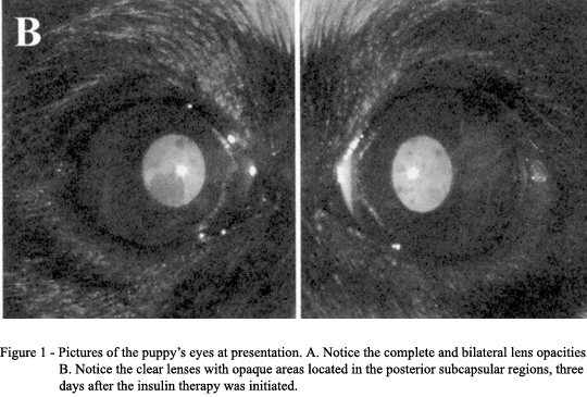
A complete ophthalmic examination was performed. The dogs behavior was abnormal, due to vision impairment, which was confirmed by the menace reflex, cotton ball and obstacle course tests. The pupillary light responses were abnormal (slow and incomplete). There were not signs of conjunctivitis, congestion of scleral vessels nor abnormal ocular discharge. The Schirmer Tear Test 1a values were normal. The slit lampb examination showed normal corneas and irises and narrowing of the anterior chambers. Applanation tonometryc was O.D.: 17mmHg and O.S: 15 mmHg. The appearance of the lenses through a dilated examination was similar to sclerosis from aging process, but denser, completely opaque and associated to swelling (Figure 1A).

Blood and urinary samples examination
Blood and urinary samples were collected and submitted for analysis (glucose, complete blood count, alanine amino transferase, creatinine, alkaline phosphatase and urinalysis). Results: the patient presented hyperglycemia (fasting glucose: 362mg/dL), glycosuria (+++) and ketonuria (+++). Juvenile Diabetes mellitus and ketoacidosis were diagnosed and the therapy was promptly started.
Treatment
The animal remained interned at the veterinary hospital for four days, until his blood glucose levels reached the normal ranges. Insulin and intravenous fluid therapies were performed.
Initially, 0.2 IU/Kg of regular insulin was administered at two hours intervals (IM), until the animal presented significant lowering of the glycemic levels (< 250mg/dL) and restored voluntary feeding, which were observed after three days. Balanced saline solution was administered intravenously at a dosage of 60 mL/kg/day, uninterruptly during this period.
The ketoacidosis ended and the glicemic levels lowered, so the regular insulin was substituted by NPH insulin (bid) and the fluid therapy was maintained as previously described for one day. The animal remained at the hospital for one more day, his blood glucose level reached the normal range (112mg/dL) and he was dismissed.
The home prescription was: NPH insulin (SC, bid) and therapeutical diet (Moderate Energy Programe, Royal Canin).
Second ophthalmic examination
During the period in which the animal remained at the Hospital, it was observed that his vision had improved and the lens opacities had diminished, so he was submitted to a second ophthalmic examination, performed three days after the first one.
The dogs behavior was normal, without signs of visual impairment and with normal response to the usual vision tests. The pupillary light responses were almost normal, showing a discrete delay. The slit lamp examination showed discrete diminishment of the anterior chambers narrowing. Applanation tonometry values were within the normal range.
The dilated examination revealed clear swelling lenses, except opaque areas located in the posterior subcapsular regions of the lenses (Figure 1B).
Clinical sequential evaluation
The dogs owners were oriented about the medical care and to bring the animal regularly for doses adjustments and reexaminations, but no further presentations were realized. The veterinary team members made a telephone contact and the owners stated that they did not follow the orientations, the lenses regained complete opacification and the dog died two months after the initial presentation.
DISCUSSION
Transitory diabetic cataracts has been thought to occur in people with subclinical Diabetes mellitus with normal or only mildly increased blood glucose levels (DICKEY & DAILY, 1993) but in this canine patient the glucose levels were significantly increased.
The initial appearance of the lenses was that described by TEITELBAUM (1998), similar to nuclear sclerosis, even though much denser. This type of cataracts is thought to occur in early ages and to be non reversible by blood glucose levels stabilization. Controversially, after reduction of hyperglycemia in this puppy, the opacification was reduced and the lenses appeared as having a plaque in the posterior subcapsular region. This is the aspect usually described in cases of transitory diabetics cataracts in human beings (DICKEY & DAILY, 1993; GELVIN & THONN, 1993; BUTLER, 1994).
The onset of the transitory cataract formation was abrupt and resolved in a short period of time (three days), in agreement with previous reports in human beings (DICKEY & DAILY, 1993; GELVIN & THONN, 1993; BUTLER, 1994). The regression of the lens opacity has been associated to the normalization of blood glucose levels (GELVIN & THONN, 1993; TEITELBAUM, 1998) and also to poor diabetic control (DICKEY & DAILY, 1993). The lens opacity in this dog reduced when his blood glucose levels returned to the normal ranges and the ketoacidosis was resolved. The occurrence of ketoacidosis has also been associated with transitory cataracts formation in adolescents (PHILLIP et al., 1993).
The short period of time between the cataracts formation and the normalization of blood glucose levels may have prevented irreversible lenticular fibers damage. SHARMA & VASAVADA (2001) associated events that were also presented in this case to the probable genesis of acute diabetic cataracts: hyperglycemia, fluctuating plasma glucose levels and double osmotic shock (hyperglycemia and hypoglycemia) involved with each fluctuation. The cataracts regression was associated to good control of plasma glucose with no further gross fluctuation.
It is possible to theorize a correlation between the dogs age, lenticular structure and reestablishment of lens transparency.
The restoration of lens opacity after the puppy was dismissed was associated with the inadequate insulin administration by the owners, with consequent hyperglycemia and multiple extreme consequences, including his death.
CONCLUSIONS
Canine lenses may reassume transparency after a rapid glycemic control. The exact involved mechanisms in both human and canine diabetic cataracts are yet to be determined.
SOURCES AND MANUFACTURERS
aSchirmer Tear Strips® Schering Plough Animal Health
bSlit Lamp SL-14®Kowa Company Ltda
cTonopen XL® Mentor Medical Systems
ACKNOWLEDGEMENTS
The authors would like to thank FAPESP and CAPES for financial support.
Recebido para publicação 12.05.04
Aprovado em 01.12.04
- ATKINS, C.E. et al. Diabetes mellitus in the juvenile dog: a report of four cases. Journal American Veterinary Medical Association, Schaumburg, v.15, n.4, p.362-368, 1979.
- ATKINS, C.E. et al. Morphologic and immunocytochemical study of young dogs with Diabetes mellitus associated with pancreatic islet hypoplasia. American Journal of Veterinary Research, Schaumburg, v.49, n.9, p.1577-1581, 1988.
- BASHER, A.W.; ROBERTS, S.M. Ocular manifestations of Diabetes mellitus: diabetic cataracts in dogs. Veterinary Clinics of North America: Small Animal Practice, Philadelphia, v.25, n.3, p.661-676, 1995.
- BEAM, S. et al. A retrospective-cohort study on the development of cataracts in dogs with Diabetes mellitus: 200 cases. Veterinary Ophthalmology, London, v.2, p.169-172, 1999.
- BROWN, N.P.; BRON, A. Lens disorders: a clinical manual of cataract diagnosis Oxford : Butterworth-Heinemann, 1996. 179p.
- BUTLER, P.A. Reversible cataracts in Diabetes mellitus Journal American Optometric Association, St Louis, v.65, n.8, p.559-563, 1994.
- DICKEY, J.B.; DAILY, M.J. Transient posterior subcapsular lens opacities in Diabetes mellitus American Journal of Ophthalmology, Chicago, v.115, n.2, p.234-238, 1993.
- GELVIN, J.B.; THONN, V.A. The formation and reversal of acute cataracts in Diabetes mellitus Journal American Optometric Association, St Louis, v.64, n.7, p.471-474, 1993.
- LACKNER, P.A. et al. Age dependent lens changes in galactose-fed dogs. Experimental Eye Research, London, v.64, n.8, p.431-436, 1997.
- MINKUS, G. et al. Ductuloendocrine cell proliferation in the pancreas of two young dogs with Diabetes mellitus Veterinary Pathology, Washington, v.34, n.2, p.164-167, 1997.
- NEIGER, R. et al. Exocrine pancreatic insufficiency combined with insulin-dependent Diabetes mellitus in a juvenile German shepherd dog. Journal of Small Animal Practice, London, v.37, n.7, p.344-349, 1996.
- PHILLIP M. et al. Transient subcapsular cataract formation in a child with diabetes. Clinical Pediatrics, New York, v.32, n.11, p.684-685, 1993.
- SHARMA, P.; VASAVADA, A.R. Acute transient bilateral diabetic posterior subcapsular cataracts. Journal of Cataract and Refractive Surgery, Fairfax, v.27, n.5, p.789-794, 2001.
- TEITELBAUM, B.A. Reversible fingerprint cataract secondary to Diabetes mellitus Clinical Eye and Vision Care, Philadelphia, v.10, n.2, p.81-84, 1998.
Publication Dates
-
Publication in this collection
09 Nov 2005 -
Date of issue
June 2005
History
-
Accepted
01 Dec 2004 -
Received
12 May 2004