Abstracts
OBJECTIVE: This study aims to evaluate the effective ionizing radiation dose received by staff involved in interventional vascular procedures to establish parameters for comparison between the data obtained with an ionization chamber and with the dosimeters used for individual monitoring. MATERIALS AND METHODS: The evaluation was performed in a cardiac catheter laboratory and air kerma rate was measured in a real working environment, in the positions occupied by physicians and nurses, with and without the lead glass shield. The number of tests performed in the laboratory was taken into consideration and most common projections and radiographic parameters were selected. Conversion factors founded in the literature were applied for the effective dose estimation. RESULTS: The values found in the effective dose and in the dosimetry report were rather consistent. Additionally, it was observed that the use of the lead glass shield reduces in up to 97% the doses received by the professional and therefore it must be routinely used. CONCLUSION: The reported values are representative but cannot be assumed as a reproduction of individual monitoring conditions, although they are valid for comparison purposes and as a guidance for dosing optimization.
Occupational exposure; Dosimetry; X-rays; Hemodynamics
OBJETIVO: Avaliar a dose efetiva recebida pelos trabalhadores envolvidos na realização de procedimentos de hemodinâmica, de forma a estabelecer parâmetros comparativos entre os dados obtidos com uma câmara de ionização e pelos dosímetros usados para monitoração individual. MATERIAIS E MÉTODOS: A avaliação foi desenvolvida em um serviço de hemodinâmica, sendo que os testes constaram da medida do taxa de kerma no ar no ambiente real de trabalho, na posição ocupada por médicos e enfermeiros, com e sem a utilização de barreira plumbífera de proteção. Foi levado em consideração o número de exames realizados no serviço e foram selecionados as projeções e parâmetros radiográficos mais utilizados. Para a estimativa da dose efetiva foram utilizados fatores de conversão encontrados na literatura. RESULTADOS: Os valores encontrados em dose efetiva e de dosimetria são consistentes. Observou-se, ainda, que o uso da barreira protetora plumbífera reduz em até 97% a dose recebida pelo profissional, devendo esta ser utilizada na rotina de exames. CONCLUSÃO: Os valores apresentados são representativos, não podendo ser assumidos como reprodução das condições de monitoração individual, mas são válidos para comparações e orientação na otimização das doses.
Exposição ocupacional; Dosimetria; Raios X; Hemodinâmica
ORIGINAL ARTICLE
Evaluation of occupational exposure in hemodynamic procedures* * Study developed for the Program of Post-Graduation in Electrical Engineering and Industrial Information Technology at the Centro Federal de Educação Tecnológica do Paraná, Curitiba, PR.
Silvia C. Gusso ScreminI; Hugo R. SchelinII; João G. Tilly Jr.III
IMD Radiologist, Centro Federal de Educação Tecnológica do Paraná
IIDoctor Professor, Centro Federal de Educação Tecnológica do Paraná
IIIPhysicist in Medicine at the Clinics Hospital, Universidade Federal do Paraná
Mailing address Mailing address: Dra. Silvia C. Gusso Scremin Rua Pedro Ivo, 318 Curitiba, PR, Brasil 80010-020 E-mail: silvia@institutoforlanini.com.br
ABSTRACT
OBJECTIVE: This study aims to evaluate the effective ionizing radiation dose received by staff involved in interventional vascular procedures, to establish parameters for comparison between the data obtained with an ionization chamber and with the dosimeters used for individual monitoring.
MATERIALS AND METHODS: The evaluation was performed in a cardiac catheter laboratory and air kerma rate was measured in a real working environment, in the positions occupied by physicians and nurses, with and without the lead glass shield. The number of tests performed in the laboratory was taken into consideration and most common projections and radiographic parameters were selected. Conversion factors founded in the literature were applied for the effective dose estimation.
RESULTS: The values found in the effective dose and in the dosimetry report were rather consistent. Additionally, it was observed that the use of the lead glass shield reduces in up to 97% the doses received by the professional and therefore it must be routinely used..
CONCLUSION: The reported values are representative but cannot be assumed as a reproduction of individual monitoring conditions, although they are valid for comparison purposes and as a guidance for dosing optimization.
Keywords: Occupational exposure, Dosimetry, X-ray, Hemodynamics
INTRODUCTION
Controlling the radiation dose received by professionals in services involving ionizing radiations is extremely important, especially during hemodynamic procedures. Based on the review of imaging services individual monitoring reports, we have observed that the sector where the dose is higher is the hemodynamics sector.
In the last years, an increase in the number of types and in the complexity of interventional procedures was observed in this area. This happens because some therapeutic procedures can be performed without the need for surgery and consequently with lower risk for the patient(1).
The International Commission on Radiological Protection (ICRP) establishes that no practice involving ionizing radiation should be adopted unless its benefits outweigh its harmful effects. Therefore, the techniques involving the utilization of such a radiation should be optimized so that the doses received are the smallest possible, but compatible with the diagnostic purposes(2).
The occupational exposure in hemodynamics is associated with the occurrence of deterministic effects. The thyroid and the crystalline are examples of organs under risk of appearance of such effects, although well documented case reports are scarce(3). However, for radiological protection purposes, the stochastic effects are assumed as proportional to the effective dose that constitutes the indicator to be followed up, considering the magnitude of the doses resulting from the performance of hemodynamic procedures(4).
This study objective is to estimate the effective dose in professionals involved in the performance of hemodynamic procedures (physicians and nurses working environment), verify the dose reduction achieved with the interposition of a protection barrier and if such reduction corresponds to the data reported in the literature, and establish a protocol of professional behavior in the examination room.
MATERIALS AND METHODS
The evaluation was developed in the service of hemodynamics of a reference hospital for cardiac surgery in the metropolitan region of Curitiba, State of Paraná, where a General Electric Advantx DXL device is used.
The basic requirements considered for the development of this study were: the instruments used, the reference sample, the projections selection (the most frequent examination), the protocol, the test duration and the number of examinations per month.
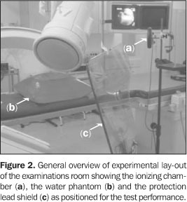
The devices used for data collection were a Radcal 180 cm³ ionization chamber with radiation monitor model 9010, water phantom, tape measure, tripod, table and protocols for data collection.
In the hemodynamics services studied the following procedures are performed: angioplasty, cardiac catheterization, brain arteriography, cholangiography, among others. The cardiac catheterization was selected for being the most frequent procedure and, considering that for each projection the equipment operates with different radiographic parameters, commonly used projections were selected. All the tests were performed following parameters automatically selected by the device for a 18cm-thick patient, and the magnification remained constant in 23.
The projections are identified with their respective parameters values, as follows:
I. Right oblique 30º Fluoroscopy mode: 75 kV and 5.2 mA; cine mode: 60 kV and 50 mA.
II. Right anterior oblique 10º and caudal 30º Fluoroscopy mode: 75 kV and 5.3 mA; cine mode: 60 kV and 51 mA.
III. Left oblique 55º caudal 34º Fluoroscopy mode: 75 kV and 3.2 mA; cine mode: 60 kV and 28 mA.
IV. Posteroanterior cranial 28º left oblique 10º Fluoroscopy mode: 75 kV and 5.3 mA; cine mode: 60 kV and 52 mA.
V. Left oblique 50º cranial 25º fluoroscopy mode: 80 kV and 5.9 mA; cine mode: 63 kV and 65 mA.
For the cardiac catheterization examination, the device is run in fluoroscopy mode for ten minutes, corresponding to two thirds of the exposure time for the catheter introduction and the realization of the examination itself. For images recording, the device runs in cine mode for five minutes, that is, one third of exposure time.
The positions routinely occupied in the examinations room by the professionals were identified as per Figure 1. The physician's position is indicated by (A) and the nurse's position is indicated by (B). Aiming at reproducing the usual irradiation conditions, the distances of (A) and (B) were measured in relation to the center of the water phantom in the same position of a patient being submitted to a cardiac catheterization.
The ionization chamber was placed on points (A) and (B), at the same height of the lapel dosimeter for a person of median height. The measurements were made according to the following schedule:
a) The ionization chamber was placed on the measurement point (A);
b) an exposure was performed in fluoroscopy mode and another in cine mode, with the reading and technical parameters (kV and mA) being noted down on a form, at projection I;
c) this procedure was repeated for the other four projections;
d) the ionization chamber was positioned on the measurement point (B);
e) steps b) and c) were repeated.
Afterwards, the test was performed with placement of a 0.5 mm lead equivalent protection lead glass shield, near the patient, between him/her and the position (A), as shown in the Figure 2.
The readings with ionization chamber were performed in air kerma rate. Once the measurements were performed for all the projections, the values obtained in each reference point, both in the fluoroscopy and cine modes were summed up, resulting in a total air kerma rate in the places of the clinical staff professionals in the room.
With the purpose of establishing a relationship between the operational quantity (air kerma rate) and the protection quantity (effective dose) so as to represent the dose affecting the professionals, the calculation was made according to the following equation:
in which:
E = effective power (mSv);
 = air kerma rate summation of projections I to V (mGy/h);
FCTp = correction factor for temperature and pressure;
ft = working time fraction/examination (h);
N = average number of examinations performed per month;
FEF= coefficient of conversion for effective dose per unit of air kerma (mSv/mGy).
For comparison purposes, the results of calculations were compared to the mean values of individual monitoring. Dose reports from a total of three services of hemodynamics in the region of Curitiba, covering a three-year period were collected, including those from the institution where the measurements were performed. Values of doses affecting each physician and each nurse were taken into consideration for calculating the mean dose for each professional.
RESULTS
Table 1 shows results in terms of air kerma rate, in mGy/h, in the points (A) and (B) for each projection, and the total value for each position during a cardiac catheterization, with and without use of radiation protection shield. The values presented are corrected for temperature, pressure and power dependence.
Each examination has an average duration of 15 minutes comprising ten minutes (0.167 hour) running in the scopy mode and five minutes (0.083 hour) in the cine mode % equally distributed among all projections. Therefore, the air kerma per examination in each reference point is, respectively, 3.246 and 0,159 mGy in the points (A) and (B), without protection shield, and 0.138 and 0.108 mGy, with protection shield.
In the institution studied, 314 examinations are performed each month. Therefore, with these data and conversion factors included in the ICRP 74(2), we can estimate the effective monthly dose in mSv. The conversion factor utilized was 0.122 mSv/mGy applied to the air kerma per examination in each reference point, respectively without and with protection shield. The Table 2 shows the values obtained.
Two physicians and seven nurses work in the hemodynamics sector being studied. With comparison purposes, the effective dose values shown in Table 2 were equally distributed among the professionals, resulting in monthly mean effective doses of 62.2 and 0,9 mSv in the points (A) and (B), respectively, without protection shield, and 2.6 and 0.6 mSv, with protection shield.
The review of the individual monitoring of physicians and nurses in the sector studied has revealed doses of 5.0 mSv and 1.0 mSv for each physician and 2.6 mSv for the nurses.
Individual monitoring data were also collected in other three hemodynamics services, covering a three-year period. Data of professionals presenting doses higher than the register level were taken into consideration and data of professionals who didn't present a reading were discarded. Twenty-two professionals presented a dose in their reports seven physicians and 15 nurses.
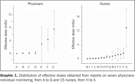
The Graphic 1 represents the individual monitoring data of these three serviced in terms of mean effective monthly dose for each group, per professional physicians to the left and nursed to the right. The mean doses as well as the standard deviation for each professional are represented in ascending order.
It is possible to observe that, for both groups, the dispersion around each individual professional mean dose increases proportionally to the mean dose. The professionals receiving lower doses (from A to D and from H to V) present lower standard deviation, and those presenting higher doses (E to G and X) present dispersion of the higher mean dose.
DISCUSSION
The technique applied by the physicians at the hemodynamics service studied for catheter insertion was the brachial artery technique that may double the dose in relation to the femoral technique.
Several oblique projections lateral, cranio-caudal and caudal-cranial are necessary for analysis of the heart arteries. So, when left projections, mainly the V projections (left oblique 35º, cranial 25º) are utilized, the physicians are near the x-ray primary beam, registering the highest doses.
The conversion factor applied for estimating the effective dose is specified for a whole-body uniform anteroposterior exposure to a monoenergetic photons beam. Unfortunately, this factor presents a strong energetic dependence in the power range utilized. As a reference, we have chosen the 20 keV power, resulting in the 0.122 mSv/mGy coefficient.
Although the air kerma rate values presented in this study are compatible with the values presented in the literature about 2 mGy/h(5) and up to 8 mGy/h near the patient, and 0.053 mSv per procedure with protective lead shield in the physician position(3), the effective doses calculated for the physicians are clearly overestimated, principally due to the exposure condition, considering that the physician is fairly near the patient and the exposure cannot be considered as uniform. On the other hand, for nurses, the values obtained are more similar to those of individual monitoring, possibly due to the choice of the measurement point position.
The radiation affecting professionals, above all, is related to the disseminated radiation. With the protection shield interposition, the dose received by the physician is significantly reduced. On the other hand, for the nursing staff, this difference is not so significant because the radiation protection shield is located near the physician. Even without the shield, the nurse is in a shadow region as a result of the interposition of the physician with his/her lead apron between the patient and his/her position in the examination room.
Each professional exposure is different principally due to the time dedicated to the activity and the distance in relation to the patient during the procedure(6). Data regarding individual monitoring of physicians and nurses in other institutions, although not being comprehensive enough to infer a general behavior, reveal that about three quarters of all dose occurrences are below the monthly dose limit (herein assumed as 1.5 mSv), in spite of the high mean doses.
The doses registered in the individual monitoring were measured in non-uniform conditions as dosimeters can be carried in different ways. There are cases reporting dosimeters under the aprons and others reporting dosimeters on the aprons, as well as the possibility of failure in the use of some dosimeters during the whole period of the professional exposure.
Some physicians present a lower hourly workload or a lower number of procedures they perform in service. Also, in a same service it is possible to observe different doses in a determined professional category due to their attitude during the performance of the examinations.
Each nurse behavior also defines dose differences since it was observed that some of them remain out of the examinations room during the use of x-rays. As a result some nurses present lower doses.
CONCLUSIONS
In this study, we have evaluated the effective ionizing radiation dose in the positioning adopted by professionals (physicians and nurses) in the hemodynamics room during the realization of cardiac catheterization and made a correlation between data obtained with ionization chamber and through individual monitoring. Also, the measurements have taken into consideration the use and non-use of a 0.5 mm lead equivalent protection shield.
Based on data researched, one has observed that, in hemodynamics, the physicians receive higher radiation doses than other professionals, as shown in Table 2, similarly to data presented in the literature(7).
The nursing staff receives doses equivalent to the doses received by physicians. However their doses could be substantially reduced as they frequently position themselves unnecessarily near the equipment and the patients.
Results of the correlation between estimate effective dose and individual monitoring are representative and valid for comparison purposes and for guidance of doses optimization.
The test performed in this study with interposition of a 0.5 mm lead equivalent protection shield, has evidenced a mean reduction in the physician exposure of about 90% during a cardiac catheterization. For the nursing staff, the exposure reduction may reach 80%.
We have sought to demonstrate that the radiation doses can be reduced if the professional knows his/her working environment, the x-rays equipment and the protection means available.
Therefore, the professionals continued education and the implementation of objective procedures for doses reduction are actions that will result in an adequate control of the radiation, by means of an internal protocol establishing the doses related to the procedures. This protocol should be discussed by the clinical staff and should be kept under strict control and updating.
Acknowledgement
Prof. Paulo Roberto Costa, Instituto de Eletrotécnica e Energia (IEE-USP).
REFERENCES
Received January 28, 2005.
Accepted after revision July 12, 2005.
- 1. Faulkner K. Radiation protection in interventional radiology. Br J Radiol 1997;70:325326.
-
2International Commission on Radiological Protection. Conversion coefficients for use in radiological protection against external radiation. ICRP Publication 74. New York: Pergamon Press, 1997.
-
3International Commission on Radiological Protection. Avoidance of radiation injuries from medical interventional procedures. ICRP Publication 85. New York: Pergamon Press, 2000.
- 4. Oliveira ML, Khoury H. Influência do procedimento radiográfico na dose de entrada na pele de pacientes em raios X pediátricos. Radiol Bras 2003;36:105109.
-
5United Nations Scientific Committee on the Effects of Atomic Radiation. Sources and effects of ionizing radiation. UNSCEAR Publication 2000. New York: United Nations Press, 2000.
- 6. Kamenopoulou V, Drikos G, Dimitriou P. Dose constraints to the individual annual doses of exposed workers in the medical sector. Eur J Radiol 2001;37:204208.
- 7. Bacelar A, Borges V, Zago AJ, Krebs EM, Hoff G, Nied L. Evaluation of the equivalent effective dose in the work team exposed to ionizated radiation at the hemodinamic unit. World Congress on Medical Physics and Biomedical Engineering; Nice, France. September, 1997:1419.
Publication Dates
-
Publication in this collection
25 May 2006 -
Date of issue
Apr 2006
History
-
Accepted
12 July 2005 -
Received
28 Jan 2005


