Abstracts
Congenital adrenal hyperplasia (CAH) is an autossomic recessive disorder caused by impaired steroidogenesis. Patients with CAH may present adrenal insufficiency with or without salt-wasting, as well as various degrees of virilization and fertility impairment, carrying a high incidence of testicular adrenal rest tumors and increased incidence of adrenal tumors. The diagnosis of CAH is made based on the adrenocortical profile hormonal evaluation and genotyping, in selected cases. Follow-up is mainly based on hormonal and clinical evaluation. Utility of imaging in this clinical setting may be helpful for the diagnosis, management, and follow-up of the patients, although recommendations according to most guidelines are weak when present. Thus, the authors aimed to conduct a narrative synthesis of how imaging can help in the management of patients with CAH, especially focused on genitography, ultrasonography, computed tomography, and magnetic resonance imaging.
Adrenal hyperplasia; congenital; magnetic resonance imaging; computed tomography; ultrasonography; diagnostic imaging
Hiperplasia congênita de suprarrenal (CAH) é uma doença autossômica recessiva causada por deficiências enzimáticas na esteroidogênese. Clinicamente, os pacientes com CAH podem apresentar insuficiência adrenal com ou sem perda de sal, vários graus de virilização e diminuição na fertilidade, alta incidência de restos adrenais testiculares e de tumores adrenais. O diagnóstico de CAH é feito baseado nos resultados da avaliação hormonal e genotípica, em casos selecionados. O seguimento dos pacientes é principalmente feito com avaliação clínica e hormonal. Métodos de diagnóstico por imagem podem ser muito úteis não só no diagnóstico como no manejo e seguimento dos pacientes com CAH. Porém, as recomendações, de acordo com a maioria dos consensos, quando existem, são escassas. Nesse contexto, com base em uma revisão sistemática, o objetivo deste artigo foi sintetizar a literatura em relação a como os métodos de diagnóstico por imagem podem ser úteis no manejo dos pacientes com CAH, com foco em genitografia, ultrassonografia, tomografia computadorizada e ressonância magnética.
Hiperplasia adrenal congênita; ressonância magnética; tomografia computadorizada; ultrassonografia; diagnóstico por imagem
INTRODUCTION
Congenital adrenal hyperplasia (CAH) is an autossomic recessive disorder caused by impaired steroidogenesis, in approximately 95% of the cases, secondary to 21-hydroxylase deficiency. Patients may present adrenal insufficiency with or without salt-wasting, as well as various degrees of virilization and fertility impairment, carrying a high incidence of testicular adrenal rest tumors (TART) and increased incidence of adrenal tumors (11 Merke DP, Bornstein SR. Congenital adrenal hyperplasia. Lancet. 2005;365(9477):2125-36.). Neonatal screening is recommended because it reduces and prevents morbidity and mortality from salt-losing crisis (22 Speiser PW, Azziz R, Baskin LS, Ghizzoni L, Hensle TW, Merke DP, et al. Congenital adrenal hyperplasia due to steroid 21-hydroxylase deficiency: an Endocrine Society clinical practice guideline. J Clin Endocrinol Metab. 2010;95(9):4133-60.). The diagnosis is made with adrenocortical profile hormonal evaluation and genotyping in selected cases (22 Speiser PW, Azziz R, Baskin LS, Ghizzoni L, Hensle TW, Merke DP, et al. Congenital adrenal hyperplasia due to steroid 21-hydroxylase deficiency: an Endocrine Society clinical practice guideline. J Clin Endocrinol Metab. 2010;95(9):4133-60.).
Utility of imaging in this clinical setting may be helpful for the diagnosis, management, and follow-up of the patients, although recommendations according to most guidelines are weak when present (22 Speiser PW, Azziz R, Baskin LS, Ghizzoni L, Hensle TW, Merke DP, et al. Congenital adrenal hyperplasia due to steroid 21-hydroxylase deficiency: an Endocrine Society clinical practice guideline. J Clin Endocrinol Metab. 2010;95(9):4133-60.). Bone mineral density evaluation in children is not recommended and adrenal imaging is suggested only for patients with an atypical course (22 Speiser PW, Azziz R, Baskin LS, Ghizzoni L, Hensle TW, Merke DP, et al. Congenital adrenal hyperplasia due to steroid 21-hydroxylase deficiency: an Endocrine Society clinical practice guideline. J Clin Endocrinol Metab. 2010;95(9):4133-60.). Conversely, CAH must be excluded in cases of adrenal incidentaloma supposedly asymptomatic or oligosymptomatic. To evaluate gonads, ultrasonography (US) is recommended for screening males from adolescence, but there is no recommendation to screen females (22 Speiser PW, Azziz R, Baskin LS, Ghizzoni L, Hensle TW, Merke DP, et al. Congenital adrenal hyperplasia due to steroid 21-hydroxylase deficiency: an Endocrine Society clinical practice guideline. J Clin Endocrinol Metab. 2010;95(9):4133-60.).
Thus, the authors aim to conduct a narrative synthesis of how imaging can help in the management of patients with CAH, especially focused on genitography (GX), US, computed tomography (CT), and magnetic resonance imaging (MRI).
SEARCH STRATEGY AND SELECTION OF ARTICLES
A systematic search was conducted in MedLine® (from 1950 to July 2013) and in Web of Science® (from 1965 to July 2013) databases for articles published in English, Spanish, Portuguese, and French. On MedLine®, the MeSH term “adrenal hyperplasia, congenital” was searched with the other imaging related MeSH terms with AND at a time using “ultrasonography”, or “magnetic resonance imaging”, or “diffusion magnetic resonance imaging”, or “tomography, X-ray computed”, or “multidetector computed tomography”, or “positron-emission tomography and computed tomography”, or “tomography scanners, X-ray computed radiography”, or “diagnostic imaging”. Web of Science® was searched for articles with the search terms “congenit* and adren* and hyperpl*” “AND imag*”. Books and other selected references cited in the most relevant retrieved articles were also reviewed.
Studies that were conducted in animals and in which only scintigraphy or nuclear imaging modalities were used as imaging modalities were not within the scope of this review. Also, studies that only used X-ray to evaluate bone age or any imaging modalities to assess bone mineral density were not extensively reviewed, as it is well established by the guidelines that bone age should be assessed annually after 2 years of age and regular evaluation of bone mineral density is not recommended (22 Speiser PW, Azziz R, Baskin LS, Ghizzoni L, Hensle TW, Merke DP, et al. Congenital adrenal hyperplasia due to steroid 21-hydroxylase deficiency: an Endocrine Society clinical practice guideline. J Clin Endocrinol Metab. 2010;95(9):4133-60.).
Imaging
Genitography
The urogenital sinus is the embryologic precursor of the bladder, urethra, and distal third of the vagina in females. Excessive androgen exposure in utero leads to virilization of the external genitalia and urogenital malformations in females with CAH (11 Merke DP, Bornstein SR. Congenital adrenal hyperplasia. Lancet. 2005;365(9477):2125-36.). Signs of urogenital sinus malformation secondary to virilization are hydrocolpos or hydrometrocolpos and only two apertures (one of them is the anus) in the perineal region associated with ambiguous genitalia (33 Chavhan GB, Parra DA, Oudjhane K, Miller SF, Babyn PS, Pippi Salle FL. Imaging of ambiguous genitalia: classification and diagnostic approach. Radiographics. 2008;28(7):1891-904.). Typically, female patients with classic CAH have ambiguous genitalia at birth. Thus an anatomic detailed image plays an important role in planning strategy for feminizing surgery. Genitography shows the urethra, the level of external sphincter, the presence or absence of the vagina, the urethrovaginal confluence, and the cervical impression of the uterus (33 Chavhan GB, Parra DA, Oudjhane K, Miller SF, Babyn PS, Pippi Salle FL. Imaging of ambiguous genitalia: classification and diagnostic approach. Radiographics. 2008;28(7):1891-904.) (Figure 1).
Newborn with “non palpable testicles”. Ultrasonography (A-C) showed enlarged and cerebriform pattern of the adrenal glands (arrows in A). Sagittal view of the pelvis through the abdominal wall (B): the uterus (“ut”) and the presence of hydrocolpus (“hc”) are well depicted. The ovaries (not shown) were also present. Sagittal ultrasonography view of the pelvis through the perineum (C) was not sufficient to show with high confidence the confluence of the urethra and the vagina, which was better viewed on magnetic resonance imaging (MRI) (E, sagittal T2-weighetd MRI of the pelvis) and genitography (F). The arrows in (E) and (F) are pointing the confluence of the vagina and the urethra (“u”). Axial T2-weighted MRI of the pelvis showing prostate tissue (dashed arrows in D) in this patient. The pattern of the adrenal glands and presence of mullerian derivatives allowed institution of therapy while laboratory tests were done to confirm congenital adrenal hyperplasia in this XX neonate. MRI and genitography were requested for planning feminizing surgery.
Genitography associated with voiding cistography has also the ability to show upper genitourinary abnormalities, present in 21%-80% (44 Nabhan ZM, Eugster EA. Upper-tract genitourinary malformations in girls with congenital adrenal hyperplasia. Pediatrics. 2007;120(2):e304-7.) of the patients with CAH, mostly seen in girls.
Although some authors concluded that genitography did not add information to endoscopic (55 Vanderbrink BA, Rink RC, Cain MP, Kaefer M, Meldrum KK, Misseri R, et al. Does preoperative genitography in congenital adrenal hyperplasia cases affect surgical approach to feminizing genitoplasty? J Urol. 2010;184(4 Suppl):1793-8.) or surgical findings, many advocate its use as part of routine investigation in female patients with CAH (44 Nabhan ZM, Eugster EA. Upper-tract genitourinary malformations in girls with congenital adrenal hyperplasia. Pediatrics. 2007;120(2):e304-7.), particularly in those infants with ambiguous genitalia.
Ultrasonography
US is the modality of choice to image abdominal and pelvic organs in children and fetuses. It is widely available, versatile, and portable, with lack of ionizing radiation, there is no need for sedation, and provides high-resolution images in any required plane.
The adrenal glands in young children and fetuses are well depicted with US, especially in the neonate period. Sonographic abnormalities of the adrenal glands in CAH are similar in pre- and postnatal periods. The most prevalent sign is bilateral enlarged glands with width measurements of one limb ≥ 4 mm (66 Sivit CJ, Hung W, Taylor GA, Catena LM, Brown-Jones C, Kushner DC. Sonography in neonatal congenital adrenal hyperplasia. AJR Am J Roentgenol. 1991;156(1):141-3.). In addition, adrenal size was positively correlated with plasma concentration of dehydroepiandrosterone sulphate (77 Hauffa BP, Menzel D, Stolecke H. Age-related changes in adrenal size during the first year of life in normal newborns, infants and patients with congenital adrenal hyperplasia due to 21-hydroxylase deficiency: comparison of ultrasound and hormonal parameters. Eur J Pediatr. 1988;148(1):43-9.). Also, a coiled or cerebriform pattern (88 Avni EF, Rypens F, Smet MH, Galetty E. Sonographic demonstration of congenital adrenal hyperplasia in the neonate: the cerebriform pattern. Pediatr Radiol. 1993;23(2):88-90.,99 Hernanz-Schulman M, Brock JW, 3rd, Russell W. Sonographic findings in infants with congenital adrenal hyperplasia. Pediatr Radiol. 2002;32(2):130-7.) is specific for this condition (Figure 1A). This finding should prompt a thorough investigation in male fetus and whenever a corticoid therapy is initiated serial US will show decrease in size of the adrenal glands (1010 Saada J, Grebille AG, Aubry MC, Rafii A, Dumez Y, Benachi A. Sonography in prenatal diagnosis of congenital adrenal hyperplasia. Prenat Diagn. 2004;24(8):627-30.). In infants, the morphology was shown to be of valuable importance while waiting for laboratory results in patients investigated for suspicious of the disease, enabling earlier diagnosis and treatment (99 Hernanz-Schulman M, Brock JW, 3rd, Russell W. Sonographic findings in infants with congenital adrenal hyperplasia. Pediatr Radiol. 2002;32(2):130-7.). However, it is very important to notice that normal adrenal glands do not exclude the possibility of CAH (66 Sivit CJ, Hung W, Taylor GA, Catena LM, Brown-Jones C, Kushner DC. Sonography in neonatal congenital adrenal hyperplasia. AJR Am J Roentgenol. 1991;156(1):141-3.).
Although the clinical features of CAH are usually present, testicular adrenal
rest tumor (TART) may be the only clinical finding at presentation (1111 Avila NA, Premkumar A, Shawker TH, Jones JV, Laue L, Cutler GB,
Jr. Testicular adrenal rest tissue in congenital adrenal hyperplasia: findings
at Gray-scale and color Doppler US. Radiology.
1996;198(1):99-104.). Based on microscopic studies,
TARTs are reported to be present in all males with CAH (1212 Shanklin DR, Richardson AP Jr, Rothstein G. Testicular hilar
nodules in adrenogenital syndrome. The nature of the nodules. Am J Dis Child.
1963;106:243-50.). On US, it has been documented with
a prevalence of up to 94% (1313 Stikkelbroeck NM, Otten BJ, Pasic A, Jager GJ, Sweep CG, Noordam
K, et al. High prevalence of testicular adrenal rest tumors, impaired
spermatogenesis, and Leydig cell failure in adolescent and adult males with
congenital adrenal hyperplasia. J Clin Endocrinol Metab.
2001;86(12):5721-8.). The
most common sonographic features are bilateral spikelike appearance
intratesticular hypoechoic masses with no sound attenuation, surrounding the
mediastinal testis (1111 Avila NA, Premkumar A, Shawker TH, Jones JV, Laue L, Cutler GB,
Jr. Testicular adrenal rest tissue in congenital adrenal hyperplasia: findings
at Gray-scale and color Doppler US. Radiology.
1996;198(1):99-104.). On color
Doppler they are hypo or avascular (1414 Stikkelbroeck NM, Suliman HM, Otten BJ, Hermus AR, Blickman JG,
Jager GJ. Testicular adrenal rest tumours in postpubertal males with congenital
adrenal hyperplasia: sonographic and MR features. Eur Radiol.
2003;13(7):1597-603.) and there is no deviation or changes in caliber of the
vessels that course the lesions (1111 Avila NA, Premkumar A, Shawker TH, Jones JV, Laue L, Cutler GB,
Jr. Testicular adrenal rest tissue in congenital adrenal hyperplasia: findings
at Gray-scale and color Doppler US. Radiology.
1996;198(1):99-104.,1414 Stikkelbroeck NM, Suliman HM, Otten BJ, Hermus AR, Blickman JG,
Jager GJ. Testicular adrenal rest tumours in postpubertal males with congenital
adrenal hyperplasia: sonographic and MR features. Eur Radiol.
2003;13(7):1597-603.). However, TART may
also appear as heterogeneous or hyperechoic nodules (1111 Avila NA, Premkumar A, Shawker TH, Jones JV, Laue L, Cutler GB,
Jr. Testicular adrenal rest tissue in congenital adrenal hyperplasia: findings
at Gray-scale and color Doppler US. Radiology.
1996;198(1):99-104.,1414 Stikkelbroeck NM, Suliman HM, Otten BJ, Hermus AR, Blickman JG,
Jager GJ. Testicular adrenal rest tumours in postpubertal males with congenital
adrenal hyperplasia: sonographic and MR features. Eur Radiol.
2003;13(7):1597-603.,1515 Avila NA, Shawker TS, Jones JV, Cutler GB Jr, Merke DP. Testicular
adrenal rest tissue in congenital adrenal hyperplasia: serial sonographic and
clinical findings. AJR Am J Roentgenol. 1999;172(5):1235-8.), and even as an epididymal nodule
(1111 Avila NA, Premkumar A, Shawker TH, Jones JV, Laue L, Cutler GB,
Jr. Testicular adrenal rest tissue in congenital adrenal hyperplasia: findings
at Gray-scale and color Doppler US. Radiology.
1996;198(1):99-104.). TART echogenicity is
related to the size of the lesions, being hypoechoic in lesions smaller than
2 cm and heterogeneous or hyperechoic in lesions larger than 2 cm (1414 Stikkelbroeck NM, Suliman HM, Otten BJ, Hermus AR, Blickman JG,
Jager GJ. Testicular adrenal rest tumours in postpubertal males with congenital
adrenal hyperplasia: sonographic and MR features. Eur Radiol.
2003;13(7):1597-603.). These hyperechoic areas may
represent fibrosis or calcifications (1414 Stikkelbroeck NM, Suliman HM, Otten BJ, Hermus AR, Blickman JG,
Jager GJ. Testicular adrenal rest tumours in postpubertal males with congenital
adrenal hyperplasia: sonographic and MR features. Eur Radiol.
2003;13(7):1597-603.). In addition, larger lesions may not be confined to the
mediastinal testis and smaller lesions are more often seen unilaterally
(1414 Stikkelbroeck NM, Suliman HM, Otten BJ, Hermus AR, Blickman JG,
Jager GJ. Testicular adrenal rest tumours in postpubertal males with congenital
adrenal hyperplasia: sonographic and MR features. Eur Radiol.
2003;13(7):1597-603.). On follow-up, TART can vary
in size (1515 Avila NA, Shawker TS, Jones JV, Cutler GB Jr, Merke DP. Testicular
adrenal rest tissue in congenital adrenal hyperplasia: serial sonographic and
clinical findings. AJR Am J Roentgenol. 1999;172(5):1235-8.), but there is no
correlation between hormonal control or hormonal markers and TART (1515 Avila NA, Shawker TS, Jones JV, Cutler GB Jr, Merke DP. Testicular
adrenal rest tissue in congenital adrenal hyperplasia: serial sonographic and
clinical findings. AJR Am J Roentgenol. 1999;172(5):1235-8.
16 Reisch N, Scherr M, Flade L, Bidlingmaier M, Schwarz HP,
Müller-Lisse U, et al. Total adrenal volume but not testicular adrenal rest
tumor volume is associated with hormonal control in patients with 21-hydroxylase
deficiency. J Clin Endocrinol Metab. 2010;95(5):2065-72.-1717 Reisch N, Flade L, Scherr M, Rottenkolber M, Pedrosa Gil F,
Bidlingmaier M, et al. High prevalence of reduced fecundity in men with
congenital adrenal hyperplasia. J Clin Endocrinol Metab.
2009;94(5):1665-70.). TARTs are thought to be responsible for testicular
parenchymal damage that contributes to reduced fertility (1717 Reisch N, Flade L, Scherr M, Rottenkolber M, Pedrosa Gil F,
Bidlingmaier M, et al. High prevalence of reduced fecundity in men with
congenital adrenal hyperplasia. J Clin Endocrinol Metab.
2009;94(5):1665-70.,1818 Claahsen-van der Grinten HL, Otten BJ, Hermus AR, Sweep FC,
Hulsbergen-van de Kaa CA. Testicular adrenal rest tumors in patients with
congenital adrenal hyperplasia can cause severe testicular damage. Fertil
Steril. 2008;89(3):597-601.). It can be found even in young children with a prevalence of
21% (1919 Martinez-Aguayo A, Rocha A, Rojas N, et al. Testicular adrenal
rest tumors and Leydig and Sertoli cell function in boys with classical
congenital adrenal hyperplasia. J Clin Endocrinol Metab.
2007;92(12):4583-9.) and it is suggested that
gonadal dysfunction is already present before puberty (1919 Martinez-Aguayo A, Rocha A, Rojas N, et al. Testicular adrenal
rest tumors and Leydig and Sertoli cell function in boys with classical
congenital adrenal hyperplasia. J Clin Endocrinol Metab.
2007;92(12):4583-9.). Therefore, early detection of testicular lesions
is advised (2020 Claahsen-van der Grinten HL, Otten BJ, Stikkelbroeck MM, Sweep FC,
Hermus AR. Testicular adrenal rest tumours in congenital adrenal hyperplasia.
Best Pract Res Clin Endocrinol Metab. 2009;23(2):209-20.) to improve treatment
and prevent longstanding gonadal impairment function. Thus, some authors
advocate that not only adolescents should undergo US (2121 Dieckmann K, Lecomte P, Despert F, Maurage C, Sirinelli D, Rolland
JC. [Congenital adrenal hyperplasia and testicular hypertrophy]. Arch Pediatr.
1995;2(12):1167-72.) (Figure
2).
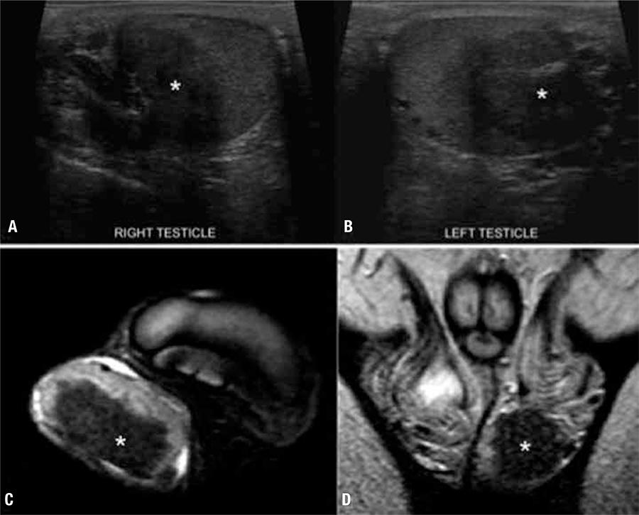
Testicular adrenal rest tumor (TART) in a 14 year-old male, with simple virilizing congenital adrenal hyperplasia. On ultrasonography (A-B), there is a round heterogeneous, predominantly hypoechoic nodule (*) within both testicles, in the region of the mediastinal testis. On magnetic resonance imaging (C, axial; D, coronal) the TARTs are hypointense on T2-weighted images.
High prevalence of impaired fertility is not restricted to men as it was reported also in women with CAH (1313 Stikkelbroeck NM, Otten BJ, Pasic A, Jager GJ, Sweep CG, Noordam K, et al. High prevalence of testicular adrenal rest tumors, impaired spermatogenesis, and Leydig cell failure in adolescent and adult males with congenital adrenal hyperplasia. J Clin Endocrinol Metab. 2001;86(12):5721-8.,1717 Reisch N, Flade L, Scherr M, Rottenkolber M, Pedrosa Gil F, Bidlingmaier M, et al. High prevalence of reduced fecundity in men with congenital adrenal hyperplasia. J Clin Endocrinol Metab. 2009;94(5):1665-70.). The prevalence of polycystic ovaries is increased in women with classical and nonclassical CAH (2222 New MI. Nonclassical congenital adrenal hyperplasia and the polycystic ovarian syndrome. Ann N Y Acad Sci. 1993;687:193-205.). Bilateral enlarged ovaries (2323 Forsbach G, Guitron-Cantu A, Vazquez-Lara J, Mota-Morales M, Diaz-Mendoza ML. Virilizing adrenal adenoma and primary amenorrhea in a girl with adrenal hyperplasia. Arch Gynecol Obstet. 2000;263(3):134-6.), bilateral ovarian cysts, and ovarian adrenal rest tumors (OART) (2424 Russo G, Paesano P, Taccagni G, Del Maschio A, Chiumello G. Ovarian adrenal-like tissue in congenital adrenal hyperplasia. N Engl J Med. 1998;339(12):853-4.) may occur and can also be depicted by US. OART may present as hypoechoic nodules on US (2424 Russo G, Paesano P, Taccagni G, Del Maschio A, Chiumello G. Ovarian adrenal-like tissue in congenital adrenal hyperplasia. N Engl J Med. 1998;339(12):853-4.,2525 Tiosano D, Vlodavsky E, Filmar S, Weiner Z, Goldsher D, Bar-Shalom R. Ovarian adrenal rest tumor in a congenital adrenal hyperplasia patient with adrenocorticotropin hypersecretion following adrenalectomy. Horm Res Paediatr. 2010;74(3):223-8.), similar to TART.
The most common cause of disorders of sex differentiation in the perinatal period is CAH. A clue to the prenatal diagnosis of CAH is usually the presence of ambiguous genitalia that may be present by the second trimester. The most often presentation is of an enlarged clitoris, but more complex abnormalities can be seen (2626 Sivan E, Koch S, Reece EA. Sonographic prenatal diagnosis of ambiguous genitalia. Fetal Diagn Ther. 1995;10(5):311-4.).
In postnatal period, to assess internal anatomy of the pelvis, US is the first choice (Figure 1C). It is easily performed and must include images of the pelvis, scrotum, inguinal, perineal, renal, and adrenal regions (2727 Garel L. Abnormal sex differentiation: who, how and when to image. Pediatr Radiol. 2008;38 Suppl 3:S508-11.). The main purpose of the pelvic sonography is to depict accurately the size and morphology of the Mullerian structures, the uterus, the vagina, and the gonads (33 Chavhan GB, Parra DA, Oudjhane K, Miller SF, Babyn PS, Pippi Salle FL. Imaging of ambiguous genitalia: classification and diagnostic approach. Radiographics. 2008;28(7):1891-904.). In addition to the adrenal glands findings described above, the presence of a uterus in a patient with ambiguous genitalia indicates that the diagnosis is mostly likely CAH (99 Hernanz-Schulman M, Brock JW, 3rd, Russell W. Sonographic findings in infants with congenital adrenal hyperplasia. Pediatr Radiol. 2002;32(2):130-7.). Ultrasound evaluation of the pelvic structures is not only performed for diagnosis but also as part of the preoperative approach for surgery, often in conjunction with other exams, such as genitography and MRI (33 Chavhan GB, Parra DA, Oudjhane K, Miller SF, Babyn PS, Pippi Salle FL. Imaging of ambiguous genitalia: classification and diagnostic approach. Radiographics. 2008;28(7):1891-904.). US provides adequate information about the vagina and urogenital sinus for preoperative decision-making (2828 Chertin B, Hadas-Halpern I, Fridmans A, Kniznik M, Abu-Arafeh W, Zilberman M, et al. Transabdominal pelvic sonography in the preoperative evaluation of patients with congenital adrenal hyperplasia. J Clin Ultrasound. 2000;28(3):122-4.).
Many other abnormalities in patients with CAH can also be demonstrated by US: cardiac dysfunction that reverses with therapy (2929 Minette MS, Hoyer AW, Pham PP, DeBoer MD, Reller MD, Boston BA. Cardiac function in congenital adrenal hyperplasia: a pattern of reversible cardiomyopathy. J Pediatr. 2013;162(6):1193-8, 8 e1.), vascular dysfunction and increased carotid intima media thickness (3030 Wasniewska M, Balsamo A, Valenzise M, Manganaro A, Faggioli G, Bombaci S, et al. Increased large artery intima media thickness in adolescents with either classical or non-classical congenital adrenal hyperplasia. J Endocrinol Invest. 2013;36(1):12-5.), skeletal and midface malformations associated with P450 oxidoreductase deficiency in prenatal diagnosed fetuses (3131 Reisch N, Idkowiak J, Hughes BA, Ivison HE, Abdul-Rahman OA, Hendon LG, et al. Prenatal diagnosis of congenital adrenal hyperplasia caused by P450 oxidoreductase deficiency. J Clin Endocrinol Metab. 2013;98(3):E528-36.), hydrops of placental stem villi in a 46,XX fetus (3232 Furuhashi M, Oda H, Nakashima T. Hydrops of placental stem villi complicated with fetal congenital adrenal hyperplasia. Arch Gynecol Obstet. 2000;264(2):101-4.), association with increased nuchal translucency detected in the prenatal period (3333 Fincham J, Pandya PP, Yuksel B, Loong YM, Shah J. Increased first-trimester nuchal translucency as a prenatal manifestation of salt-wasting congenital adrenal hyperplasia. Ultrasound Obstet Gynecol. 2002;20(4):392-4.), bilateral ovarian steroid cell tumor in a girl with CAH 11 beta-hydroxylase deficiency (3434 Baş F, Saka N, Darendeliler F, Tuzlali S, Ilhan R, Bundak R, et al. Bilateral ovarian steroid cell tumor in congenital adrenal hyperplasia due to classic 11beta-hydroxylase deficiency. J Pediatr Endocrinol Metab. 2000;13(6):663-7.), and adrenal rest tissue extending from the lower pole of the kidney (3535 Souverijns G, Peene P, Keuleers H, Vanbockrijck M. Ectopic localisation of adrenal cortex. Eur Radiol. 2000;10(7):1165-8.).
Computed tomography
Adrenocortical tumors in patients with CAH are not rare. A prevalence of up
to 83% of adrenocortical masses in homozygote patients is reported (3636 Jaresch S, Kornely E, Kley HK, Schlaghecke R. Adrenal
incidentaloma and patients with homozygous or heterozygous congenital adrenal
hyperplasia. J Clin Endocrinol Metab. 1992;74(3):685-9.). Despite this high frequency,
adrenocortical tumors in this setting are most likely to be benign, as
malignant lesions are rare (3636 Jaresch S, Kornely E, Kley HK, Schlaghecke R. Adrenal
incidentaloma and patients with homozygous or heterozygous congenital adrenal
hyperplasia. J Clin Endocrinol Metab. 1992;74(3):685-9.). In
many reports CT scans showed nodules (3636 Jaresch S, Kornely E, Kley HK, Schlaghecke R. Adrenal
incidentaloma and patients with homozygous or heterozygous congenital adrenal
hyperplasia. J Clin Endocrinol Metab. 1992;74(3):685-9.
37 Giacaglia LR, Mendonca BB, Madureira G, Melo KF, Suslik CA,
Arnhold IJ, et al. Adrenal nodules in patients with congenital adrenal
hyperplasia due to 21-hydroxylase deficiency: regression after adequate hormonal
control. J Pediatr Endocrinol Metab. 2001;14(4):415-9.-3838 Harinarayana CV, Renu G, Ammini AC, Khurana ML, Ved P, Karmarkar
MG, et al. Computed tomography in untreated congenital adrenal hyperplasia.
Pediatr Radiol. 1991;21(2):103-5.) that may regress
with adequate therapy (3737 Giacaglia LR, Mendonca BB, Madureira G, Melo KF, Suslik CA,
Arnhold IJ, et al. Adrenal nodules in patients with congenital adrenal
hyperplasia due to 21-hydroxylase deficiency: regression after adequate hormonal
control. J Pediatr Endocrinol Metab. 2001;14(4):415-9.), adenomas
(2323 Forsbach G, Guitron-Cantu A, Vazquez-Lara J, Mota-Morales M,
Diaz-Mendoza ML. Virilizing adrenal adenoma and primary amenorrhea in a girl
with adrenal hyperplasia. Arch Gynecol Obstet.
2000;263(3):134-6.), myelolipomas (3939 Mermejo LM, Elias Junior J, Saggioro FP, Tucci Junior S, Castro
Md, Moreira AC, et al. Giant adrenal myelolipoma associated with 21-hydroxylase
deficiency: unusual association mimicking an androgen-secreting adrenocortical
carcinoma. Arq Bras Endocrinol Metabol. 2010;54(4):419-24.), and the typical pattern of diffuse
enlargement (3838 Harinarayana CV, Renu G, Ammini AC, Khurana ML, Ved P, Karmarkar
MG, et al. Computed tomography in untreated congenital adrenal hyperplasia.
Pediatr Radiol. 1991;21(2):103-5.) with a heterogeneous
enhancement (Figure 3).
Adrenal glands in three different patients with congenital adrenal hyperplasia (CAH). Enlarged adrenal glands (arrows) can be seen on computed tomography (A) and magnetic resonance imaging (B), in different patients. On “A”, the left adrenal gland had nodular margins (dashed arrow). On “B” the left adrenal gland (dashed arrow) is larger than the right adrenal gland, which was within normal limits. Another adult patient with abdominal pain in which ultrasonography (not shown) depicted an adrenal mass. Computed tomography (C) and magnetic resonance imaging (D-E) showed a right adrenal myelolipoma (circles). CAH was confirmed posteriorly. Axial contrast-enhanced computed tomography (A); coronal T1-weighted magnetic resonance imaging (B); axial computed tomography (C) and T1-in-phase (D) and out-of-phase (E) magnetic resonance imaging.
Positron emission tomography with CT scan (PET-CT) was used in 3 case reports. In one, PET-CT was used to evaluate an adrenal mass in an untreated patient and showed a mass proved to be an adrenocortical tumor of uncertain prognosis (4040 Chevalier N, Carrier P, Piche M, Chevallier A, Wagner K, Tardy V, et al. Adrenocortical incidentaloma with uncertain prognosis associated with an inadequately treated congenital adrenal hyperplasia. Ann Endocrinol (Paris). 2010;71(1):56-9.). In the other two, PET-CT depicted OARTs (2525 Tiosano D, Vlodavsky E, Filmar S, Weiner Z, Goldsher D, Bar-Shalom R. Ovarian adrenal rest tumor in a congenital adrenal hyperplasia patient with adrenocorticotropin hypersecretion following adrenalectomy. Horm Res Paediatr. 2010;74(3):223-8.,4141 Crocker MK, Barak S, Millo CM, Beall SA, Niyyati M, Chang R, et al. Use of PET/CT with cosyntropin stimulation to identify and localize adrenal rest tissue following adrenalectomy in a woman with congenital adrenal hyperplasia. J Clin Endocrinol Metab. 2012;97(11):E2084-9.), interestingly, in one of these reports both MRI and CT could not shown this finding. On the other hand, adrenal rest tumors have already been described on conventional CT as a soft tissue masse in the ovary, OART (4242 Al-Ahmadie HA, Stanek J, Liu J, Mangu PN, Niemann T, Young RH. Ovarian ‘tumor’ of the adrenogenital syndrome: the first reported case. Am J Surg Pathol. 2001;25(11):1443-50.), and in the perirenal region (4343 Claahsen-van der Grinten HL, Duthoi K, Otten BJ, d’Ancona FC, Hulsbergen-vd Kaa CA, Hermus AR. An adrenal rest tumour in the perirenal region in a patient with congenital adrenal hyperplasia due to congenital 3beta-hydroxysteroid dehydrogenase deficiency. Eur J Endocrinol. 2008;159(4):489-91.).
Magnetic resonance imaging
Studies based on MRI are in accordance with others performed with CT (3636 Jaresch S, Kornely E, Kley HK, Schlaghecke R. Adrenal incidentaloma and patients with homozygous or heterozygous congenital adrenal hyperplasia. J Clin Endocrinol Metab. 1992;74(3):685-9.) demonstrating a high prevalence of adrenal nodules (73%) in adults with CAH (1616 Reisch N, Scherr M, Flade L, Bidlingmaier M, Schwarz HP, Müller-Lisse U, et al. Total adrenal volume but not testicular adrenal rest tumor volume is associated with hormonal control in patients with 21-hydroxylase deficiency. J Clin Endocrinol Metab. 2010;95(5):2065-72.). Moreover, adrenal MRI imaging in CAH patients showed findings such as normal or diffuse enlarged adrenal glands (4444 Azziz R, Kenney PJ. Magnetic resonance imaging of the adrenal gland in women with late-onset adrenal hyperplasia. Fertil Steril. 1991;56(1):142-4.), adrenal nodules (1616 Reisch N, Scherr M, Flade L, Bidlingmaier M, Schwarz HP, Müller-Lisse U, et al. Total adrenal volume but not testicular adrenal rest tumor volume is associated with hormonal control in patients with 21-hydroxylase deficiency. J Clin Endocrinol Metab. 2010;95(5):2065-72.,3737 Giacaglia LR, Mendonca BB, Madureira G, Melo KF, Suslik CA, Arnhold IJ, et al. Adrenal nodules in patients with congenital adrenal hyperplasia due to 21-hydroxylase deficiency: regression after adequate hormonal control. J Pediatr Endocrinol Metab. 2001;14(4):415-9.,4545 Chervin RA, Danilowicz K, Pitoia F, Gomez RM, Bruno OD. [A study of 34 cases of adrenal incidentaloma]. Medicina (B Aires). 2007;67(4):341-50.), and myelolipomas (4646 Nagai T, Imamura M, Honma M, Murakami M, Mori M. 17alpha-hydroxylase deficiency accompanied by adrenal myelolipoma. Intern Med. 2001;40(9):920-3.,4747 Umpierrez MB, Fackler S, Umpierrez GE, Rubin J. Adrenal myelolipoma associated with endocrine dysfunction: review of the literature. Am J Med Sci. 1997;314(5):338-41.), which is consistent with US and CT findings (Figure 3). The size of the adrenal glands and presence of nodules may relate to hormonal control status; a significant correlation between adrenal and nodule sizes and hormonal parameters has been described (1616 Reisch N, Scherr M, Flade L, Bidlingmaier M, Schwarz HP, Müller-Lisse U, et al. Total adrenal volume but not testicular adrenal rest tumor volume is associated with hormonal control in patients with 21-hydroxylase deficiency. J Clin Endocrinol Metab. 2010;95(5):2065-72.). Moreover, these morphological adrenal features are more prevalent in patients with a poor hormonal control status (4848 Bachega TAM J, Madureira G, Suslik CA, Gomes GC, Secaf E, Bloise W, et al. Adrenal image studies in patients with congenital adrenal hyperplasia due to 21-hydroxylase deficiency. 7th Annual Meeting of the Sociedad Latinoamericana de Endocrinologia Pediatrica. Itapanema, Brasil. 1993. p. 678.), even as the prevalence of adrenal masses increases with adrenal volume (1616 Reisch N, Scherr M, Flade L, Bidlingmaier M, Schwarz HP, Müller-Lisse U, et al. Total adrenal volume but not testicular adrenal rest tumor volume is associated with hormonal control in patients with 21-hydroxylase deficiency. J Clin Endocrinol Metab. 2010;95(5):2065-72.), which may regress with adequate treatment (4949 Mokshagundam S, Surks MI. Congenital adrenal hyperplasia diagnosed in a man during workup for bilateral adrenal masses. Arch Intern Med. 1993;153(11):1389-91.).
Excellent soft-tissue contrast, spatial resolution, and capability of multiplanar imaging make MRI more sensitive than other imaging modalities to evaluate the pelvis. MRI is indicated when US fails to adequately demonstrate the morphology, size, and relationship between Mullerian duct derivatives in virilized female infants (33 Chavhan GB, Parra DA, Oudjhane K, Miller SF, Babyn PS, Pippi Salle FL. Imaging of ambiguous genitalia: classification and diagnostic approach. Radiographics. 2008;28(7):1891-904.,2727 Garel L. Abnormal sex differentiation: who, how and when to image. Pediatr Radiol. 2008;38 Suppl 3:S508-11.) (Figure 1). It is the primary imaging modality when evaluation of pelvic organs and morphology in older children, adolescents, and adults are needed. In addition, although rare, prostatic tissue has been shown in females with CAH with a prevalence of up to 15% (5050 Paulino Mda C, Steinmetz L, Menezes Filho HC, Kuperman H, Della Manna T, Vieira JG, et al. [Search of prostatic tissue in 46,XX congenital adrenal hyperplasia]. Arq Bras Endocrinol Metabol. 2009;53(6):716-20.).
The prevalence of ectopic adrenal rest tumors in the testicles showed by MRI is high, ranging from around 29% (3232 Furuhashi M, Oda H, Nakashima T. Hydrops of placental stem villi complicated with fetal congenital adrenal hyperplasia. Arch Gynecol Obstet. 2000;264(2):101-4.) to 94% (1414 Stikkelbroeck NM, Suliman HM, Otten BJ, Hermus AR, Blickman JG, Jager GJ. Testicular adrenal rest tumours in postpubertal males with congenital adrenal hyperplasia: sonographic and MR features. Eur Radiol. 2003;13(7):1597-603.). Despite this high incidence, presence of TART did not show any correlation with short hormonal parameters in adults (1616 Reisch N, Scherr M, Flade L, Bidlingmaier M, Schwarz HP, Müller-Lisse U, et al. Total adrenal volume but not testicular adrenal rest tumor volume is associated with hormonal control in patients with 21-hydroxylase deficiency. J Clin Endocrinol Metab. 2010;95(5):2065-72.). On MRI, TARTs are typically isointense relatively to parenchyma on T1-weighted images, hypointense on T2-weighted images (1414 Stikkelbroeck NM, Suliman HM, Otten BJ, Hermus AR, Blickman JG, Jager GJ. Testicular adrenal rest tumours in postpubertal males with congenital adrenal hyperplasia: sonographic and MR features. Eur Radiol. 2003;13(7):1597-603.,5151 Avila NA, Premkumar A, Merke DP. Testicular adrenal rest tissue in congenital adrenal hyperplasia: comparison of MR imaging and sonographic findings. AJR Am J Roentgenol. 1999;172(4):1003-6.), and present well-defined margins (1414 Stikkelbroeck NM, Suliman HM, Otten BJ, Hermus AR, Blickman JG, Jager GJ. Testicular adrenal rest tumours in postpubertal males with congenital adrenal hyperplasia: sonographic and MR features. Eur Radiol. 2003;13(7):1597-603.). After intravenous injection of gadolinium they have a significant enhancement (5151 Avila NA, Premkumar A, Merke DP. Testicular adrenal rest tissue in congenital adrenal hyperplasia: comparison of MR imaging and sonographic findings. AJR Am J Roentgenol. 1999;172(4):1003-6.). Although MRI has the same sensitivity as US in detecting TART (5151 Avila NA, Premkumar A, Merke DP. Testicular adrenal rest tissue in congenital adrenal hyperplasia: comparison of MR imaging and sonographic findings. AJR Am J Roentgenol. 1999;172(4):1003-6.), when testis sparing surgery is considered MRI is recommended due to its better depiction of the tumor margins (1414 Stikkelbroeck NM, Suliman HM, Otten BJ, Hermus AR, Blickman JG, Jager GJ. Testicular adrenal rest tumours in postpubertal males with congenital adrenal hyperplasia: sonographic and MR features. Eur Radiol. 2003;13(7):1597-603.). Adrenal rest tissue was also documented in the retroperitoneum encasing the aorta with regression of the size after glucocorticoid treatment (5252 Storr HL, Barwick TD, Snodgrass GA, Booy R, Morel Y, Reznek RH, et al. Hyperplasia of adrenal rest tissue causing a retroperitoneal mass in a child with 11 beta-hydroxylase deficiency. Horm Res. 2003;60(2):99-102.).
Concerning brain changes in patients with CAH, MRI showed white matter abnormalities (5353 Sinforiani E, Livieri C, Mauri M, Bisio P, Sibilla L, Chiesa L, et al. Cognitive and neuroradiological findings in congenital adrenal hyperplasia. Psychoneuroendocrinology. 1994;19(1):55-64.), smaller amygdalas (5454 Rose AB, Merke DP, Clasen LS, Rosenthal MA, Wallace GL, Vaituzis AC, et al. Effects of hormones and sex chromosomes on stress-influenced regions of the developing pediatric brain. Ann N Y Acad Sci. 2004;1032:231-3.), and temporal lobe atrophy in young population (5555 Nass R, Heier L, Moshang T, Oberfield S, George A, New MI, et al. Magnetic resonance imaging in the congenital adrenal hyperplasia population: increased frequency of white-matter abnormalities and temporal lobe atrophy. J Child Neurol. 1997;12(3):181-6.). White matter abnormalities may also be secondary to electrolytic complications of the disease (5656 Lee S, Sanefuji M, Watanabe K, Uematsu A, Torisu H, Baba H, et al. Clinical and MRI characteristics of acute encephalopathy in congenital adrenal hyperplasia. J Neurol Sci. 2011;306(1-2):91-3.). Pituitary abnormalities (5757 Speiser PW, Heier L, Serrat J, New MI, Nass R. Failure of steroid replacement to consistently normalize pituitary function in congenital adrenal hyperplasia: hormonal and MRI data. Horm Res. 1995;44(6):241-6.) and hypothalamic hamartoma (5858 Pasquino AM, Pucarelli I, Cambiaso P, Cappa M. Precocious puberty with hypothalamic hamartoma and non classical form of congenital adrenal hyperplasia. Report of two cases. Minerva Pediatr. 2009;61(5):561-4.) were described in CAH patients in anecdotal case reports. Functional MRI demonstrated different patterns of activation in emotional memory comparing CAH patients with controls (5959 Mazzone L, Mueller SC, Maheu F, VanRyzin C, Merke DP, Ernst M. Emotional memory in early steroid abnormalities: an FMRI study of adolescents with congenital adrenal hyperplasia. Dev Neuropsychol. 2011;36(4):473-92.), and a virilized amygdala function in females affected (6060 Ernst M, Maheu FS, Schroth E, Hardin J, Golan LG, Cameron J, et al. Amygdala function in adolescents with congenital adrenal hyperplasia: a model for the study of early steroid abnormalities. Neuropsychologia. 2007;45(9):2104-13.).
CONCLUSIONS
Although the diagnosis of CAH is based on hormonal dosages and genetic analysis, imaging still has an important role in the management of these patients regarding a proper clinical setting. In addition to radiographs evaluating bone age included as a tool in the clinical follow-up, genitography, US, MRI, CT, and other imaging modalities add important information for diagnosis, follow-up, and surgical planning (Table 1).
The detection of TART is essential to be done as early as possible, as the patients can be monitored and treated more intensively, in order to prevent fertility impairment and testicles damage (2020 Claahsen-van der Grinten HL, Otten BJ, Stikkelbroeck MM, Sweep FC, Hermus AR. Testicular adrenal rest tumours in congenital adrenal hyperplasia. Best Pract Res Clin Endocrinol Metab. 2009;23(2):209-20.). US figures as the modality of choice for this purpose (2020 Claahsen-van der Grinten HL, Otten BJ, Stikkelbroeck MM, Sweep FC, Hermus AR. Testicular adrenal rest tumours in congenital adrenal hyperplasia. Best Pract Res Clin Endocrinol Metab. 2009;23(2):209-20.,5151 Avila NA, Premkumar A, Merke DP. Testicular adrenal rest tissue in congenital adrenal hyperplasia: comparison of MR imaging and sonographic findings. AJR Am J Roentgenol. 1999;172(4):1003-6.). Therefore, it should be used routinely and not only from adolescence. While laboratorial results are pending, US may show a specific coiled pattern and/or enlargement of the adrenal glands (66 Sivit CJ, Hung W, Taylor GA, Catena LM, Brown-Jones C, Kushner DC. Sonography in neonatal congenital adrenal hyperplasia. AJR Am J Roentgenol. 1991;156(1):141-3.), enabling early treatment of neonates with suspicious of CAH. Furthermore, US is the first modality to evaluate neonates and young infants with ambiguous genitalia (2727 Garel L. Abnormal sex differentiation: who, how and when to image. Pediatr Radiol. 2008;38 Suppl 3:S508-11.), a feature frequently present in virilized females. As a tool to help early detection of risk for cardiovascular diseases, US may be used to evaluate carotid vessels (3030 Wasniewska M, Balsamo A, Valenzise M, Manganaro A, Faggioli G, Bombaci S, et al. Increased large artery intima media thickness in adolescents with either classical or non-classical congenital adrenal hyperplasia. J Endocrinol Invest. 2013;36(1):12-5.).
MRI is a problem solving for detailed depiction of pelvic structures when US in not sufficient, mainly in cases of ambiguous genitalia (33 Chavhan GB, Parra DA, Oudjhane K, Miller SF, Babyn PS, Pippi Salle FL. Imaging of ambiguous genitalia: classification and diagnostic approach. Radiographics. 2008;28(7):1891-904.,2727 Garel L. Abnormal sex differentiation: who, how and when to image. Pediatr Radiol. 2008;38 Suppl 3:S508-11.). It is recommended to evaluate TART before testes sparing surgery (1414 Stikkelbroeck NM, Suliman HM, Otten BJ, Hermus AR, Blickman JG, Jager GJ. Testicular adrenal rest tumours in postpubertal males with congenital adrenal hyperplasia: sonographic and MR features. Eur Radiol. 2003;13(7):1597-603.). Also, MRI can evaluate in detail adrenal nodules. Due to its lack of radiation and better soft tissue contrast, MRI may be considered as the method of choice to follow-up patients, as adrenal changes on MRI correlate to hormonal parameters (1616 Reisch N, Scherr M, Flade L, Bidlingmaier M, Schwarz HP, Müller-Lisse U, et al. Total adrenal volume but not testicular adrenal rest tumor volume is associated with hormonal control in patients with 21-hydroxylase deficiency. J Clin Endocrinol Metab. 2010;95(5):2065-72.). CT is mainly used to evaluate the adrenal glands but with the drawback of ionizing radiation. To assess pelvic anatomy, genitography is used for planning feminizing surgery (33 Chavhan GB, Parra DA, Oudjhane K, Miller SF, Babyn PS, Pippi Salle FL. Imaging of ambiguous genitalia: classification and diagnostic approach. Radiographics. 2008;28(7):1891-904.).
For radiologists and sonographers, the detection of enlarged adrenal glands, adrenal gland nodules, and coiled adrenal glands in any imaging study should prompt raise the possibility of CAH. Also, they must keep in mind the possibility of CAH in cases of ambiguous genitalia, testis lesions, ovarian solid nodules, abnormal retroperitoneum solid tissues, and even white matter abnormalities seen in brain MRI.
REFERENCES
-
1Merke DP, Bornstein SR. Congenital adrenal hyperplasia. Lancet. 2005;365(9477):2125-36.
-
2Speiser PW, Azziz R, Baskin LS, Ghizzoni L, Hensle TW, Merke DP, et al. Congenital adrenal hyperplasia due to steroid 21-hydroxylase deficiency: an Endocrine Society clinical practice guideline. J Clin Endocrinol Metab. 2010;95(9):4133-60.
-
3Chavhan GB, Parra DA, Oudjhane K, Miller SF, Babyn PS, Pippi Salle FL. Imaging of ambiguous genitalia: classification and diagnostic approach. Radiographics. 2008;28(7):1891-904.
-
4Nabhan ZM, Eugster EA. Upper-tract genitourinary malformations in girls with congenital adrenal hyperplasia. Pediatrics. 2007;120(2):e304-7.
-
5Vanderbrink BA, Rink RC, Cain MP, Kaefer M, Meldrum KK, Misseri R, et al. Does preoperative genitography in congenital adrenal hyperplasia cases affect surgical approach to feminizing genitoplasty? J Urol. 2010;184(4 Suppl):1793-8.
-
6Sivit CJ, Hung W, Taylor GA, Catena LM, Brown-Jones C, Kushner DC. Sonography in neonatal congenital adrenal hyperplasia. AJR Am J Roentgenol. 1991;156(1):141-3.
-
7Hauffa BP, Menzel D, Stolecke H. Age-related changes in adrenal size during the first year of life in normal newborns, infants and patients with congenital adrenal hyperplasia due to 21-hydroxylase deficiency: comparison of ultrasound and hormonal parameters. Eur J Pediatr. 1988;148(1):43-9.
-
8Avni EF, Rypens F, Smet MH, Galetty E. Sonographic demonstration of congenital adrenal hyperplasia in the neonate: the cerebriform pattern. Pediatr Radiol. 1993;23(2):88-90.
-
9Hernanz-Schulman M, Brock JW, 3rd, Russell W. Sonographic findings in infants with congenital adrenal hyperplasia. Pediatr Radiol. 2002;32(2):130-7.
-
10Saada J, Grebille AG, Aubry MC, Rafii A, Dumez Y, Benachi A. Sonography in prenatal diagnosis of congenital adrenal hyperplasia. Prenat Diagn. 2004;24(8):627-30.
-
11Avila NA, Premkumar A, Shawker TH, Jones JV, Laue L, Cutler GB, Jr. Testicular adrenal rest tissue in congenital adrenal hyperplasia: findings at Gray-scale and color Doppler US. Radiology. 1996;198(1):99-104.
-
12Shanklin DR, Richardson AP Jr, Rothstein G. Testicular hilar nodules in adrenogenital syndrome. The nature of the nodules. Am J Dis Child. 1963;106:243-50.
-
13Stikkelbroeck NM, Otten BJ, Pasic A, Jager GJ, Sweep CG, Noordam K, et al. High prevalence of testicular adrenal rest tumors, impaired spermatogenesis, and Leydig cell failure in adolescent and adult males with congenital adrenal hyperplasia. J Clin Endocrinol Metab. 2001;86(12):5721-8.
-
14Stikkelbroeck NM, Suliman HM, Otten BJ, Hermus AR, Blickman JG, Jager GJ. Testicular adrenal rest tumours in postpubertal males with congenital adrenal hyperplasia: sonographic and MR features. Eur Radiol. 2003;13(7):1597-603.
-
15Avila NA, Shawker TS, Jones JV, Cutler GB Jr, Merke DP. Testicular adrenal rest tissue in congenital adrenal hyperplasia: serial sonographic and clinical findings. AJR Am J Roentgenol. 1999;172(5):1235-8.
-
16Reisch N, Scherr M, Flade L, Bidlingmaier M, Schwarz HP, Müller-Lisse U, et al. Total adrenal volume but not testicular adrenal rest tumor volume is associated with hormonal control in patients with 21-hydroxylase deficiency. J Clin Endocrinol Metab. 2010;95(5):2065-72.
-
17Reisch N, Flade L, Scherr M, Rottenkolber M, Pedrosa Gil F, Bidlingmaier M, et al. High prevalence of reduced fecundity in men with congenital adrenal hyperplasia. J Clin Endocrinol Metab. 2009;94(5):1665-70.
-
18Claahsen-van der Grinten HL, Otten BJ, Hermus AR, Sweep FC, Hulsbergen-van de Kaa CA. Testicular adrenal rest tumors in patients with congenital adrenal hyperplasia can cause severe testicular damage. Fertil Steril. 2008;89(3):597-601.
-
19Martinez-Aguayo A, Rocha A, Rojas N, et al. Testicular adrenal rest tumors and Leydig and Sertoli cell function in boys with classical congenital adrenal hyperplasia. J Clin Endocrinol Metab. 2007;92(12):4583-9.
-
20Claahsen-van der Grinten HL, Otten BJ, Stikkelbroeck MM, Sweep FC, Hermus AR. Testicular adrenal rest tumours in congenital adrenal hyperplasia. Best Pract Res Clin Endocrinol Metab. 2009;23(2):209-20.
-
21Dieckmann K, Lecomte P, Despert F, Maurage C, Sirinelli D, Rolland JC. [Congenital adrenal hyperplasia and testicular hypertrophy]. Arch Pediatr. 1995;2(12):1167-72.
-
22New MI. Nonclassical congenital adrenal hyperplasia and the polycystic ovarian syndrome. Ann N Y Acad Sci. 1993;687:193-205.
-
23Forsbach G, Guitron-Cantu A, Vazquez-Lara J, Mota-Morales M, Diaz-Mendoza ML. Virilizing adrenal adenoma and primary amenorrhea in a girl with adrenal hyperplasia. Arch Gynecol Obstet. 2000;263(3):134-6.
-
24Russo G, Paesano P, Taccagni G, Del Maschio A, Chiumello G. Ovarian adrenal-like tissue in congenital adrenal hyperplasia. N Engl J Med. 1998;339(12):853-4.
-
25Tiosano D, Vlodavsky E, Filmar S, Weiner Z, Goldsher D, Bar-Shalom R. Ovarian adrenal rest tumor in a congenital adrenal hyperplasia patient with adrenocorticotropin hypersecretion following adrenalectomy. Horm Res Paediatr. 2010;74(3):223-8.
-
26Sivan E, Koch S, Reece EA. Sonographic prenatal diagnosis of ambiguous genitalia. Fetal Diagn Ther. 1995;10(5):311-4.
-
27Garel L. Abnormal sex differentiation: who, how and when to image. Pediatr Radiol. 2008;38 Suppl 3:S508-11.
-
28Chertin B, Hadas-Halpern I, Fridmans A, Kniznik M, Abu-Arafeh W, Zilberman M, et al. Transabdominal pelvic sonography in the preoperative evaluation of patients with congenital adrenal hyperplasia. J Clin Ultrasound. 2000;28(3):122-4.
-
29Minette MS, Hoyer AW, Pham PP, DeBoer MD, Reller MD, Boston BA. Cardiac function in congenital adrenal hyperplasia: a pattern of reversible cardiomyopathy. J Pediatr. 2013;162(6):1193-8, 8 e1.
-
30Wasniewska M, Balsamo A, Valenzise M, Manganaro A, Faggioli G, Bombaci S, et al. Increased large artery intima media thickness in adolescents with either classical or non-classical congenital adrenal hyperplasia. J Endocrinol Invest. 2013;36(1):12-5.
-
31Reisch N, Idkowiak J, Hughes BA, Ivison HE, Abdul-Rahman OA, Hendon LG, et al. Prenatal diagnosis of congenital adrenal hyperplasia caused by P450 oxidoreductase deficiency. J Clin Endocrinol Metab. 2013;98(3):E528-36.
-
32Furuhashi M, Oda H, Nakashima T. Hydrops of placental stem villi complicated with fetal congenital adrenal hyperplasia. Arch Gynecol Obstet. 2000;264(2):101-4.
-
33Fincham J, Pandya PP, Yuksel B, Loong YM, Shah J. Increased first-trimester nuchal translucency as a prenatal manifestation of salt-wasting congenital adrenal hyperplasia. Ultrasound Obstet Gynecol. 2002;20(4):392-4.
-
34Baş F, Saka N, Darendeliler F, Tuzlali S, Ilhan R, Bundak R, et al. Bilateral ovarian steroid cell tumor in congenital adrenal hyperplasia due to classic 11beta-hydroxylase deficiency. J Pediatr Endocrinol Metab. 2000;13(6):663-7.
-
35Souverijns G, Peene P, Keuleers H, Vanbockrijck M. Ectopic localisation of adrenal cortex. Eur Radiol. 2000;10(7):1165-8.
-
36Jaresch S, Kornely E, Kley HK, Schlaghecke R. Adrenal incidentaloma and patients with homozygous or heterozygous congenital adrenal hyperplasia. J Clin Endocrinol Metab. 1992;74(3):685-9.
-
37Giacaglia LR, Mendonca BB, Madureira G, Melo KF, Suslik CA, Arnhold IJ, et al. Adrenal nodules in patients with congenital adrenal hyperplasia due to 21-hydroxylase deficiency: regression after adequate hormonal control. J Pediatr Endocrinol Metab. 2001;14(4):415-9.
-
38Harinarayana CV, Renu G, Ammini AC, Khurana ML, Ved P, Karmarkar MG, et al. Computed tomography in untreated congenital adrenal hyperplasia. Pediatr Radiol. 1991;21(2):103-5.
-
39Mermejo LM, Elias Junior J, Saggioro FP, Tucci Junior S, Castro Md, Moreira AC, et al. Giant adrenal myelolipoma associated with 21-hydroxylase deficiency: unusual association mimicking an androgen-secreting adrenocortical carcinoma. Arq Bras Endocrinol Metabol. 2010;54(4):419-24.
-
40Chevalier N, Carrier P, Piche M, Chevallier A, Wagner K, Tardy V, et al. Adrenocortical incidentaloma with uncertain prognosis associated with an inadequately treated congenital adrenal hyperplasia. Ann Endocrinol (Paris). 2010;71(1):56-9.
-
41Crocker MK, Barak S, Millo CM, Beall SA, Niyyati M, Chang R, et al. Use of PET/CT with cosyntropin stimulation to identify and localize adrenal rest tissue following adrenalectomy in a woman with congenital adrenal hyperplasia. J Clin Endocrinol Metab. 2012;97(11):E2084-9.
-
42Al-Ahmadie HA, Stanek J, Liu J, Mangu PN, Niemann T, Young RH. Ovarian ‘tumor’ of the adrenogenital syndrome: the first reported case. Am J Surg Pathol. 2001;25(11):1443-50.
-
43Claahsen-van der Grinten HL, Duthoi K, Otten BJ, d’Ancona FC, Hulsbergen-vd Kaa CA, Hermus AR. An adrenal rest tumour in the perirenal region in a patient with congenital adrenal hyperplasia due to congenital 3beta-hydroxysteroid dehydrogenase deficiency. Eur J Endocrinol. 2008;159(4):489-91.
-
44Azziz R, Kenney PJ. Magnetic resonance imaging of the adrenal gland in women with late-onset adrenal hyperplasia. Fertil Steril. 1991;56(1):142-4.
-
45Chervin RA, Danilowicz K, Pitoia F, Gomez RM, Bruno OD. [A study of 34 cases of adrenal incidentaloma]. Medicina (B Aires). 2007;67(4):341-50.
-
46Nagai T, Imamura M, Honma M, Murakami M, Mori M. 17alpha-hydroxylase deficiency accompanied by adrenal myelolipoma. Intern Med. 2001;40(9):920-3.
-
47Umpierrez MB, Fackler S, Umpierrez GE, Rubin J. Adrenal myelolipoma associated with endocrine dysfunction: review of the literature. Am J Med Sci. 1997;314(5):338-41.
-
48Bachega TAM J, Madureira G, Suslik CA, Gomes GC, Secaf E, Bloise W, et al. Adrenal image studies in patients with congenital adrenal hyperplasia due to 21-hydroxylase deficiency. 7th Annual Meeting of the Sociedad Latinoamericana de Endocrinologia Pediatrica. Itapanema, Brasil. 1993. p. 678.
-
49Mokshagundam S, Surks MI. Congenital adrenal hyperplasia diagnosed in a man during workup for bilateral adrenal masses. Arch Intern Med. 1993;153(11):1389-91.
-
50Paulino Mda C, Steinmetz L, Menezes Filho HC, Kuperman H, Della Manna T, Vieira JG, et al. [Search of prostatic tissue in 46,XX congenital adrenal hyperplasia]. Arq Bras Endocrinol Metabol. 2009;53(6):716-20.
-
51Avila NA, Premkumar A, Merke DP. Testicular adrenal rest tissue in congenital adrenal hyperplasia: comparison of MR imaging and sonographic findings. AJR Am J Roentgenol. 1999;172(4):1003-6.
-
52Storr HL, Barwick TD, Snodgrass GA, Booy R, Morel Y, Reznek RH, et al. Hyperplasia of adrenal rest tissue causing a retroperitoneal mass in a child with 11 beta-hydroxylase deficiency. Horm Res. 2003;60(2):99-102.
-
53Sinforiani E, Livieri C, Mauri M, Bisio P, Sibilla L, Chiesa L, et al. Cognitive and neuroradiological findings in congenital adrenal hyperplasia. Psychoneuroendocrinology. 1994;19(1):55-64.
-
54Rose AB, Merke DP, Clasen LS, Rosenthal MA, Wallace GL, Vaituzis AC, et al. Effects of hormones and sex chromosomes on stress-influenced regions of the developing pediatric brain. Ann N Y Acad Sci. 2004;1032:231-3.
-
55Nass R, Heier L, Moshang T, Oberfield S, George A, New MI, et al. Magnetic resonance imaging in the congenital adrenal hyperplasia population: increased frequency of white-matter abnormalities and temporal lobe atrophy. J Child Neurol. 1997;12(3):181-6.
-
56Lee S, Sanefuji M, Watanabe K, Uematsu A, Torisu H, Baba H, et al. Clinical and MRI characteristics of acute encephalopathy in congenital adrenal hyperplasia. J Neurol Sci. 2011;306(1-2):91-3.
-
57Speiser PW, Heier L, Serrat J, New MI, Nass R. Failure of steroid replacement to consistently normalize pituitary function in congenital adrenal hyperplasia: hormonal and MRI data. Horm Res. 1995;44(6):241-6.
-
58Pasquino AM, Pucarelli I, Cambiaso P, Cappa M. Precocious puberty with hypothalamic hamartoma and non classical form of congenital adrenal hyperplasia. Report of two cases. Minerva Pediatr. 2009;61(5):561-4.
-
59Mazzone L, Mueller SC, Maheu F, VanRyzin C, Merke DP, Ernst M. Emotional memory in early steroid abnormalities: an FMRI study of adolescents with congenital adrenal hyperplasia. Dev Neuropsychol. 2011;36(4):473-92.
-
60Ernst M, Maheu FS, Schroth E, Hardin J, Golan LG, Cameron J, et al. Amygdala function in adolescents with congenital adrenal hyperplasia: a model for the study of early steroid abnormalities. Neuropsychologia. 2007;45(9):2104-13.
Publication Dates
-
Publication in this collection
Oct 2014
History
-
Received
21 Mar 2014 -
Accepted
17 June 2014