Zika virus infection in a non-neural cell host promotes differential expression of proteins associated with neurological conditions
Abstract
The Zika Virus (ZIKV) is a Flavivirus that caused a recent outbreak worldwide resulting in different neurological outcomes that are still poorly characterized and understood. Concerning this issue, in vitro and in vivo models are being applied to improve the molecular understanding of ZIKV infection. In this work, applying shotgun proteomics we revealed the differential ZIKV infection proteome in Vero cells, a non-neural cell model. A dramatic change resulting from infection was found including the differential expression of several proteins previously associated with brain diseases. The molecular alterations caused by this pathogen were further characterized through bioinformatics such as Gene Ontology and protein-protein interaction network of resulting differential proteome. Our findings identified molecular markers that were differentially expressed during ZIKV infection and had been previously linked to neurological conditions and infections caused by ZIKV and/or SARS-CoV-2. The results presented in this article highlight molecular markers associated with neurological dysfunctions, demonstrating that ZIKV infection can dysregulate neural-specific genes, even in non-neural cells.
Key words
Zika virus; protein-protein interaction; infection; proteomics
INTRODUCTION
Zika virus (ZIKV) is a positive strand ribonucleic acid (RNA) virus that belongs to the Flaviviridae family (Sirohi et al. 2016). ZIKV as well as other flaviviruses depend on the host’s cellular machinery for their replication, maturation and release of virions. After infection, viruses leave molecular fingerprints that impact host cells and tissues usually triggering pathological states linked to their specific tropism and virulence potential. ZIKV presents a strong tropism for neural cells being described as pathogens able to experimentally infect neural progenitor cells, neurons, mesenchymal stem cells, astrocytes, among others (Gharbaran & Somenarain 2017). This pathogenic feature is obviously linked to all pathological issues and clinical outcomes implicated in ZIKV infection cases up to date, such as microcephaly, Guillain-Barré Syndrome and neurodevelopmental delay (Hendrixson & Newland 2018). However, since ZIKV is a recently discovered global pathogen, the molecular alterations involved in ZIKV-related diseases are far from being understood.
Proteomics approaches have been widely used to identify and quantify proteins in several biological phenomena, reflecting cellular metabolic states and identifying specific molecular processes. Differential proteomics is one of these approaches and has been proven to be a powerful tool to characterize and survey host pathogen interaction processes and infectious diseases (Chen et al. 2017). Regarding ZIKV infection, although its potential in the characterization of molecular alterations is promoted in infection, only a few works have been published until now (Rosa et al. 2020), especially using Vero cells (Glover et al. 2019, Srivastava et al. 2020, Campbell et al. 2020). Anyway, through proteomics, infected human Mesenchymal Stem Cells (hMSC) have presented specific molecular markers of brain diseases differentially expressed. In human neural progenitor cells (NPC) was verified the effect of ZIKV inducing microcephaly and molecular pathways up-regulation, conducting to NPCs cell death and irregular differentiation, besides to cause an expanded down-regulation synapse related protein and the wane of synapse density (Rosa-Fernandes et al. 2019). According to these studies, the potential of proteomic characterization of infected cell models can contribute to improvements in the understanding of the molecular puzzle of ZIKV infection. Previously, our research group compared the infection of Vero cells by two different strains of ZIKV, one Brazilian clinical strain from the 2015 outbreak, and an African reference strain aiming to establish the difference in immune system response and evasion (Tatara et al. 2023). It is important to note that Vero cells are a non-human cell line which is considered a good host model for ZIKV infection studies (Glover et al. 2019, Ramos et al. 2018).
In this article, our hypothesis is that ZIKV infection alters the expression of neural specific genes even in non-neural cell models, highlighting the virus’s potential to influence neurological gene networks independently of cellular context. Accordingly, we aimed to evaluate the impact of a ZIKV clinical strain on gene expression at the protein level in a non neural cell model. Additionally, we correlated the results with the potential dysregulation of neuro-associated genes, independent of the host cell type. Our findings identified molecular markers that were differentially expressed during ZIKV infection in Vero cells and had been previously linked to neurological conditions and infections caused by ZIKV and/or SARS CoV-2. These results highlight molecular markers associated with neurological dysfunctions, demonstrating that ZIKV infection can dysregulate neural-specific genes, even in non-neural cells, underscoring the broad impact of the virus on gene expression beyond cell type constraints in vitro.
MATERIALS AND METHODS
ZIKV
The ZIKV strain 17 SM used in this work was kindly provided by Dr. Davis Fernandes Ferreira of Universidade Federal do Rio de Janeiro (UFRJ). The strain was isolated in 2016, in São Paulo city, Brazil, from a 33 year old male patient, whose reported symptoms included a 2 days long fever, accompanied by headache and retro-orbital pain during days 2 to 3 of infection and a maculopapular rash, with itching and arthralgia on the fourth day (Tatara et al. 2023). The 17 SM ZIKV strain was inoculated in the kidney cells of African green monkey (Vero cells - ATCC CRL-1586) and had the expression of titers as plaque-forming units (PFU). The viral stocks were kept at -80°C until use.
Infection assays
To infect the Vero cells, a multiplicity of infection (MOI) of 1. The adsorption period was 2 hours at 37°C in the T75 culture flasks. Afterwards, the procedures occurred as previously described (Beys-da-Silva et al. 2019): replacing the inoculum with fresh DMEM supplemented with penicillin (20 units/mL), streptomycin (20 mg/mL), and being maintained at 37°C in a 5% CO2 atmosphere. After two days of the infection, occurred the removal of the medium and the addition of PBS buffer, to then scrape off the cells. They were pelleted (using a centrifuge in cycles of 5 minutes at 2000 rpm) and re-washed in PBS. For the control group, mock-infected cells were utilized. Each analytical group had its experiments performed in three independent biological replicates.
Sample preparation for mass spectrometry analysis
After the second wash, the infected and mock-infected Vero cells were suspended in 100 μL of the extraction buffer, composed of 8 M urea, 50 mM ammonium bicarbonate (ABC), 50 mM HEPES, protease inhibitor cocktail and EDTA (Thermo Scientific, Rockford, IL) at pH 7.5. The next step was the sonication process, in which the samples remained inside the sonication for 10 minutes, followed by 3 cycles of 20 seconds each, using a tip sonicator (intervals of 1 min on ice after each sonication round) (Beys-da-Silva et al. 2019). The remaining cell lysate was centrifuged at 14,000xg for 10 minutes to remove cellular debris, and the protein concentration of the supernatant was then determined using bicinchoninic acid (BCA) protein assay (Thermo Scientific). After that, the proteins were reduced and alkylated with 5 mM of tris(2-carboxyethyl)phosphine (TCEP) and 15 mM iodoacetamide (IAA), respectively (Hope et al. 2023). Then, the urea was diluted to 1 M using 50 mM ABC and mass spec grade Trypsin/Lys-C mix (Promega) was used to digest the proteins overnight. Next, the samples were acidified using formic acid (FA) and then desalted with AssayMap C18 cartridges mounted on an Agilent AssayMap BRAVO liquid handling system (Hope et al. 2023). Firstly, the cartridges were conditioned with 100% acetonitrile (ACN) and 0.1% FA, then the samples were loaded and washed with 0.1% FA, and the peptides were eluted with 60% ACN and 0.1% FA. Finally, the organic solvent was diluted to allow peptide quantification using a Nanodrop spectrophotometer (Thermo Scientific), and then the remaining of the sample was dried in a SpeedVac concentrator.
LC-MS/MS
The LC-MS/MS analysis for the sample peptides used a Proxeon EASY nanoLC system linked to a Fusion Lumos mass spectrometer (Thermo Fisher Scientific). Then, for the peptide separation, it was used a C18 reverse-phase column (25 cm x 75 μm packed with Waters BEH 1.7 μm particles) at a flow rate of 270 nL/min at 60°C using a 201-min gradient: 1% to 6% B in 1 min, 6% to 25% B in 130 min, 25% to 35% B in 35 min, and 35% to 45% B in 35 min (A=FA, 0.1% B=80% ACN: 0.1% FA). The analysis of the obtained peptides by tandem MS was performed in a data-dependent acquisition (DDA) mode. To acquire the precursor peptide spectra, MS1 survey scan at a resolution of 120000 (mass range: 350-1500 m/z; AGC target: 1 e6; maximum injection time: 100 ms; duty cycle: 3 s; exclusion time: 30 s) was used. Then, the selected precursor peptides were fragmented by HCD and obtained at a resolution of 15000 (isolation window: 1.3 m/z; NCE: 30; AGC target 1 e5; maximum injection time: 22 ms). Generated raw data files were collected by Thermo Xcalibur software.
Data analysis
The MaxQuant software version 1.5.5.1 was used to analyze all mass spectra. The obtained MS/MS spectra were searched against the Homo sapiens Uniprot protein sequence database (downloaded in January 2017) and GPM cRAP sequences (common protein contaminants). Precursor mass tolerance was set to 20 ppm for the initial search, in which the mass recalibration was completed, and for the main search it was set to 4.5 ppm. Product ions were searched with a 0.5 Da mass tolerance. The maximum precursor ion charge state used for the search was 7. As a fixed modification, the carbamidomethylation of cysteine was searched, and as variable modifications, the oxidation of methionine and acetylation of protein N-terminal were searched. Trypsin was set in a specific mode as the enzyme and had two missed cleavages as the maximum allowed for searching. For spectrum and protein identification, the target decoy-based false discovery rate (FDR) was set to 1%.
To the statistical analysis of the label-free intensity data, R programming language using our in-house workflow was utilized, which includes limma and MSstats Bioconductor packages. Briefly, a peptide sequence of a given charge state and potential amino acid modifications, that is, features intensities were log2-transformed and less-normalized across every sample to account for possible systematic errors. The MSstatsBioconductor package was utilized in testing for differential abundance, based on a linear mixed-effects model. We avoided missing value imputation prior to the statistical test, however, in order to capture proteins completely missing in one condition but detected in other conditions, we performed a post-statistical test imputation of the log2FC, and p-value as follows. The imputed Log2FC was calculated as the sum of intensities for all peptides uniquely identified for a given protein across all replicates within the detected condition; the summed intensity value was then divided by 3.3. To calculate the imputed p-value and adjusted p-value, 0.05 or 0.1, respectively, were divided by the number of replicates in which the specific protein was confidently identified. Thus, the imputed log2FC gives a notion of the protein intensity within the pulldowns, as the imputed p-value and adjusted p-value give a report about the confidence of identification in terms of reproducibility of detection across replicates of a given condition.
Pathway mapping and functional annotation of the differential proteome
The FASTA files with the identified protein sequences were converted to the best match, above 70% identity, of corresponding human homologue sequence (Additional File, available at https://github.com/alinef12/I-10.1590-0001-3765202520240849) and given, separately to Blast2GO tool (version 5.1.12) (http://www.blast2go.com) as input for BLAST Mol Neurobiol annotation (Götz et al. 2008). As part of the Blast2GO analysis, proteins were assigned an enzyme code number (EC) and mapped to relevant Kyoto Encyclopedia of Genes and Genomes (KEGG) pathways, allowed the visualization of enzymatic functions in the context of metabolic pathways in which these proteins participate. The DAVID (Database for Annotation, Visualization, and Integrated Discovery) Bioinformatic database (https://david.ncifcrf.gov/) was used in the categorization of the identified proteins in different levels, including biological process, molecular function and cellular component (Gaudet et al. 2011), according to GO terms (http://www.geneontology.org), that were statistically significant (pvalue<0.05). The GO terms are an explicit evolutionary framework to deduce protein annotations from a wide collection of genomes attained through experimental annotations using a semiatomated approach.
Correlation and assignment of identified proteins to brain diseases and interactome analysis
Identified proteins and those related to the pathogenesis of the human brain and cardiovascular diseases were compared to aid in the investigation of which differentially expressed proteins identified in the samples were related to previously described clinical phenotypes and other brain pathologies using the MalaCards database of human diseases and DisGeNET (Tables SI and SII) (Piñero et al. 2015). Each protein identified against all proteins correlated to those selected diseases was searched manually on MalaCards. Another tool used for correlating proteins and affected pathways was Reactome (https://reactome.org/) and the software Ingenuity Pathways Analysis (IPA) (Krämer et al. 2014).
The analysis was performed through a protein-protein network of the proteins assigned for ZIKV infection, submitting the corresponding protein IDs to the STRING software (version 10.5) (http://stringdb.org) (Szklarczyk et al. 2017). Each association stored in STRING between proteins received a score (i.e., the Edge weights in each network), which represent confidence scores and are comprehended in a scale between 0 and 1. The proteins were represented as nodes and numerically quantified by a node score, which indicate the estimation of a likelihood that a given interaction is biologically meaningful, specific and reproducible, given the supporting evidence. The interactions between proteins were represented as continuous lines, showing their direct interactions (physical), for which the supporting evidence is divided into one or more evidence channels, depending on the origin and type of evidence. Every edge was supported by at least one reference, either from literature or from canonical information stored in the STRING database. In order to visualize the relationship network regarding the protein-protein interactions, the Cytoscape software (version 3.6.1) (Shannon et al. 2003) was applied, and, to calculate the degree related with the predicted regulatory relevance of each node, the Centiscape 2.2 application, available within of the software, was utilized (Scardoni et al. 2014).
RESULTS
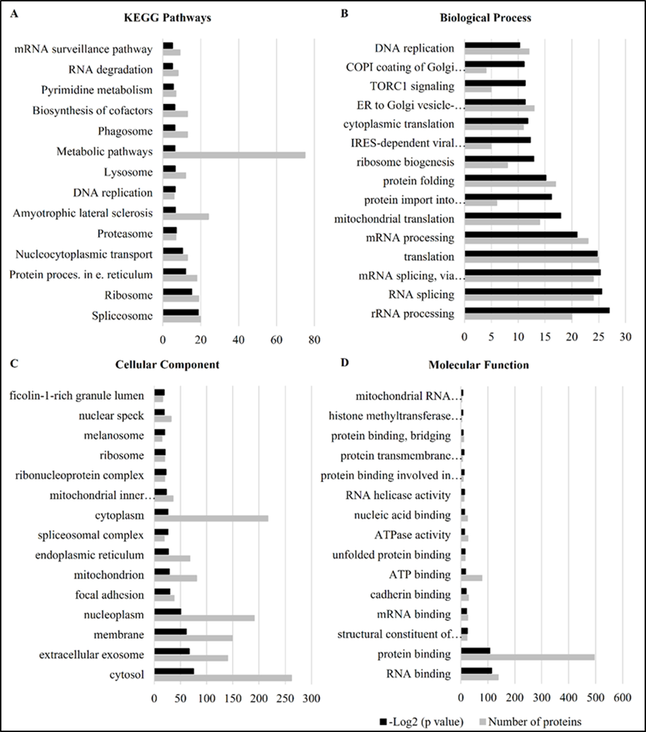
The proteome from ZIKV infected Vero cells showed a total of 2,718 identified proteins being 609 considered differentially expressed (p<0.05). Comparing those differentially expressed proteins with the control mock-infected cells, 350 proteins were found to be up regulated and 259 were down-regulated. In total, 146 pathways were found with at least one differentially expressed protein which was identified in the ZIKV infection condition. The General Metabolic pathway was the most affected one in number of proteins according to the results (Fig. 1a). In the biological process, the highest percentage of assigned protein was found in translation, followed by mRNA splicing, via spliceosome and RNA splicing (Fig. 1b). At the cellular component level, the largest number of proteins was associated with cytosol, cytoplasm and nucleoplasm (Fig. 1c). The largest number of proteins of the statistically significant total number was visualized in molecular functions (Fig. 1d), being 247 of the up regulated proteins belonging to the protein binding functional group.
Functional characterization of differential proteome of Vero cells infected with ZIKV. A. KEGG pathways, in the number of proteins. GO assignment of proteins in B. Biological Process, C. Cellular Component and D. Molecular Function. On bars in red color are located the number of proteins. The bars in black color represent the –log2 p-value. (*regulation of mRNA stability; *ER to Golgi vesicle-mediated transport; *COPI coating of Golgi vesicle; *IRES-dependent viral translational initiation; *protein import into mitochondrial outer membrane; *mRNA splicing, via spliceosome; *mitochondrial inner membrane;)
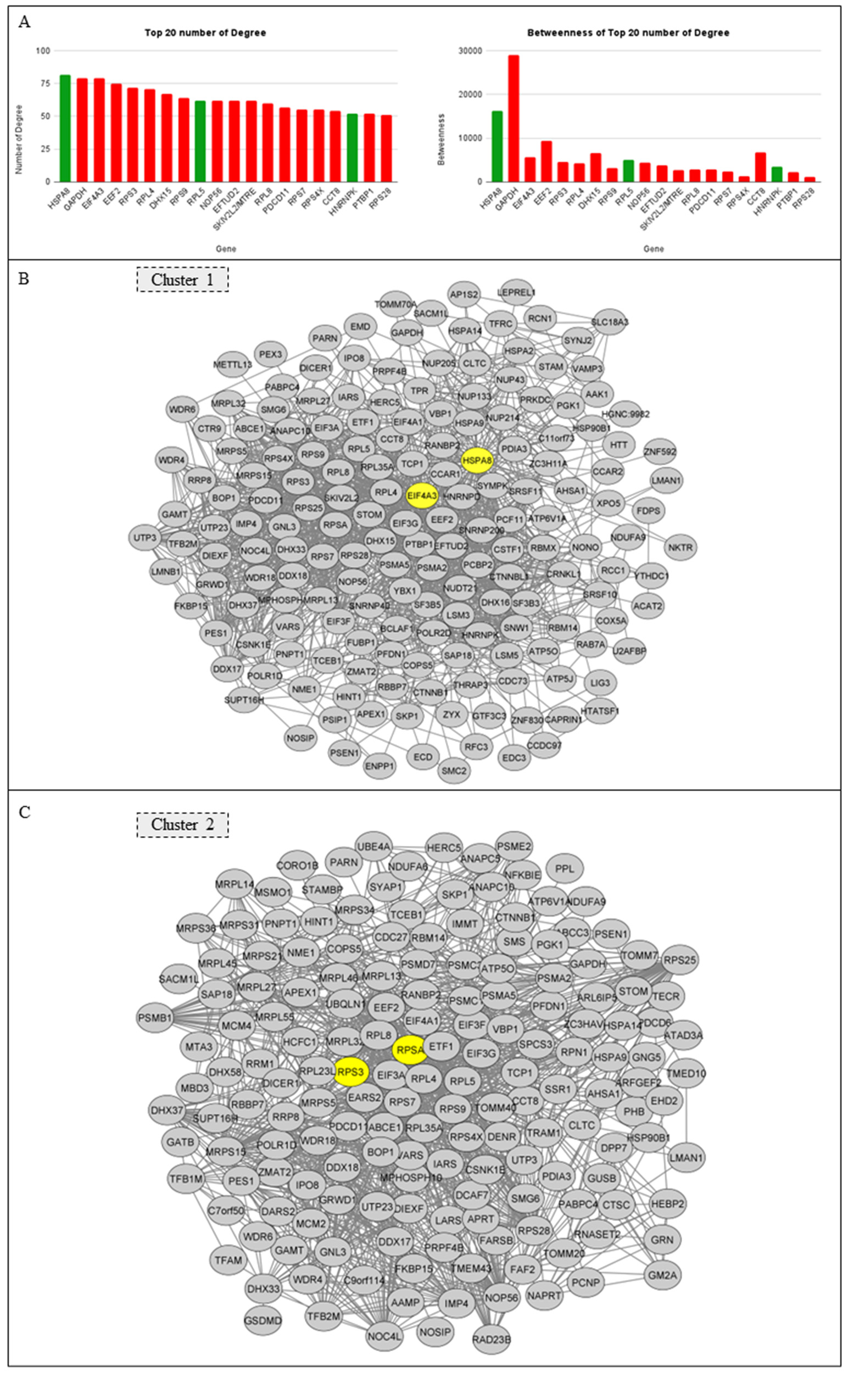
Heat shock cognate 71 kDa protein (HSPA8/Hsc70 – member of heat shock protein 70 family (Hsp70)), with 82 interactions, present in cluster 1 (Table SIII), is the protein with the highest number of interactions in cluster 1 (Fig. 2a), this chaperone exerts quality control and orientates proteins to distinct biogenesis routes and facilitates protein folding, among many other functions (Shan 2023). Eukaryotic initiation factor 4A-III (EIF4A3) is the protein with the second highest number of interactions in the cluster 1, with 79 degrees (Fig. 2a). Still in cluster 1, the Pre-mRNA-splicing factor ATP-dependent RNA helicase DHX15 (DHX15), 116 kDa U5 small nuclear ribonucleoprotein component (EFTUD2), Exosome RNA helicase MTR4 (MTREX), Heterogeneous nuclear ribonucleoprotein K (HNRNPK) and Polypyrimidine tract-binding protein 1 (PTBP1) also present a considerable number of degrees. It is important to note that these proteins play a central hub in the cell and are able to interact with a major number of molecules.
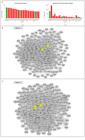
Degrees and betweenness selected from the interactome with the differentially expressed proteins and their respective clusters. A. Graph presenting the Top 20 number of Degree and Betweenness of differentially expressed proteins (according to MCODE Cytoscape program). B. Protein-protein interaction networks of Cluster 1 with the central nodes highlighted in yellow. C. Protein-protein interaction networks of Cluster 2 with the central nodes highlighted in yellow.
In the analysis of the protein-protein interaction networks (p <0.05) highlighted in yellow are the main proteins of cluster 1 and 2 (the most significant clusters in number of protein) (Fig. 2b and 2c). An expressive number of proteins were found in cluster 2 (Fig. 2c), presenting as central members Elongation factor 2 (eEF2), with 75 degrees and 40S ribosomal protein S7 (RPS7), T-complex protein 1 subunit theta (CCT8) and 40S ribosomal protein S28 (RPS28). Glyceraldehyde-3-phosphate dehydrogenase (GAPDH), 40S ribosomal protein S3 (RPS3), 60S ribosomal protein L4 (RPL4), 40S ribosomal protein S9 (RPS9), 60S ribosomal protein L5 (RPL5) were the most significant proteins considering degree and betweenness (Table SIII).
Another approach to verify the protein-protein interactions was considering the analysis of MalaCards and Disgenet. Those two tools were used to check the diseases or conditions associated with the proteins identified in our dataset (in the top 20 major number of degrees). Our focus was the neurological and mental disorders, neurodegenerative and cardiocirculatory conditions (Tables SI and SII). When analyzing the disease markers of top 20 major numbers of degrees it was possible to observe that most of the altered proteins in question were associated with diseases of the nervous system, cardiovascular system, congenital malformations, hematopoietic diseases, among others (Table SIV).
The KEGG pathway was verified one by one of the top 20 number of degrees and the largest number of proteins within this parameter are involved in ribosome pathways. It’s possible to observe the most relevant proteins considering the interaction and the pathways involved in Table SV. The HSPA8 and GAPDH were the proteins involved in the majority of pathways, 12 and 9 respectively. In addition, it is important to highlight the relevance of the protein GAPDH in metabolic routes such as carbon metabolism, amino acid biosynthesis and glycolysis/gluconeogenesis and HSPA8 in pathways such as the spliceosome.
Another important result to be mentioned is the most affected pathways within the proteomic alteration evidenced. The most affected metabolic pathways (66 proteins) considering the number of proteins were carbohydrate metabolism and lipid metabolism. It is also important to mention the alteration in the spliceosome (21 proteins) and ribosome (19 proteins) pathways (Tables SVI and SVII).
DISCUSSION
Proteomics is a powerful approach for prognosis, early disease diagnosis and monitoring disease development (Aslam et al. 2017). Through proteomics, it is possible to analyze the cellular response to infections and its molecular outcomes, as well as predict the possible consequences resulting from these molecular alterations. Regarding ZIKV infection, several proteins have been described implicated in infection under different experimental conditions (Rosa et al. 2020). Most studies apply, as expected, nervous systems-related host models due to clinical neurological consequences of ZIKV infection. Although neural-related host models choice is obvious, our hypothesis is that ZIKV infection dysregulates the expression of genes associated with central nervous system functions, regardless of the cell type. Applying Vero cells and differential proteomics we could identify a new set of proteins linked to molecular alterations related to clinical impairments resulting from ZIKV.
Molecular alterations in infected cell by ZIKV
Considering proteomic analysis through observation of protein-protein- interaction (PPI) networks in our ZIKV infection proteome, the main cluster is centralized in the protein Eukaryotic initiation factor 4A-III, a RNA helicase ATP-dependent, found up-regulated in ZIKV infected cells. This protein acts as nucleotide binding protein, RNA helicase, RNA stem loop, selenocysteine insertion sequence binding and translation regulator (Andreou & Klostermeier 2013). This protein plays a role in cellular response to brain-derived neurotrophic factor stimulus, embryonic cranial skeleton morphogenesis, gene expression (transcription and translation processes), regulation of translation at postsynapse and modulating synaptic transmission. RNA helicases play essential regulatory roles in cells and participate in all aspects of RNA metabolism (Jankowsky 2011).
In our analysis, a significant change in EIF4A3 was observed, as well as a change in RNA-binding protein 8A, although not statistically significant. EIF4A3 is important to virus replication, helping the export of amended host mRNAs from the nucleus and its depletion results in decrease of virus replication (Ziehr et al. 2016). In our findings this protein was increased suggesting a propitious condition for viral replication and establishment of infection. Such molecular alterations may indicate possible impacts on the developmental processes of EJC and consequent impairment in the embryonic development of the central nervous system, beyond potential alterations in neural plasticity in adults. Thus, ZIKV infection in Vero cells alters the expression of specific proteins molecularly related with phenotypes in ZIKV infection cases in humans and in experimental infection of animal models (Janssens et al. 2018). The core proteins of EJC are involved in the central nervous system development, especially in embryonic neurogenesis. The alteration of these components also affects the adult brain and is linked to conditions such as abnormal human behaviors, especially intellectual disability and synaptic plasticity (Bartkowska et al. 2018).
RNA helicases, as EIF4A3, in ZIKV infection it is important understanding the crucial role that these enzymes promote in cell metabolism, more specifically in neural plasticity, given that EIF4A3 is part of the minimally stable core of the EJC along with protein MAGOH (Barker-Haliski et al. 2012). Hsp70 acts in viral infection as a crucial protein for ZIKV life cycle and the decrease of this protein is related with lower viral production (Khachatoorian et al. 2018). The HSPA9, another heat shock protein, is involved in energy metabolism within mitochondria, protein polyubiquitination, associated with the complex protein degradation system and consequent cell detoxification and many others involvement in countless biological processes (Ebrahimi et al. 2021).
The Heat shock cognate 71 kDa protein (HSPA8/Hsc70/Hsp70) found down-regulated in the proteome was the second protein in number of interactions in the main protein cluster. This protein is a molecular chaperone expressed and involved in numerous cellular processes, for instance stress-response, folding and transportation of newly synthesized polypeptides, activation of proteolysis of misfolded proteins and formation and dissociation of protein complexes. The Hsp70 acts in the nervous system and is potentially associated with the pathogenesis of multiple system atrophy, according to immunohistochemical results with human autopsied brains (Kawamoto et al. 2007). HSPA8/Hsp70 was also previously associated with TAR DNA-binding protein 43 (TDP-43), the major pathological protein in sporadic amyotrophic lateral sclerosis (ALS) (Mackenzie & Rademakers 2008). Coyne et al. (2014) showed that an overexpression of TDP-43 causes a depletion in HSPA8/Hsp70 synaptic levels and this fact leads to abnormal interaction between the TDP-43 and the chaperone, leading ultimately to defects in endocytosis and conducting to synaptic defects present in ALS. Loeffler et al. (2016) associated decreased levels of Hsp70 with increased oxidative stress age-related in cerebrospinal fluid (CSF), which may be associated with Parkinson’s Disease (PD). All the conditions mentioned above have shown a reduction in HSPA8 expression, consistent with the findings of our study. Furthermore, molecular markers of ALS and PD were found differentially expressed in a previously reported ZIKV infection proteome, using the same Brazilian clinical strain of ZIKV used for this article, of hMSC (Beys-da-Silva et al. 2019).
Another protein previously linked with brain diseases found differentially expressed in our proteome and central in resulting interactome was the Stress-70 protein (HSPA9), a chaperone which plays an important role in mitochondrial iron-sulfur cluster (ISC) biogenesis, interacts with and stabilizes ISC cluster assembly proteins corresponding to the genes FXN, NFU1, NFS1 and ISCU (Shan & Cortopassi 2016). The HSPA9 was already implicated in neurodegenerative diseases, such as Alzheimer’s disease (AD) and PD, mainly because it is involved in energy metabolism within mitochondria, being certain dysfunctions in this organelle associated with such diseases (Müller et al. 2010). The 26S proteasome regulatory subunit 4 (PSMC1) protein, down-regulated in ZIKV-infected Vero cells, is a component of the 26S proteasome, a multiprotein complex involved in the ATP-dependent degradation of ubiquitinated proteins. Among several roles in biological processes, this protein is related to negative regulation of neuron death, protein polyubiquitination and many others. Ebrahimi-Fakhari et al. (2012) reported the association of the concept of dysfunctional degradation in PD, which is supported by previous studies in knockout models, as they show deep neurodegeneration and protein inclusion bodies in mice that miss essential components of the autophagylysosomal pathway (ALP) or the ubiquitin-proteasome system (UPS/PSMC1) (Ebrahimi et al. 2021). However, although this gene is associated with the complex protein degradation system and consequent cell detoxification, they were unable to establish relationships between UPS alterations and the risk of developing PD. Even so, the possibility of association between variations in the corresponding gene and other dysfunctions that cause PD phenotypes is not ruled out (Gómez-Garre et al. 2012).
Another member of the chaperone family is HSPA2, which is involved in binding processes and antigen presentation to the immune system (Rowland et al. 2018). This chaperone is also implicated in myelination function and fast cognitive decline. Panitch et al. (2021) found that HSPA2 levels were linked to tau protein levels and consequent deposition of amyloid plaques in brain regions. Along with the previously reported chaperones and the conditions associated with them, another molecule that needs to be mentioned is HSPA9, whose overexpression is associated with decreased apoptosis in some cell models (Peng et al. 2013). This protein, as well as those mentioned above, has been reported to influence the process of accumulation of defective proteins (protein aggregates) involved in neurodegenerative diseases. The location of HSPA9 inside the mitochondria is a predictor of implication in conditions of neurological damage. In this regard, increased reactive oxygen species and mitochondrial dysfunction in axons affect neuronal functioning (Flachbartová & Kovacech 2013). The increase of HSPA9 may predict the possible damage to come in this case. Finally, the heat shock protein HSPA14 is still poorly understood, but as a member of the chaperone family it will also have implications. As it is a mammalian ribosome-associated complex with MPP11, it acts as a Th1 adjuvant through activation of dendritic cells, and a deregulation in this protein will cause damage to the host cell (Wu et al. 2011).
Ribosomal Protein SA (RPSA), a component of the 40S ribosomal subunit (Villas-Boas et al. 2016), is implicated in cell adhesion, viral transcription, translation, among others roles (Zidane et al. 2012) another significant protein in our results. Other relevant protein in Cluster 2 was 40S ribosomal protein S3 (RPS3), a component of the 40S small ribosomal subunit also implicated in its maturation. This protein has been implicated in several molecular processes including DNA repair, apoptotic process, positive regulation of NIK/NF-kappaB signaling, response to oxidative stress, viral transcription and many others (Poncová et al. 2019).
RPSA is selectively expressed in the hippocampus (Smagin et al. 2016) and was found up-regulated in the infected cells. Together with the specific roles that have been previously associated with this protein are cell surface receptors for laminin, which is important for the differentiation, the migration and the adhesion of the cell, along with synapse stabilization (Omar et al. 2017). It is also important to note that this protein interacts with ZNF804A, a known schizophrenia risk gene (Zhang et al. 2011). Zhou et al. (2018) found that RPSA overexpression stimulated neuronal migration and ameliorated the migration defect caused by ZFP804A (homolog of ZNF804A) knockdown in the cortical plate, also suggest that both RPSA silencing and overexpression decreased translational efficiency. However, they also proposed that 30S ribosomal protein S1, acting as a direct interacting protein of ZNF804A, can rescue ZFP804A-mediated migration defect, but cannot repair the neuronal progenitor cells proliferation.
Previously, the overexpression of RPS3 protein, found up-regulated in Vero cells infected with ZIKV, was associated inducing neuronal apoptosis by cooperating with Transcription factor E2F1 and causing up-regulation of proapoptotic BH3- only proteins, Bim
and death protein 5/harakiri (Dp5/Hrk) (Lee et al. 2010). Ahn et al. (2013) previously postulated, the overexpression of RPS3 could induce apoptosis through the activation of caspases 8 and 3 and DNA fragmentation. It suggests that cell death would be caused by a high affinity of RPS3 to damaged DNA sites such as 7,8-dihydro8-oxoguanine and may strongly bind to the damaged DNA site and interfere with DNA repair, which is done by other repair enzymes, subsequently leading to cell death (Ahn et al. 2013).
Furthermore, there is a set of differentially expressed proteins, that have been correlated with neurological conditions, such as ALS, schizophrenia, attention deficit hyperactivity disorder (ADHD), depression, PD, and autism spectrum disorder (ASD), among others, in studies using in vitro and in vivo models as presented in the Additional File (Aishworiya et al. 2023, Bao et al. 2024, Park et al. 2021, Shen et al. 2023, Zhuang et al. 2023). Regarding the effects caused on the neurological system by viral infections, among those proteins one was already implicated in an in vitro Zika infection model. Other 10 proteins were reported implicated in SARS-CoV-2 infection, that must be considered since the long Covid phenotype is linked to cognitive impairment, fatigue and mood disturbances, along with other neurological issues (Rudroff 2024). Concerning PTBP1 protein, it was already reported to be implicated in local translation in axons, nociceptor neuron regeneration and both thermal and mechanical sensation in an in vivo model (Alber et al. 2023). Also, this protein was previously detected up-regulated in Zika infection of primary mouse astrocytes as we found in our results (Shereen et al. 2021). All those proteins previously described in viral infections (Table I) might be considered as an indirect validation of our results and the specific impact that Zika may have on dysregulation of specific neural genes independently of the infection model, since we are using a non-neural cell as host.
Set of differentially expressed proteins previously identified in Zika and/or SARS-CoV-2 infection and correlated with neurological conditions.
Most impacted pathways
The metabolic pathways were the most affected in the number of proteins. Within the metabolic pathways, carbohydrate and lipid metabolism were the most impacted. Here it is important to cite the involvement of GAPDH in pathways such as carbon metabolism, biosynthesis of amino acids and glycolysis/gluconeogenesis. When the host is exposed to pathogenic microorganisms, such as viruses, the first metabolic response of cells is to produce energy to supply the environment and then be able to synthesize molecules that will improve the combat of an infection (Ebrahimi et al. 2021). The GAPDH (glycolytic enzyme) plays a crucial role in the energetic routes and their excess has been associated in the exacerbated response such as in SARS-CoV-2 infection (Ebrahimi et al. 2021), in which the exacerbated production of GAPDH will cause a greater synthesis of oxidized nicotinamide adenine dinucleotide (NAD+), with the induction of oxidative phosphorylation. Other enzymes involved in glycolysis were found in our results, such as Phosphoglycerate kinase 1 (PGK1) and ADP-dependent glucokinase (ADPGK). The importance of these related proteins in metabolic pathways, particularly GAPDH, is both the induction of an increase in energy demand and the possible regulation of Warburg effect (Rosa et al. 2019), in which cells use the anaerobic pathway even with the availability of oxygen. producing lactic acid. Considering that GAPDH, PGK1 and ADPGK were up-regulated in our results, it is inferred that the cells may be in a glycolytic and, possibly, anaerobic state (Liberti et al. 2020). Moreover, these data suggest a decrease in oxidative phosphorylation, as well as the induction of defense cell activation, like T cells, promoting the generation of reactive species (Kamiński et al. 2012, Zhang et al. 2020).
In addition to its importance in metabolic pathways, GAPDH has also been linked to increased susceptibility to infection by viruses such as dengue and hepatitis C (from the same family as ZIKV) (Raj et al. 2017). Raj et al. (2017) found that GAPDH silencing induces a decrease in viral infectivity in Huh-7 cells (Human hepatoma cells). Considering that in our results GAPDH is up-regulated, cells may be more susceptible to infection by ZIKV. Gale e al. (2019) also evidenced the importance of GAPDH in viruses, noting that the inhibition of this enzyme in cells infected with ZIKV can lead to reduced plaque formation. However, more studies are required to establish the crucial role of this protein in the infection of ZIKV and other flaviviruses. As well as in other viruses such as in Sars-CoV-2 infection, that may lead to impact coincident routes.
The second most affected pathway in the number of proteins was Spliceosome. HSPA8, DHX15 and EIF4A3 are relevant in this pathway and appear to play a key role in the response to ZIKV infection and its consequences. As HSPA8 is a component of the PRP19-CDC5L complex that forms parts of the spliceosome, its alteration ends up involving this pathway. Sirtori et al. (2020) associated that the reduction of this protein leads to the accumulation of alpha synuclein and other toxic substances to neurons, which can lead to neurodegenerative diseases, such as PD and AD. A key protein in the spliceosome pathway is DHX15, acting on the release of introns and disassembly of the spliceosome, which may be important in the development of tissues such as brain and ocular, in the model used by McElderry et al. (2019). The up-regulation of DHX15 may be also related to an incidence of greater inflammatory response in these tissues and, consequently, a likely increase in the chance of alterations occurring in the developmental phase.
Within the spliceosome pathway, it is worth to notice the involvement of chaperones HSPA8, HSPA9, HSPA2 (Heat Shock Protein Family A (Hsp70) Member 2) and HSPA14 (Heat Shock Protein Family A (Hsp70) Member 14), which were differentially expressed in the proteome. To enter the host cell, the virus makes use of chaperones in its favor for biosynthesis, endocytosis and assembly processes, indirectly facilitating viral replication (Pujhari et al. 2019). HSPA8 is one of the key proteins in the infection of Vero cells by ZIKV, as can be seen in the results of our work, as well as to enter in Huh7.5 and HEK cells (Pujhari et al. 2019). Therefore, it is essential to understand its role for both infectivity and molecular outcomes that ultimately lead to physiological changes and to pathogenesis.
The third most affected pathway in the number of proteins was the ribosomal pathway. Proteins related to this pathway are extremely important, considering that when listing the top 20 in number of degrees and betweenness, 8 of the proteins are involved in it. Of these 8, 7 are up-regulated. Therefore, it is important to mention that when ribosomal stress occurs, the nucleolus ruptures and leads to apoptosis in neural progenitor cells. These facts will lead to changes and damage in the nervous system, which may ultimately be associated with pathologies such as microcephaly, autism, and intellectual disorders (Slomnicki et al. 2017).
CONCLUSIONS
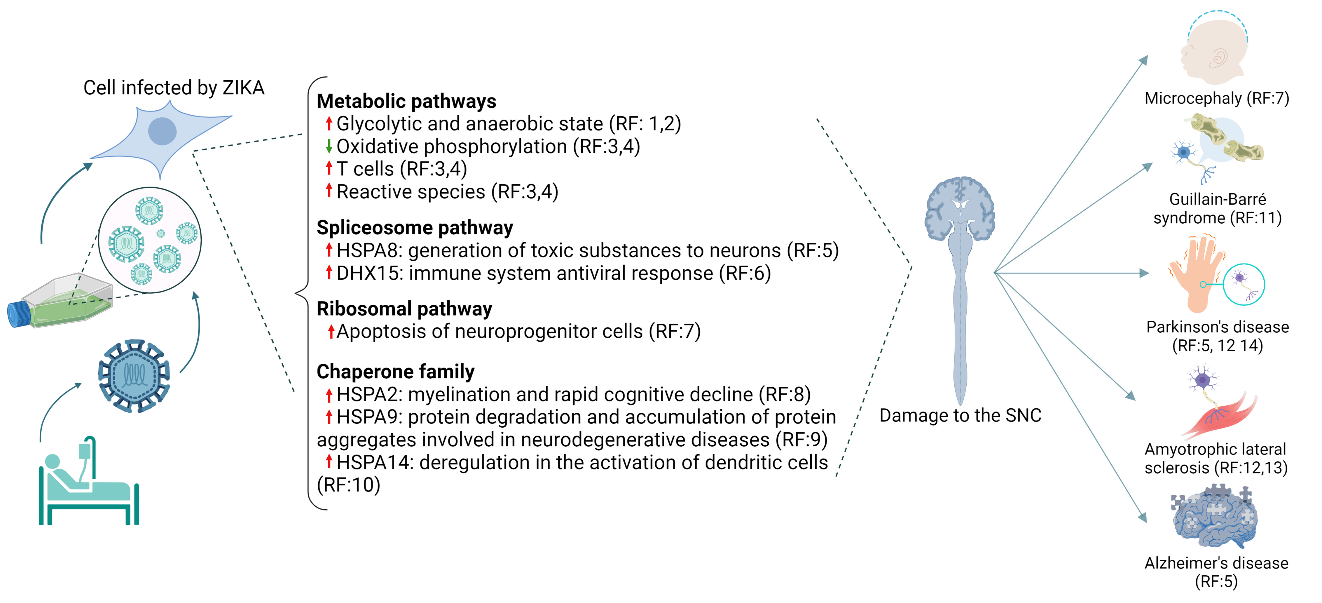
ZIKV is capable of infecting Vero cells, a non-neural infection model, causing significant alterations in the expression of proteins that are previously implicated in neuro-alterations. Among those molecular changes, pathways such as the metabolic and spliceosome can cause damage to the neurological system. In our data, the disturbance promoted by ZIKV infection in vitro, showed proteins differentially expressed that have already been reported in previous works with human cells being molecularly linked to disorders beyond microcephaly and Guillain-Barré Syndrome, clinical phenotypes of Zika disease, such as AD, PD, ALS, and others (Fig. 3). The disturbance of these proteins even in a non-neural cell model can be related to the intense inflammatory process and damage caused by viral infection. Some of these proteins could be decisive both in this antiviral response, and in the propensity to these conditions visualized (Wang et al. 2020). As central nodes we detected HSPA8 being a key protein having involvement in the stress response and DHX15 being highlighted in the antiviral response of the innate immune system.
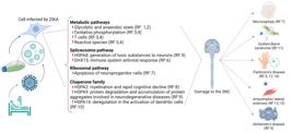
Infection by the Zika virus (ZIKV) triggers differential protein expression and metabolic pathways associated with central nervous system (CNS) disorders in a non-neural cellular model. References: 1- Rosa et al. 2019, 2- Liberti et al. 2020, 3- Kamiński et al. 2012, 4- Zhang et al. 2020, 5- Sirtori et al. 2020, 6- Wang et al. 2020, 7- Slomnicki et al. 2017, 8- Panitch et al. 2021, 9- Peng et al. 2013, 10- Wu et al. 2011, 11- Hendrixson & Newland 2018, 12- Mackenzie & Rademakers 2008, 13- Beys-da-Silva et al. 2019, 14- Gómez-Garre et al. 2012.
In addition, our results identified a potential influence of chaperones towards changes resulting from the virus infection, which needs to be further investigated, given that the possible association with neuroinflammatory state may result in diseases like AD, PD, ALS and other neurodegenerative pathologies long-term. Moreover, although using a non-neural model cell such as Vero, ZIKV still can impact the expression of proteins previously described in neurological diseases that are still not found as a ZIKV clinical phenotype. The results presented here highlight a set of dysregulated proteins and genes that require further validation, including infection studies using other non-neural cell models and in vivo models targeting tissues and organs beyond the neurological system. Despite this limitation, our findings provide a valuable foundation for guiding future studies on the long-term consequences of Zika outbreaks, both in clinical surveys and in experimental models focused on neurological outcomes.
SUPPLEMENTARY MATERIAL
Acknowledgements
This work was supported by the Ministry of Health and the Brazilian funding agencies, Coordenação de Aperfeiçoamento Pessoal de Nível Superior (CAPES), Conselho Nacional de Desenvolvimento Científico e Tecnológico (CNPq), Edital MCTIC/FNDCT-CNPq/MEC-CAPES/MS-Decit/No 14/2016, project 440763/2016-9, CNPq project 406181/2021-7 and CNPq 310061/2021-0 to W.O.B.S. Conflict of interests: The authors declare that they have no conflict of interest.
References
- AHN EH ET AL. 2013. PEP-1-ribosomal protein S3 protects dopaminergic neurons in an MPTP-induced Parkinson’s disease mouse model. Free Radic Biol Med 55: 36-45.
- AISHWORIYA R ET AL. 2023. Adaptive, behavioral, and cognitive outcomes in individuals with fragile X syndrome with varying autism severity. Int J Dev Neurosci 83: 715-727.
- ALBER S ET AL. 2023 PTBP1 regulates injury responses and sensory pathways in adult peripheral neurons. Sci Adv 9: eadi0286.
- ANDREOU AZ & KLOSTERMEIER D. 2013. The DEAD-box helicase eIF4A: paradigm or the odd one out? RNA Biol 10: 19-32.
- ASLAM B, BASIT M, NISAR MA, KHURSHID M & RASOOL MH. 2017. Proteomics: Technologies and Their Applications. J Chromatogr Sci 55: 182-196.
- BAO H, ZHANG Q, LI Y & NIE C. 2024. CircDLGAP4 overexpression ameliorates neuronal injury in Parkinson’s disease by binding to EIF4A3 and increasing HMGA2 expression. J Biochem Mol Toxicol 38: e23530.
- BARKER-HALISKI ML, PASTUZYN ED & KEEFE KA. 2012. Expression of the core exon junction complex factor eukaryotic initiation factor 4A3 is increased during spatial exploration and striatally-mediated learning. Neuroscience 226: 51-61.
- BARTKOWSKA K, TEPPER B, TURLEJSKI K & DJAVADIAN RL. 2018. Roles of the exon junction complex components in the central nervous system: a mini review. Rev Neurosci 29: 817-824.
- BEYS-DA-SILVA WO ET AL. 2019. Zika Virus Infection of Human Mesenchymal Stem Cells Promotes Differential Expression of Proteins Linked to Several Neurological Diseases. Mol Neurobiol 56: 4708-4717.
- BI Y, LI T, ZHANG S, YANG Y & DONG M. 2024. Bioinformatics-based analysis of the dialog between COVID-19 and RSA. Heliyon 10: e30371.
- CAMPBELL VL ET AL. 2020. Proteome-Wide Zika Virus CD4 T Cell Epitope and HLA Restriction Determination. Immunohorizons 4: 444-453.
- CHEN B, ZHANG D, WANG X, MA W, DENG S, ZHANG P, ZHU H, XU N & LIANG S. 2017. Proteomics progresses in microbial physiology and clinical antimicrobial therapy. Eur J Clin Microbiol Infect Dis 36: 403-413.
- COYNE AN, SIDDEGOWDA BB, ESTES PS, JOHANNESMEYER J, KOVALIK T, DANIEL SG, PEARSON A, BOWSER R & ZARNESCU DC. 2014. Futsch/MAP1B mRNA is a translational target of TDP-43 and is neuroprotective in a Drosophila model of amyotrophic lateral sclerosis. J Neurosci 34: 15962-15974.
- EBRAHIMI KH, GILBERT-JARAMILLO J, JAMES WS & MCCULLAGH JSO. 2021. Interferon-stimulated gene products as regulators of central carbon metabolism. FEBS J 288: 3715-3726.
- EBRAHIMI-FAKHARI D, MCLEAN PJ & UNNI VK. 2012. Alpha-synuclein’s degradation in vivo: opening a new (cranial) window on the roles of degradation pathways in Parkinson disease. Autophagy 8: 281-283.
- FLACHBARTOVÁ Z & KOVACECH B. 2013 Mortalin - a multipotent chaperone regulating cellular processes ranging from viral infection to neurodegeneration. Acta Virol 57: 3-15.
- GALE TV, HORTON TM, HOFFMANN AR, BRANCO LM & GARRY RF. 2019. Host Proteins Identified in Extracellular Viral Particles as Targets for Broad-Spectrum Antiviral Inhibitors. J Proteome Res 18: 7-17.
- GAUDET P, LIVSTONE MS, LEWIS SE & THOMAS PD. 2011. Phylogenetic-based propagation of functional annotations within the Gene Ontology consortium. Brief Bioinform 12: 449-462.
- GHARBARAN R & SOMENARAIN L. 2017. Insights into the molecular roles of Zika virus in human reproductive complications and congenital neuropathologies. Pathology 49: 707-714.
- GLOVER KKM, GAO A, ZAHEDI-AMIRI A & COOMBS KM. 2019. Vero Cell Proteomic Changes Induced by Zika Virus Infection. Proteomics 19: e1800309.
- GÓMEZ-GARRE P, JESÚS S, CARRILLO F, CÁCERES-REDONDO MT, BERNAL-BERNAL I, CARBALLO M, GAO L & MIR P. 2012. PSMC1 Gene in Parkinson’s Disease. Eur Neurol 68: 193-198.
- GÖTZ S, GARCÍA-GÓMEZ JM, TEROL J, WILLIAMS TD, NAGARAJ SH, NUEDA MJ, ROBLES M, TALÓN M, DOPAZO J & CONESA A. 2008. High-throughput functional annotation and data mining with the Blast2GO suite. Nucleic Acids Res 36: 3420-3435.
- HENDRIXSON DT & NEWLAND JG. 2018. Zika Virus Infection in Children. Infect Dis Clin North Am 32: 215-224.
- HOPE JL ET AL. 2023. PSGL-1 attenuates early TCR signaling to suppress CD8+ T cell progenitor differentiation and elicit terminal CD8+ T cell exhaustion. Cell Rep 42: 112436.
- JANKOWSKY E. 2011. RNA helicases at work: binding and rearranging. Trends Biochem Sci 36: 19-29.
-
JANSSENS S, SCHOTSAERT M, KARNIK R, BALASUBRAMANIAM V, DEJOSEZ M, MEISSNER A, GARCÍA-SASTRE A & ZWAKA TP. 2018. Zika Virus Alters DNA Methylation of Neural Genes in an Organoid Model of the Developing Human Brain. mSystems. https://doi.org/10.1128/mSystems.00219-17
» https://doi.org/10.1128/mSystems.00219-17 - JIANG X, LV J, ZHOU R & XIANG X. 2021. The effect of cofilin 1 expression and phosphorylation dynamics on cell fate determination and neuron maturation in neural stem cells. Neurosci Lett 764: 136292.
- KAMIŃSKI MM, SAUER SW, KAMIŃSKI M, OPP S, RUPPER T, GRIGARAVIČIUS P, GRUDNIK P, GRÖNE HJ, KRAMMER PH & GÜLOW K. 2012. T cell activation is driven by an ADP-dependent glucokinase linking enhanced glycolysis with mitochondrial reactive oxygen species generation. Cell Rep 2: 1300-1315.
- KAWAMOTO HK, HELLER JB, HELLER MM, URREGO A, GABBAY JS, WASSON KL & BRADLEY JP. 2007. Craniofrontonasal dysplasia: a surgical treatment algorithm. Plast Reconstr Surg 120: 1943-1956.
- KHACHATOORIAN R, COHN W, BUZZANCO A, RIAHI R, ARUMUGASWAMI V, DASGUPTA A, WHITELEGGE JP & FRENCH SW. 2018. HSP70 Copurifies with Zika Virus Particles. Virology 522: 228-233.
- KHAN WH, AHMAD R, ALAM R, KHAN N, RATHER IA, WANI MY, SINGH RKB & AHMAD A. 2024. Role of ribosomal pathways and comorbidity in COVID-19: Insight from SARS-CoV-2 proteins and host proteins interaction network analysis. Heliyon 10: e29967.
-
KIM A, LEE DY & SUNG JJ. 2024. Cdk5 inhibition in the SOD1G93A transgenic mouse model of amyotrophic lateral sclerosis suppresses neurodegeneration and extends survival. J Neurochem: https://doi.org/10.1111/jnc.16160
» https://doi.org/10.1111/jnc.16160 - KIM JE, HONG YH, KIM JY, JEON GS, JUNG JH, YOON BN, SON SY, LEE KW, KIM JI & SUNG JJ. 2017. Altered nucleocytoplasmic proteome and transcriptome distributions in an in vitro model of amyotrophic lateral sclerosis. PLoS One 12: e0176462.
- KIM SY, LEE MY, CHO KC, CHOI YS, SUNG KW, KWON OJ, KIM HS, KIM IK & JEONG SW. 2003. Alterations in mRNA Expression of Ribosomal Protein S9 in Hydrogen Peroxide Treated Neurotumor Cells and in Rat Hippocampus After Transient Ischemia. Neurochem Res 28(6): 925-931.
- KRÄMER A, GREEN J, POLLARD J JR & TUGENDREICH S. 2014. Causal analysis approaches in Ingenuity Pathway Analysis. Bioinformatics 30: 523-530.
- LAM T ET AL. 2023. Repeat expansions in NOP56 are a cause of spinocerebellar ataxia Type 36 in the British population. Brain Commun 5: fcad244.
- LEE SB, KWON IS, PARK J, LEE KH, AHN Y, LEE C, KIM J, CHOI SY, CHO SW & AHN JY. 2010. Ribosomal protein S3, a new substrate of Akt, serves as a signal mediator between neuronal apoptosis and DNA repair. J Biol Chem 285: 29457-29468.
- LIBERTI MV, ALLEN AE, RAMESH V, DAI Z, SINGLETON KR, GUO Z, LIU JO, WOOD KC & LOCASALE JW. 2020. Evolved resistance to partial GAPDH inhibition results in loss of the Warburg effect and in a different state of glycolysis. J Biol Chem 295: 111-124.
- LOEFFLER DA, KLAVER AC, COFFEY MP, AASLY JO & LEWITT PA. 2016. Age Related Decrease in Heat Shock 70-kDa Protein 8 in Cerebrospinal Fluid Is Associated with Increased Oxidative Stress. Front Aging Neurosci 8: 178.
- MACKENZIE IRA & RADEMAKERS R. 2008. The role of transactive response DNA binding protein-43 in amyotrophic lateral sclerosis and frontotemporal dementia. Curr Opin Neurol 21: 693-700.
- MCELDERRY J ET AL. 2019. Splicing factor DHX15 affects tp53 and mdm2 expression via alternate splicing and promoter usage. Hum Mol Genet 28: 4173-4185.
- MÜLLER WE, ECKERT A, KURZ C, ECKERT GP & LEUNER K. 2010. Mitochondrial dysfunction: common final pathway in brain aging and Alzheimer’s disease--therapeutic aspects. Mol Neurobiol 41: 159-171.
- OMAR MH, KERRISK CAMPBELL M, XIAO X, ZHONG Q, BRUNKEN WJ, MINER JH, GREER CA & KOLESKE AJ. 2017. CNS Neurons Deposit Laminin α5 to Stabilize Synapses. Cell Rep 21: 1281-1292.
- PAN Y ET AL. 2023. Gelation of cytoplasmic expanded CAG RNA repeats suppresses global protein synthesis. Nat Chem Biol 19: 1372-1383.
- PANITCH R ET AL. 2021. Integrative brain transcriptome analysis links complement component 4 and HSPA2 to the APOE ε2 protective effect in Alzheimer disease. Mol Psychiatry 26: 6054-6064.
- PARK CY, ZHOU J, WONG AK, CHEN KM, THEESFELD CL, DARNELL RB & TROYANSKAYA OG. 2021. Genome-wide landscape of RNA-binding protein target site dysregulation reveals a major impact on psychiatric disorder risk. Nat Genet 53: 166-173.
- PENG C, YANG P, CUI Y, HE M, LIANG L & DI Y. 2013. HSPA9 overexpression inhibits apoptin-induced apoptosis in the HepG2 cell line. Oncol Rep 29: 2431-2437.
- PIÑERO J, QUERALT-ROSINACH N, BRAVO À, DEU-PONS J, BAUER-MEHREN A, BARON M, SANZ F & FURLONG LI. 2015. DisGeNET: a discovery platform for the dynamical exploration of human diseases and their genes. Database 2015: bav028.
- PONCOVÁ K, WAGNER S, JANSEN ME, BEZNOSKOVÁ P, GUNIŠOVÁ S, HERMANNOVÁ A, ZEMAN J, DONG J & VALÁŠEK LS. 2019. uS3/Rps3 controls fidelity of translation termination and programmed stop codon readthrough in co-operation with eIF3. Nucleic Acids Res 47: 11326-11343.
- PUJHARI S, BRUSTOLIN M, MACIAS VM, NISSLY RH, NOMURA M, KUCHIPUDI SV & RASGON JL. 2019. Heat shock protein 70 (Hsp70) mediates Zika virus entry, replication, and egress from host cells. Emerg Microbes Infect 8: 8-16.
- QUINCOZES-SANTOS A, ROSA RL, TURETA EF, BOBERMIN LD, BERGER M, GUIMARÃES JA, SANTI L & BEYS-DA-SILVA WO. 2021. COVID-19 impacts the expression of molecular markers associated with neuropsychiatric disorders. Brain Behav Immun Health 11: 100196.
- RAJ M, LANGLEY M, MCARTHUR SJ & JEAN F. 2017. Moonlighting glycolytic enzyme glyceraldehyde-3-phosphate dehydrogenase (GAPDH) is required for efficient hepatitis C virus and dengue virus infections in human Huh-7.5.1 cells. J Gen Virol 98: 977-991.
-
RAMOS A ET AL. 2024. Nuclear GAPDH in cortical microglia mediates cellular stress induced cognitive inflexibility. Mol Psychiatry. https://doi.org/10.1038/s41380-024-02553-1
» https://doi.org/10.1038/s41380-024-02553-1 -
RAMOS DA SILVA A, CHENG F, HUANG I, JUNG JU & GAO S. 2018. Efficiencies and kinetics of infection in different cell types/lines by African and Asian strains of Zika virus. J Med Virol. https://doi.org/10.1002/jmv.25306
» https://doi.org/10.1002/jmv.25306 - ROSA RL, BERGER M, SANTI L, DRIEMEIER D, BARROS TERRACIANO P, CAMPOS AR, GUIMARÃES JA, VAINSTEIN MH, YATES JR 3RD & BEYS-DA-SILVA WO. 2019. Proteomics of Rat Lungs Infected by Cryptococcus gattii Reveals a Potential Warburg-like Effect. J Proteome Res 18: 3885-3895.
- ROSA RL, SANTI L, BERGER M, TURETA EF, QUINCOZES-SANTOS A, SOUZA DO, GUIMARÃES JA & BEYS-DA-SILVA WO. 2020. ZIKAVID-Zika virus infection database: a new platform to analyze the molecular impact of Zika virus infection. J Neurovirol 26: 77-83.
- ROSA-FERNANDES L ET AL. 2019. Zika Virus Impairs Neurogenesis and Synaptogenesis Pathways in Human Neural Stem Cells and Neurons. Front Cell Neurosci 13: 64.
-
ROWLAND K ET AL. 2018. Association of Candidate Genes with Response to Heat and Newcastle Disease Virus. Genes: https://doi.org/10.3390/genes9110560
» https://doi.org/10.3390/genes9110560 -
RUDROFF T. 2024. Long COVID in Brain Health Research: A Call to Action. Brain Sci: https://doi.org/10.3390/brainsci14060587
» https://doi.org/10.3390/brainsci14060587 - SCARDONI G, TOSADORI G, FAIZAN M, SPOTO F, FABBRI F & LAUDANNA C. 2014. Biological network analysis with CentiScaPe: centralities and experimental dataset integration. F1000Res 3: 139.
-
SHAN SO. 2023. Role of Hsp70 in Post-Translational Protein Targeting: Tail-Anchored Membrane Proteins and Beyond. Int J Mol Sci: https://doi.org/10.3390/ijms24021170
» https://doi.org/10.3390/ijms24021170 - SHAN Y & CORTOPASSI G. 2016. Mitochondrial Hspa9/Mortalin regulates erythroid differentiation via iron-sulfur cluster assembly. Mitochondrion 26: 94-103.
- SHANNON P, MARKIEL A, OZIER O, BALIGA NS, WANG JT, RAMAGE D, AMIN N, SCHWIKOWSKI B & IDEKER T. 2003. Cytoscape: a software environment for integrated models of biomolecular interaction networks. Genome Res 13: 2498-2504.
-
SHEN D ET AL. 2023. Single-Cell RNA Sequencing Analysis of Microglia Dissected the Energy Metabolism and Revealed Potential Biomarkers in Amyotrophic Lateral Sclerosis. Mol Neurobiol: https://doi.org/10.1007/s12035-023-03806-w
» https://doi.org/10.1007/s12035-023-03806-w - SHEREEN MA, BASHIR N, SU R, LIU F, WU K, LUO Z & WU J. 2021. Zika virus dysregulates the expression of astrocytic genes involved in neurodevelopment. PLoS Negl Trop Dis 15: e0009362.
- SIROHI D, CHEN Z, SUN L, KLOSE T, PIERSON TC, ROSSMANN MG & KUHN RJ. 2016. The 3.8 Å resolution cryo-EM structure of Zika virus. Science 352: 467-470.
- SIRTORI R, RIVA C, FERRARESE C & SALA G. 2020. HSPA8 knock-down induces the accumulation of neurodegenerative disorder-associated proteins. Neurosci Lett 736: 135272.
- SLOMNICKI LP, CHUNG DH, PARKER A, HERMANN T, BOYD NL & HETMAN M. 2017. Ribosomal stress and Tp53-mediated neuronal apoptosis in response to capsid protein of the Zika virus. Sci Rep 7: 16652.
- SMAGIN DA, KOVALENKO IL, GALYAMINA AG, BRAGIN AO, ORLOV YL & KUDRYAVTSEVA NN. 2016. Dysfunction in Ribosomal Gene Expression in the Hypothalamus and Hippocampus following Chronic Social Defeat Stress in Male Mice as Revealed by RNA-Seq. Neural Plast 2016: 3289187.
- SRIVASTAVA M ET AL. 2020. Chemical proteomics tracks virus entry and uncovers NCAM1 as Zika virus receptor. Nat Commun 11: 3896.
- SUZUKI M, TEZUKA K, HANDA T, SATO R, TAKEUCHI H, TAKAO M, TANO M & UCHIDA Y. 2022. Upregulation of ribosome complexes at the blood-brain barrier in Alzheimer’s disease patients. J Cereb Blood Flow Metab 42: 2134-2150.
- SZKLARCZYK D ET AL. 2017. The STRING database in 2017: quality-controlled protein protein association networks, made broadly accessible. Nucleic Acids Res 45: D362-D368.
- TANDON R, LEVEY AI, LAH JJ, SEYFRIED NT & MITCHELL CS. 2023. Machine learning selection of most predictive brain proteins suggests role of sugar metabolism in Alzheimer’s disease. J Alzheimers Dis 92: 411-424.
- TATARA JM ET AL. 2023. Differential proteomics of Zika virus (ZIKV) infection reveals molecular changes potentially involved in immune system evasion by a Brazilian strain of ZIKV. Arch Virol 168: 70.
- VILLAS-BOAS F, BAGULHO A, JERÓNIMO A, TENENTE R & REAL C. 2016. Data on intracellular localization of RPSA upon alteration of its redox state. Data Brief 6: 311-315.
- WAMAN VP ET AL. 2024. Predicting human and viral protein variants affecting COVID-19 susceptibility and repurposing therapeutics. Sci Rep 14: 14208.
- WANG M, GU Y, LI Q, FENG B, LV X, ZHANG H, KONG Q, DONG Z, TIAN X & ZHANG Y. 2024. The Traf2 and NcK interacting kinase inhibitor NCB-0846 suppresses seizure activity involving the decrease of GRIA1. Genes Dis 11: 100997.
- WANG Z, LI Y, YANG X, ZHAO J, CHENG Y & WANG J. 2020. Mechanism and Complex Roles of HSC70 in Viral Infections. Front Microbiol 11: 1577.
- WU CY, LIN CT, WU MZ & WU KJ. 2011. Induction of HSPA4 and HSPA14 by NBS1 overexpression contributes to NBS1-induced in vitro metastatic and transformation activity. J Biomed Sci 18: 1.
- YOSHIDA Y ET AL. 2018. Elevation of autoantibody level against PDCD11 in patients with transient ischemic attack. Oncotarget 9: 8836-8848.
- ZHANG L, ZHANG Y, WANG R, LIU X, ZHAO J, TSUDA M & LI Y. 2022. SARS-CoV-2 infection of intestinal epithelia cells sensed by RIG-I and DHX-15 evokes innate immune response and immune cross-talk. Front Cell Infect Microbiol 12: 1035711.
- ZHANG R, VALENZUELA RK, LU S, MENG L, GUO T, DU X, KANG W & MA J. 2011. Is the conserved mammalian region of ZNF804A locus associated with schizophrenia? A population-based genetics analysis. Schizophr Res 133: 159-164.
- ZHANG Y, CAI H, LIAO Y, ZHU Y, WANG F & HOU J. 2020. Activation of PGK1 under hypoxic conditions promotes glycolysis and increases stem cell‑like properties and the epithelial‑mesenchymal transition in oral squamous cell carcinoma cells via the AKT signalling pathway. Int J Oncol 57: 743-755.
- ZHOU Y, DONG F, LANZ TA, REINHART V, LI M, LIU L, ZOU J, XI HS & MAO Y. 2018. Interactome analysis reveals ZNF804A, a schizophrenia risk gene, as a novel component of protein translational machinery critical for embryonic neurodevelopment. Mol Psychiatry 23: 952-962.
- ZHUANG LP, GAO WJ, FANG LL, ZENG GR, YE QY, DAI XM, ZHANG J & CHEN XC. 2023. HnRNPK is involved in stress-induced depression-like behavior via ERK-BDNF pathway in mice. Neurochem Int 169: 105589.
- ZIDANE N, OULD-ABEIH MB, PETIT-TOPIN I & BEDOUELLE H. 2012. The folded and disordered domains of human ribosomal protein SA have both idiosyncratic and shared functions as membrane receptors. Biosci Rep 33: 113-124.
- ZIEHR B, LENARCIC E, CECIL C & MOORMAN NJ. 2016. The eIF4AIII RNA helicase is a critical determinant of human cytomegalovirus replication. Virology 489: 194-201.