Plastic particles in three Brazilian Federal Conservation Units: are aquatic matrices really protected?
Abstract
This study investigated, for the first time, plastic particle contamination in abiotic and biotic matrices of three Brazilian Federal Conservation Units. We analyzed small freshwater bodies (lagoons and puddles), characterizing the abundance, size, shape, and color of plastic particles in three matrices: water, sediment, and tadpoles. Nine species of Neotropical anuran tadpoles were examined. Microplastic particles were identified based on visual criteria under optical microscopy. Plastic particles were detected in all matrices, with varying abundances. Both microplastics (MPs) and mesoplastics were observed, with MPs < 0.5 mm predominating, mainly in the form of blue fibers. Five tadpole species showed MP contamination: Dendropsophus novaisi, Scinax pachycrus, Leptodactylus caatingae, Leptodactylus macrosternum, and Physalaemus cuvieri, with no mesoplastics detected in tadpoles. The results indicate considerable, though not extreme, exposure to plastic particles, likely associated with human sources adjacent to the protected areas. The presence of these particles even in legally protected environments highlights the silent threat of plastic pollution and the urgent need for effective public policies. This study contributes to the understanding of plastic particle contamination in freshwater ecosystems and their bioaccumulation in tadpoles, reinforcing the importance of sustainable management in these environments.
Key words
Lagoons; Sediment; Tadpoles; Microplastics; Mesoplastics
The occurrence of plastic particles in aquatic ecosystems is a pressing environmental concern of global relevance. Inappropriate disposal of single-use plastics contributes substantially to the contamination of both terrestrial and aquatic environments (Oliveira et al. 2019, Ferreira et al. 2019). Once introduced into aquatic systems, plastics undergo fragmentation through photodegradation, weathering, physical abrasion, and biological processes, generating smaller particles that persist in the environment (Vidal et al. 2023) Based on size, plastic debris is categorized as nanoplastics (1–1000 nm), microplastics (1 μm–5 mm), and mesoplastics (5–25 mm) (Talbot & Chang 2022, Liu & Zheng 2025). These contaminants have been documented across a wide range of environmental compartments, from coastal zones to the deep sea, raising major concerns due to their persistence, ubiquity, and ecological risks (Bergmann et al. 2015, Boyle & Örmeci 2020, Araújo et al. 2021, Zhang et al. 2021, Morales-Espinoza et al. 2025).
Despite their small size, plastic particles can fragment further, generating secondary microplastics (Araújo et al. 2021). Their tiny dimensions facilitate ingestion and bioaccumulation by aquatic organisms (Cole et al. 2013). Once ingested, they may cross biological barriers and accumulate in organs such as gills, liver, and intestines, triggering harmful effects including cellular damage and mutagenesis (Lu et al. 2016).
The ingestion of plastic particles has been widely documented in aquatic organisms, with several fish species found to contain them in their stomach contents (Ferrari & Hepp 2021, Cimmaruta et al. 2022, Kiliç & Yücel 2022). Such ingestion can lead to intestinal obstruction, while microplastics may also damage the digestive tract mechanically or via the release and transport of adsorbed toxic substances, in severe cases resulting in mortality (Teuten et al. 2007, Moore 2008).
Research on plastic contamination in sediments, water, and organisms has focused predominantly on marine systems, whereas freshwater ecosystems remain comparatively understudied, reflecting the more recent growth of limnological research in this area (Vidal et al. 2023). In Brazil, a synthesis by Amparo et al. (2023) showed that 22% of microplastic studies were carried out in São Paulo, with records concentrated in biota (53%), followed by sediments (23%), marine environments (14%), and only 6% in freshwater systems.
Tadpoles play an essential role in freshwater food webs, shaping population dynamics at higher trophic levels and mediating energy transfer between aquatic and terrestrial ecosystems (Antoniazzi et al. 2020, Gonçalves et al. 2023). Depending on the species, tadpoles may be herbivores, omnivores, carnivores, or detritivores, using their buccopharyngeal structures to feed (Altig et al. 2007, Schiesari 2006). During this process, they may incidentally ingest microplastic particles.
Microplastics represent an emerging threat to amphibian health, with potential to cause morphological, cytotoxic, and behavioral changes, as well as accumulation in vital organs (Araújo et al. 2020a, Hu et al. 2022). Reported impacts include impaired growth, reproduction, immune function, and development, in addition to malformations and reduced escape responses (Tussellino et al. 2015, Ruthsatz et al. 2023). In Physalaemus cuvieri, vascular and inflammatory damage linked to microplastic exposure has been demonstrated (Araújo et al. 2020b). Alarmingly, less than 0.5% of amphibian species have been evaluated for microplastic contamination (Rahman et al. 2024), raising concern given their ecological vulnerability and non-selective feeding habits (Szkudlarek et al. 2023).
At the global scale, Liu & Zheng (2025) outlined the migration, transformation, and degradation pathways of plastic particles, highlighting their sources, typologies, and trajectories across terrestrial, atmospheric, and aquatic compartments. In freshwater systems, microplastics are continually redistributed through urban runoff and industrial and domestic effluents, with rainfall and surface flows enhancing their dispersion. This dynamic directly exposes aquatic biota, including amphibians and plankton, to persistent contamination.
Hu et al. (2018) underscored the paucity of data on microplastics in small waterbodies such as ponds, puddles, and wetlands, despite increasing attention to freshwater ecosystems. These habitats are crucial for biodiversity and ecosystem services, supporting species highly sensitive to contamination, including amphibians. In Brazil, such habitats are common within Federal Conservation Units (CUs) (Brasil 2000), yet no study has thus far addressed the presence of plastic particles in both abiotic and biotic matrices within these legally protected areas.
This study therefore aimed, for the first time, to investigate plastic particle contamination (microplastics and mesoplastics) in abiotic and biotic matrices from Brazilian Federal Conservation Units located in Northeastern Brazil. Specifically, we characterized the abundance, size, shape, and color of plastic particles in water, sediments, and the gastrointestinal tracts of nine tadpole species in puddles and ponds.
By offering a novel perspective on freshwater ecosystems within CUs, our findings highlight the originality of this study. The detection of plastic contamination in these aquatic habitats and the bioaccumulation of particles in tadpoles provides valuable insights for informing public policies aimed at the sustainable management of protected environments.
MATERIALS AND METHODS
Study area
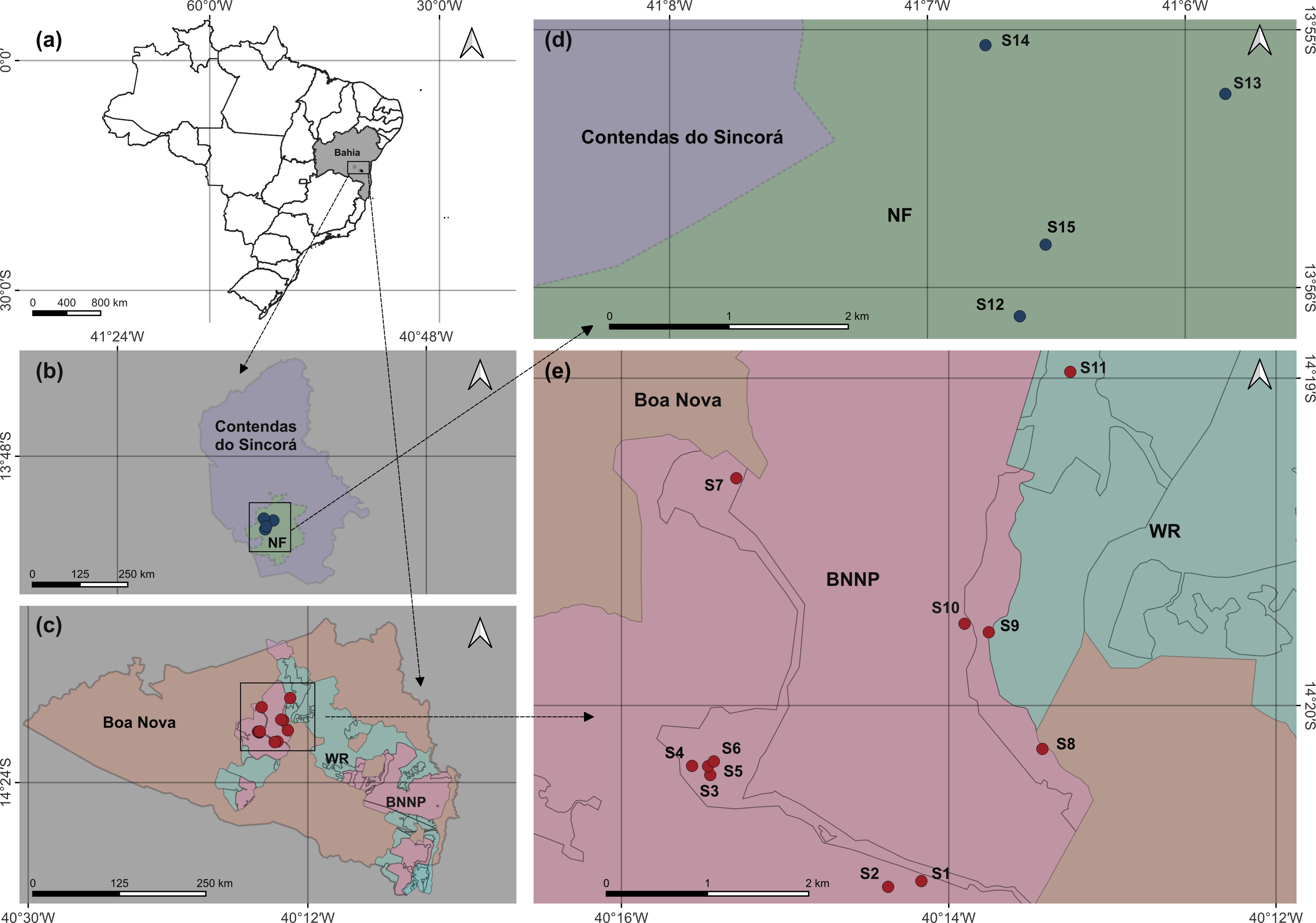
The study was conducted in three Brazilian Federal Conservation Units located in the southwestern region of Bahia State: Boa Nova National Park (BNNP), Boa Nova Wildlife Refuge (WR), and Contendas do Sincorá National Forest (NF) (Figure 1). These areas were selected because they represent legally protected ecosystems subject to varying levels of anthropogenic pressure, encompassing two distinct biomes: the Atlantic Forest and the Caatinga.
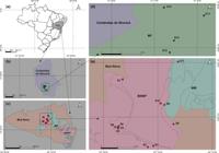
(a) Location of Brazil, highlighting the state of Bahia and the municipalities of Contendas do Sincorá and Boa Nova, the focus of this study. (b) Detail of the municipality of Contendas do Sincorá, showing the boundaries of the Contendas do Sincorá National Forest (NF) and its sampling sites. (c) Detail of the municipality of Boa Nova, highlighting Boa Nova National Park (BNNP) and Boa Nova Wildlife Refuge (WR), with the location of their respective sampling points. (d) Enlargement of the four sampling sites (S12–S15) within the Contendas do Sincorá National Forest (NF). (e) Enlargement of the 11 sampling sites (S1–S11) located within the BNNP and WR.
Boa Nova National Park (12,065 ha) and Boa Nova Wildlife Refuge (15,024 ha) were established in June 2010 and exhibit a fragmented spatial configuration, protecting an ecotonal zone between the Caatinga and Atlantic Forest biomes. The predominant vegetation includes Seasonal Semi-deciduous Forest and Dense Ombrophilous Forest, at elevations ranging from 600 to 1,000 m, approximately 130 km from the coast, within an Atlantic Forest enclave in the Drought Polygon (Brandão 2014, WikiParques 2025). The region experiences highly variable interannual rainfall, averaging 800–1,200 mm annually, concentrated from November to March (Brasil 2018). Its hydrography comprises intermittent and perennial water bodies, notably the Preto and Tabocas rivers, tributaries of the Contas River basin (Brasil 2017).
Despite their geographical proximity, the protected areas in Boa Nova are under intense anthropogenic pressure, including wildlife trafficking, hunting, illegal logging, extensive livestock grazing, land conflicts, and unregulated tourism. These activities constitute potential vectors of environmental degradation, threatening the ecological integrity of these protected ecosystems.
Contendas do Sincorá National Forest (BNNP), created in 1999, covers 11,000 ha in the municipality of Contendas do Sincorá, Bahia. This unit protects various Caatinga physiognomies, including arboreal, shrubby, and riparian forests, aiming at sustainable management and local biodiversity conservation (Brasil 2006). The area has an average annual precipitation of ~600 mm, concentrated between November and April, and is traversed by intermittent streams such as Garapa and Goiabeira. Main anthropogenic pressures include hunting and wood extraction for charcoal production, negatively impacting ecosystems and water resources.
It is worth noting that all sampling points were lentic bodies of water, that is, puddles and ponds.
Ethical and Legal Aspects
This study was conducted under collection permits issued by the Chico Mendes Institute for Biodiversity Conservation (ICMBio) (processes nº 97136-1 and nº 89834-2) and approved by the Ethics Committee on Animal Use of the Federal University of Bahia – CEUA/IMS/CAT-UFBA (protocol nº 132/2025).
Abiotic Matrix Sampling (Water and Sediment)
Sampling was carried out at 15 sites (S1–S15) (Figure 1), including 10 sites within Boa Nova National Park (S1–S10), one in the Boa Nova Wildlife Refuge (S11), and four in in NF (S12–S15). Sampling occurred on three separate occasions during March 2025 to minimize biases associated with point-in-time collection and to ensure greater representativeness of the samples; due to low plastic particle abundance per collection, results were pooled across the three sampling events.
Water samples were collected following Hu et al. (2018). All containers and instruments were pre-washed with microfiltered water (1.2 µm pore size; 47 mm diameter) and sealed (Karaoğlu & Gul 2020). Subsurface water (20 L) was filtered through a 20 µm plankton net, stored in autoclaved glass bottles, and fixed in 4% formalin. Nets were rinsed with ultrapure water between samples to prevent contamination (Prata et al. 2019).
In the laboratory, water samples were filtered under vacuum using 1.2 µm glass fiber filters, dried at 60 °C for 24 h, and analyzed under a stereomicroscope. Sediment samples (0–5 cm depth, ~0.5 kg, 15 cm from the water edge) were collected in triplicate using stainless steel spatulas and stored in aluminum foil bags. Plastic particles were extracted via NaCl hypersaturated flotation (Oliveira et al. 2023), treated with 40 mL 40% H₂O₂ at 60 °C for 48 h, and filtered through 1.2 µm glass fiber filters. Filters were air-dried and stored in glass Petri dishes (Hanke et al. 2013).
At each site, in situ measurements included water temperature (°C), pH, electrical conductivity (µS cm⁻¹), total dissolved solids (ppm), salinity (%), and dissolved oxygen (mg L⁻¹) using portable multiparameter probes and oximeters. Water transparency (cm) was assessed with a Secchi disk, and depth (cm) with a 3 m graduated tape. One liter of water was collected for laboratory chlorophyll a (Chl. a) analysis (mg L⁻¹) following the acetone extraction trichromatic method (Jeffrey & Humphrey 1975).
Biotic Matrix Sampling (Tadpoles)
Tadpoles were sampled simultaneously with water and sediment collection using aluminium sieves of varying mesh sizes, placed in autoclaved glass jars with ambient water. Specimens were euthanized with 2% lidocaine hydrochloride gel and frozen at –20 °C. In the laboratory, individuals were weighed (P, ±1 mg) and total length measured (C, ±1 mm) with a digital caliper. Developmental stage (ED) was determined using Gosner (1960) after thawing (Araújo et al. 2020a).
Each tadpole was rinsed, dissected, and immersed in 30 mL 10% H₂O₂ at 60 °C for 24 h, then vacuum-filtered through 1.2 µm glass fiber filters. Filters were dried at 65 °C for 2 h and stored in Petri dishes for analysis (Karami et al. 2017, Silva 2020).
A total of 80 tadpoles were analyzed, belonging to the families Hylidae and Leptodactylidae, across nine species: Boana semilineata, Dendropsophus minutus, D. novaisi, Julianus camposseabrai, Scinax pachycrus, Leptodactylus caatingae, Leptodactylus macrosternum, Physalaemus cuvieri, and Physalaemus cf. kroyeri. Morphometric data and occurrence records are presented in Table I.
Morphometric data and occurrence of tadpoles analyzed in this study, from from BNNP - BN, WR -BN and FN - CS N.: Number of specimens sampled; DS: Development stage according to Gosner (1960), W: Weight in grams; L: Length in mm; S1 to S15: Sampling sites.
Plastic Particle Characterization
All filters were visually inspected for the presence of microplastics. Potential microplastic particles were carefully isolated and recorded using an Olympus Axiostar Plus light microscope. Visual identification followed established criteria, including particle size, shape, color, structural homogeneity, and rigidity, as well as deformation when gently pressed with a heated metal needle, as recommended by Silva (2020). No chemical validation (e.g., FTIR or Raman spectroscopy) was performed; therefore, particle classification was based exclusively on visual criteria.
Length and width were measured using an ocular micrometer, and particles were classified into five size classes: <0.5 mm, 0.5–1 mm, 1–2 mm, 2–5 mm (microplastics), and >5 mm (mesoplastics) (Hu et al. 2022, Liu & Zheng 2025). Photomicrographs were captured using the microscope’s photomicroscope.
Particles were categorized as fibers (elongated), fragments (irregular), or granules (spherical) (Yurtsever & Yurtsever 2019, Hu et al. 2018, Qu et al. 2017), and colored as white-transparent, red (including orange and pink), blue, green, black, purple, silver, or yellow.
Contamination Control
All reagents were pre-filtered through 1.2 µm glass fiber filters. Equipment and containers were washed with filtered water and kept covered until use. Laboratory doors remained closed and air-conditioning off to minimize airborne contamination. Samples were protected with aluminum foil, and filters were stored in sealed Petri dishes. Work surfaces were regularly cleaned with 70% ethanol, and personnel limited to two individuals at a time.
Procedural blanks were processed alongside real samples using ultrapure water, and airborne contamination was monitored with exposed wet glass membrane filters. Blanks were visually inspected following Hu et al. (2018). Procedural blanks were analyzed to assess potential contamination; particles observed in blanks were recorded qualitatively, and no numerical correction was applied to the final results. Cotton lab coats and nitrile gloves were used during all procedures.
Data Analysis
Sampling sites were characterized by abiotic variables using Principal Component Analysis (PCA). PCA was applied as an exploratory ordination technique to evaluate environmental gradients and identify patterns of similarity among sampling sites based on limnological variables. They were transformed using the logarithmic function log (x + 1), with the aim of reducing data asymmetry and minimizing the influence of extreme values, as well as allowing the inclusion of zero values.
Aquatic bodies were classified according to their trophic status following Lamparelli (2004). The trophic state index was used as an integrative descriptor of system productivity and nutrient enrichment, with the purpose of supporting the interpretation of limnological heterogeneity and exploring potential relationships between trophic conditions and the occurrence of plastic particles, rather than serving as a direct assessment of water quality.
Plastic particle abundance was calculated per sampling unit: water (MP L⁻¹), sediment (MP g⁻¹), and tadpoles (MP per individual and per gram of biomass). Due to the low abundance of mesoplastics, presented data combine micro- and mesoplastics. No mesoplastics were detected in tadpoles.
RESULTS
Environmental Characteristics of Sampling Sites
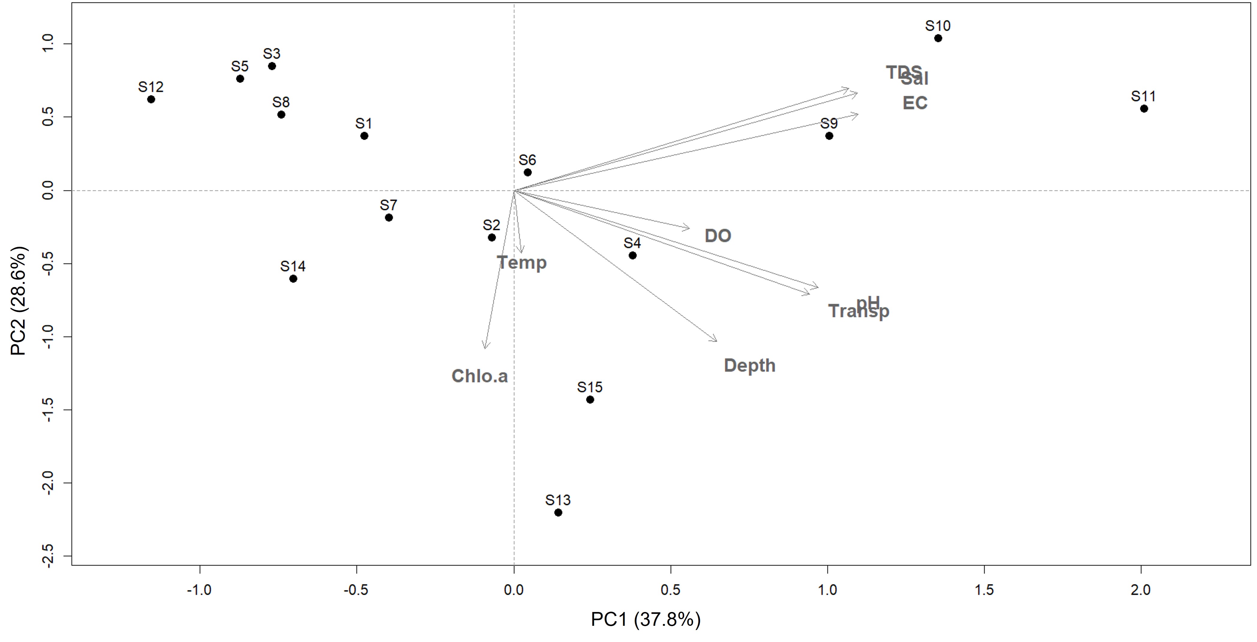
The PCA ordination (PC1 = 37.8%; PC2 = 28.6%) revealed environmental gradients structuring the sampling sites, highlighting similarities and differences among water bodies based on abiotic conditions. Sites S9, S10, and S11 were characterized by high electrical conductivity, total dissolved solids, and salinity. In contrast, sites S13 and S14 displayed distinct features, including shallower depth and higher chlorophyll a concentrations. Sites S3, S5, S8, and S12 clustered under intermediate environmental conditions, suggesting lower influence of salinity and conductivity. Overall, site distribution highlights the environmental heterogeneity among the sampled locations (Figure 2).
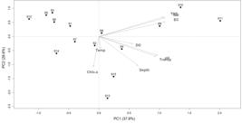
Spatial variation of abiotic variables of the sampling sites of the Conservation Units summarized by principal component analysis (PCA). DO: dissolved oxygen, TDS: total dissolved solids, EC: electrical conductivity, Temp: temperature, Chlo.a: chlorophyll a, Transp: transparency, sal: salinity.
Considering the abiotic variables within each Protected Area (Table II), water samples from BNNP - BN (S1–S10) showed a pH range from acidic to alkaline and temperatures between 21.3 and 33.2 °C. Electrical conductivity was high (31–3600 µS cm⁻¹), with total dissolved solids ranging from 15 to 1800 ppm. Aquatic bodies were generally shallow (5–110 cm), with high transparency (5–65 cm), low salinity (0–0.2%), and low dissolved oxygen (0.2–8.8 mg L⁻¹). Chlorophyll a varied from 0 to 13.63 µg L⁻¹.
Limnological characterization of sampling sites (S1 to S15) in federal Conservation Units. Oligo: Oligotrophic; Ultra: Ultraoligotrophic; Eutro: Eutrophic, Meso: Mesotrophic.
Regarding trophic status, sites were classified as ultraoligotrophic, oligotrophic, mesotrophic, or eutrophic. Only S8 was eutrophic, S10 mesotrophic, and the majority of sites ultraoligotrophic (Table II).
Water from WR - BN (S11) was characterized as a shallow aquatic body (20–80 cm depth) with pH ranging from acidic to alkaline and temperatures of 25.9–35.8 °C. Electrical conductivity was high (31–10,293 µS cm⁻¹) and total dissolved solids ranged from 15 to 1,860 ppm. The water exhibited high transparency (20–50 cm), low salinity (0–0.2%), low dissolved oxygen (4.2–9.4 mg·L⁻¹), and low chlorophyll a concentrations (0.75–0.80 µg·L⁻¹), classifying the site as ultraoligotrophic (Table II).
In contrast, water bodies from from NF - CS (S12–S14) were shallow (10–65 cm) with pH from acidic to slightly alkaline and higher temperatures (25.3–36.4 °C). Electrical conductivity (45–204 µS cm⁻¹) and total dissolved solids (22–102 ppm) were low. Transparency ranged from 10 to 65 cm, salinity was absent (0%), dissolved oxygen was low (0.8–7.2 mg·L⁻¹), and chlorophyll a varied from 0.36 to 9.00 µg·L⁻¹. These sites were classified as ultraoligotrophic, oligotrophic, or mesotrophic (Table II).
Occurrence and Abundance of Plastic Particles Across Matrices
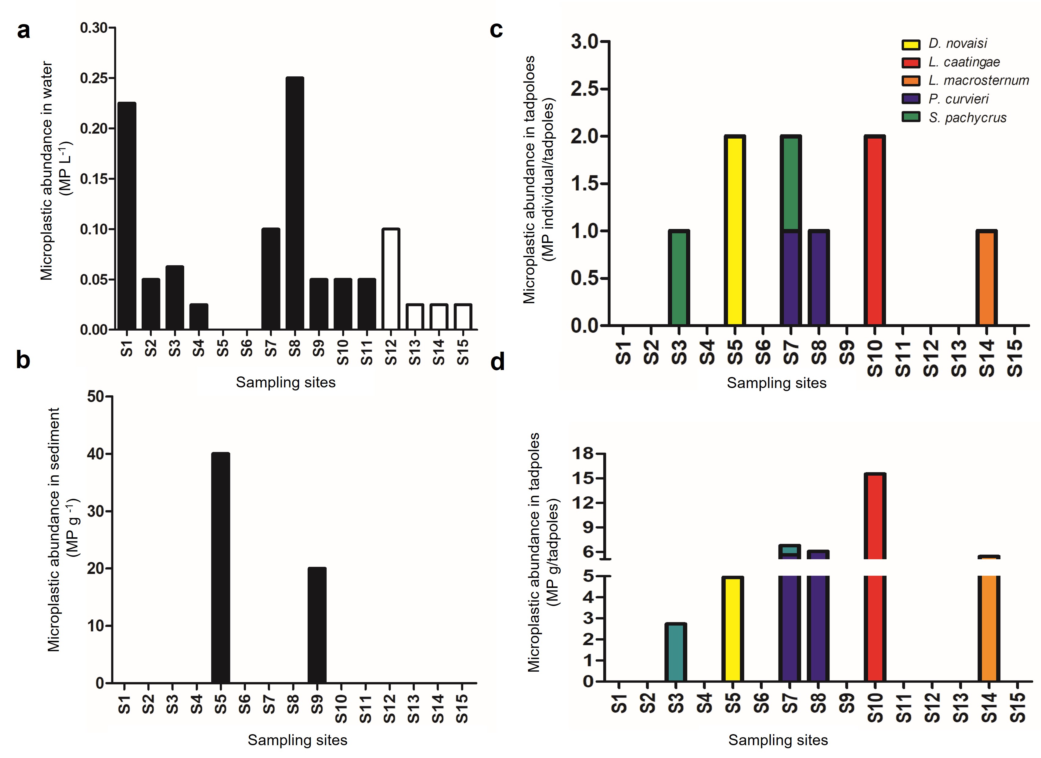
Plastic particles were detected in 14 of the 15 sampling sites, with the exception of S6. The highest occurrence was observed in water samples (13 sites; 86.7%), followed by tadpoles (6 sites; 40%) and sediment (2 sites; 13.3%) (Figure 3).
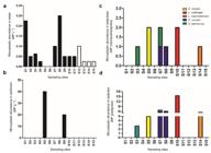
Abundance of plastic particles in three Brazilian Federal Conservation Units: BNNP - BN, WR - BN and NF-CS, Bahia, Brazil. In (a) water, (b) sediments, (c) individual/tadpole and (d) gram/tadpole.
Spatially, in the water matrix, 80% of samples from BNNP -BN were contaminated with plastic particles, while all samples from WF-BN (100%) and NF - CS (100%) were contaminated. In the sediment matrix, contamination was detected in only 20% of BNNP -BN samples and was absent in WF-BN and from NF - CS. Regarding the biotic matrix (tadpoles), approximately 50% of BNNP -BN samples contained plastic particles, followed by 25% of from NF - CS samples, while no contamination was detected in tadpoles from WF-BN.
Among the 80 tadpoles analyzed, representing nine species, microplastics were detected in nine individuals (11.25%) from five species: Dendropsophus novaisi, Leptodactylus caatingae, Leptodactylus macrosternum, Physalaemus cuvieri, and Scinax pachycrus.
In the water matrix, site S8 exhibited the highest absolute abundance of plastic particles (0.25 MP L⁻¹), followed by S1 (0.22 MP L⁻¹) (Figure 3a). In sediment, the highest abundance was recorded at S5 (40 MP g⁻¹), followed by S9 (20 MP g⁻¹) (Figure 3b). Among tadpoles, D. novaisi, L. caatingae, and S. pachycrus showed the highest per-individual abundances (2 MP per tadpole) (Figure 3c), with L. caatingae exhibiting the highest abundance per gram of biomass (15.52 MP g⁻¹) (Figure 3d).
Microplastic Size, Shape, and Color Across Matrices
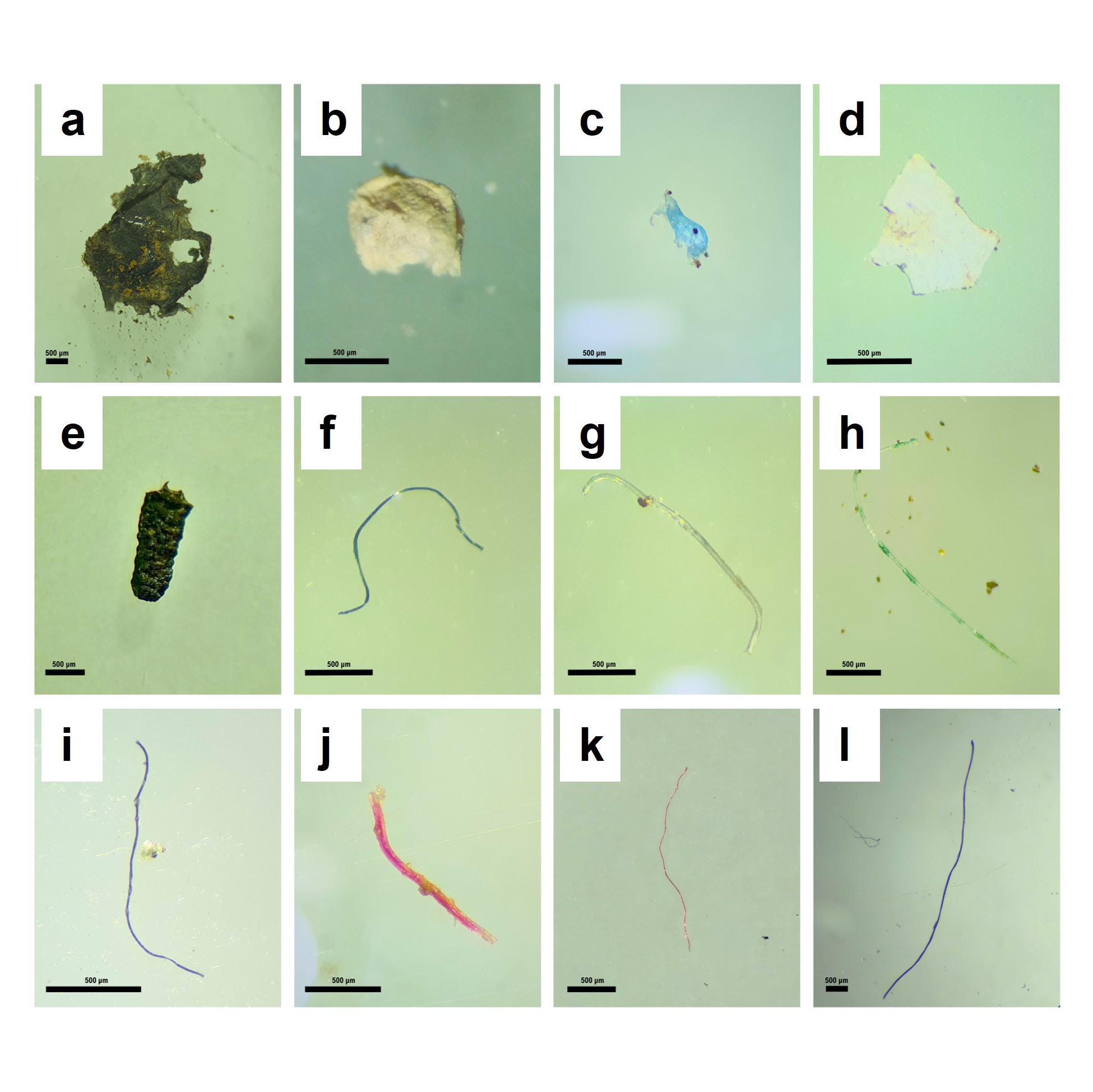
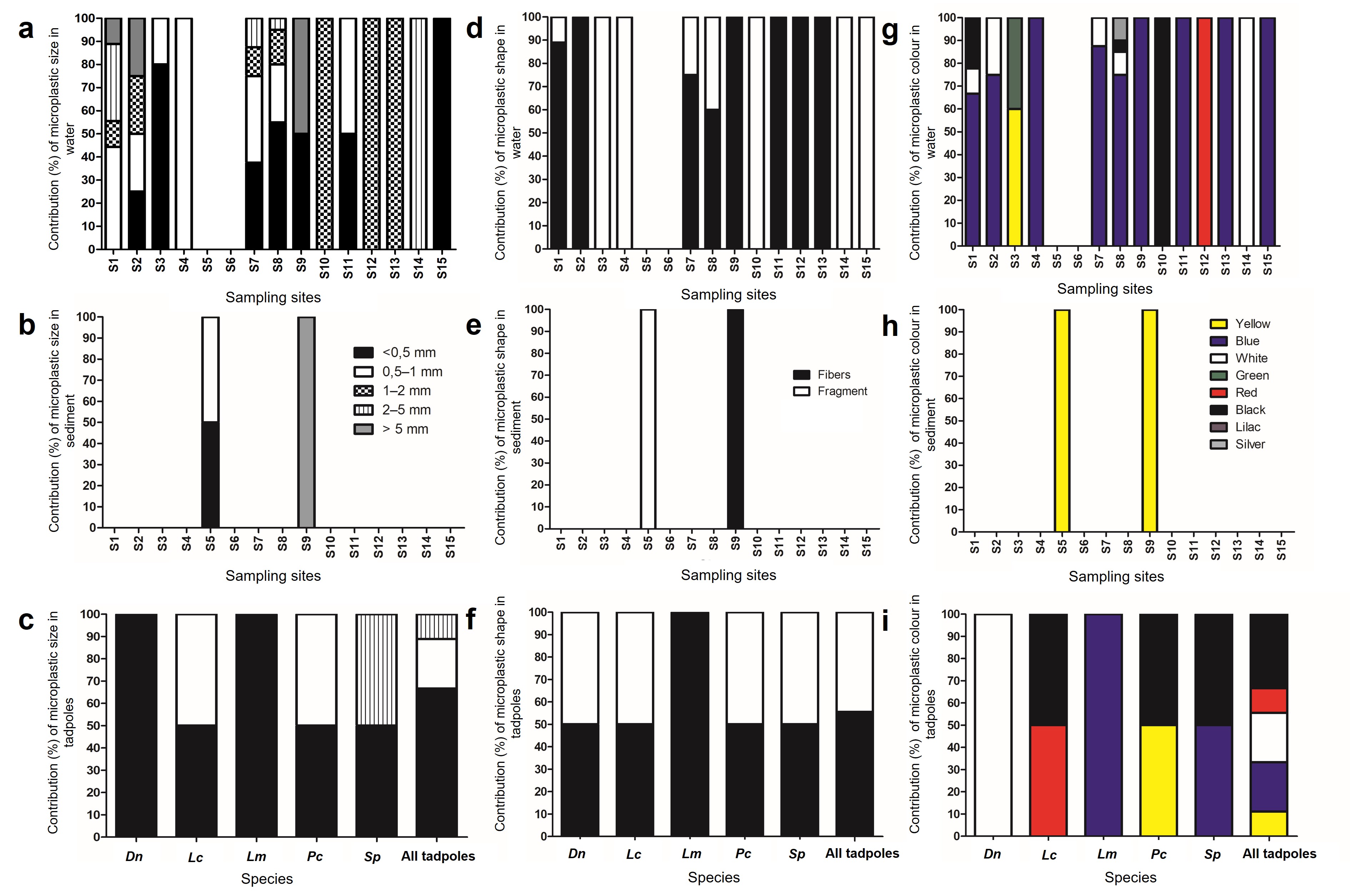
Plastic particles of varying sizes, shapes, and colors were detected in both abiotic and biotic matrices (Figures 4 and 5). Mesoplastics (>5 mm) were observed in water from sites S1, S2, and S9, comprising 10%, 25%, and 50% of particles, respectively (Figures 5a–b). In sediment from S9, all detected particles were classified as mesoplastics.
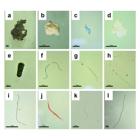
a-l: Microplastics and mesoplastics recorded in the water, sediment and tadpoles in the federal Conservation Units, BNNP - BN, WR - BN and NF-CS.
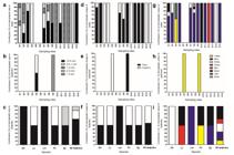
Contribution of size (a-c), shape (d-f) and color (g-i) of plastic particles expressed as a percentage in each abiotic and biotic matrix in the sampling sites (S1 to S15) of the Brazilian Federal Conservation Units. The top line corresponds to water samples, the middle line to sediments and the bottom line to species tadpoles. Dn: D. novaisi; Lc: L. caatingae; Lm: L. macrosternum; Pc: P. cuvieri and Sp: S. pachycrus.
In water samples, microplastics (MPs) < 0.5 mm predominated, followed by 1–2 mm and 0.5–1.0 mm size classes (Figure 5a). In sediment from site S5, 50% of MPs measured < 0.5 mm and 50% 0.5–1.0 mm (Figure 5b). In tadpoles, particles < 0.5 mm predominated in D. novaisi and L. macrosternum (100%), whereas L. caatingae and P. cuvieri showed equal proportions of < 0.5 mm and 0.5–1.0 mm. Particles of 2–5 mm were detected in S. pachycrus (50%) (Figure 5c). Overall, 66.7% of MPs in tadpoles were < 0.5 mm, 22.2% were 0.5–1.0 mm, and 11.1% were 2–5 mm. No mesoplastics were detected.
Fibers were the predominant type in water samples from the protected areas (Fig. 5d), whereas fragments dominated in sediment from site S5 (Figure 5e). In tadpoles, fibers accounted for 55.6% of MPs and fragments for 44.4% (Figure 5f). In L. macrosternum, only fibers were observed (100%), while other species showed roughly equal proportions of fibers and fragments.
Regarding color, blue particles predominated in water samples (Figure 5g), while only yellow MPs were detected in sediment (Figure 5h). In tadpoles, five colors were identified: blue, yellow, white/transparent, red, and black (Figure 5i). D. novaisi contained exclusively white/transparent particles; L. macrosternum, blue; S. pachycrus, blue and black; P. cuvieri, yellow and black; and L. caatingae, red and black. Overall, black MPs predominated (33.3%), followed by blue and white/transparent (22.2% each), with red and yellow each representing 11.1% (Figure 5i).
DISCUSSION
The results of the Principal Component Analysis (PCA), combined with limnological variables and plastic particle contamination data, suggest that in some sampling sites, limnological variability may directly influence the spatial distribution of these particles in both abiotic and biotic matrices. However, this relationship is not evident across all sites. For instance, site S8 showed the highest abundance of plastic particles, along with the highest chlorophyll-a concentration, indicative of a eutrophic state. Conversely, sites S13 and S14, characterized by low electrical conductivity and classified as ultraoligotrophic and mesotrophic, respectively, based on chlorophyll-a concentrations, exhibited some of the lowest abundances of plastic particles.
In contrast, sites S9, S10, and S11, located within the Boa Nova National Park and Wildlife Refuge, presented high values of electrical conductivity, salinity, and total dissolved solids, yet did not correspond to areas of high plastic particle abundance. These sites also displayed low chlorophyll-a concentrations, being classified as ultraoligotrophic, oligotrophic, and mesotrophic, respectively. These results only partially align with previous studies, which suggest that environments with higher ion concentrations and stronger anthropogenic influence tend to accumulate more plastic particles, either due to lower efficiency in natural retention processes or greater external inputs (Su et al. 2016).
In small and shallow lentic systems, elevated electrical conductivity and salinity are not necessarily indicative of anthropogenic disturbance (Wetzel 2001, Esteves 2011). These variables may primarily reflect local hydrogeological conditions, including geological substrate composition and groundwater inputs, combined with low water turnover and high evaporation rates, which concentrate dissolved ions in the water column (Tundisi & Tundisi 2008, Mitsch & Gosselink 2015). Such processes are particularly relevant in semi-arid and ecotonal regions and can produce pronounced spatial variability even within protected areas (Esteves 2011, Talbot & Chang 2022). Therefore, the limnological patterns observed in some sampling sites likely result from natural physicochemical controls rather than direct human inputs, highlighting the importance of considering hydrogeological and climatic drivers when interpreting conductivity and salinity data in small freshwater systems (Wagner et al. 2014, Rezania et al. 2018).
The wide variation observed in limnological variables reflects the natural environmental heterogeneity of small and shallow freshwater bodies, which are highly dynamic systems, particularly in tropical and semi-arid regions. In such environments, rapid fluctuations in temperature, electrical conductivity, dissolved oxygen, and chlorophyll a are driven by hydrological dynamics, evaporation, and local environmental conditions (Wetzel 2001, Tundisi & Tundisi 2008, Esteves 2011). The magnitude of this variability is further enhanced by site-specific factors such as differences in water depth, hydroperiodicity, groundwater contribution, and limited water renewal, which strongly influence ion concentration, oxygen dynamics, and thermal regimes. High surface-to-volume ratios increase the sensitivity of these systems to evaporation and atmospheric forcing, leading to rapid concentration of dissolved substances, particularly in semi-arid landscapes (Esteves 2011, Mitsch & Gosselink 2015). Consequently, pronounced spatial variability in physical and chemical parameters may occur even among nearby water bodies, reflecting natural physicochemical and hydrogeological controls rather than methodological inconsistencies or direct anthropogenic inputs (Wagner et al. 2014, Talbot & Chang 2022). Wagner et al. (2014) emphasized that both environmental and anthropogenic factors play significant roles in the presence and distribution of plastic pollutants. Similarly, Rezania et al. (2018) argued that high electrical conductivity, indicative of elevated ion concentrations, does not always correspond to higher plastic particle abundances. This may be related to increased water density, which can affect the buoyancy of plastic particles, leading them either to accumulate in less dense zones or to settle more rapidly depending on their composition and relative density, as well as the reduced anthropogenic influence in protected natural areas. Thus, the presence of plastic particles may not be strongly driven by site-specific factors alone, but also by environmental characteristics and transport dynamics, which vary depending on water depth, currents, and aquatic vegetation (Szkudlarek et al. 2024).
Although eutrophic and mesotrophic systems are often associated with higher anthropogenic inputs and nutrient loading, our results indicate that plastic particle abundance was not consistently higher in more productive environments, suggesting that plastic contamination in protected freshwater systems may be partially decoupled from trophic status (Wagner et al. 2014, Rezania et al. 2018, Talbot & Chang 2022). This pattern reinforces the idea that plastic particle occurrence in these systems may be more strongly influenced by external sources and transport dynamics than by local trophic conditions alone. Our findings are consistent with previous studies reporting the predominance of plastic particles in aquatic matrices (Hu et al. 2022, Karaoglu & Gül 2020). Of the 15 sampling sites, 14 were contaminated by plastic particles in at least one matrix: water, sediment, or tadpoles. However, although contamination was widespread, observed abundances were relatively low compared to studies from other regions worldwide. It is important to consider that many comparative studies were conducted in highly urbanized and impacted environments, where chronic plastic inputs are expected, whereas the present study focused on legally protected areas with comparatively lower direct anthropogenic pressure. Research such as Karaoglu & Gül (2020), Hu et al. (2018, 2022), Su et al. (2018), and Tatli et al. (2025) documented much higher concentrations of plastic particles in aquatic environments. In contrast, the levels observed in Brazilian waterbodies suggest a lower intensity of contamination, likely due to the fact that the study area is located within protected areas, where anthropogenic pressure and pollutant inputs should, in theory, be more limited.
Microplastic abundance was higher in the water column than in sediments, a pattern commonly reported in freshwater systems (Wagner et al. 2014, Li et al. 2018). Additionally, the use of NaCl solution for density separation may have underestimated denser polymers, such as PET and PVC, potentially contributing to the lower abundance of particles detected in sediments compared to the water column (Imhof et al. 2012, Hidalgo-Ruz et al. 2012).
The abundance of microplastics (MPs) in tadpoles of Dendropsophus novaisi, Leptodactylus caatingae, Leptodactylus macrosternum, Physalaemus cuvieri, and Scinax pachycrus was 0.60 ± 0.83 items per individual, ranging from 1.11 to 16.12 items per gram of body weight. These values are intermediate compared to published data. It is important to note that individuals of L. caatingae were analyzed from two sampling sites (S6 and S10); however, microplastics were detected exclusively in individuals collected at site S10. This pattern suggests that the higher microplastic load observed in this species is more likely associated with the specific limnological conditions of S10, such as relatively higher productivity and distinct physicochemical characteristics, rather than representing a species-wide tendency. This finding highlights the interaction between local environmental conditions and species-specific ecological traits in shaping microplastic exposure in small lentic systems. Hu et al. (2022) reported concentrations in Rana limnocharis, Microhyla ornata, and Microhyla heymonsi ranging from 0.489 to 2.53 items per individual (equivalent to 4.61–63.0 items/g), supporting in particular the high per-gram microplastic burden observed in L. caatingae in our study.
The high abundance of microplastics observed in L. caatingae may be related to its feeding habits and mode of habitat use. Tadpoles of this species were predominantly observed near the bottom of the water body, actively foraging on the substrate and scraping organic matter, which increases the likelihood of incidental ingestion of microplastics deposited in sediments or resuspended near the bottom. Similar patterns have been reported for tadpoles exploiting the sediment–water interface in shallow lentic systems, where particle availability and feeding behavior strongly influence microplastic ingestion (Antoniazzi et al. 2020). Therefore, the elevated microplastic load detected in L. caatingae likely reflects species-specific ecological traits interacting with local environmental availability rather than a generalized pattern across taxa. Earlier studies by Hu et al. (2018) with four tadpole species in China (Bufo gargarizans, Microhyla ornata, Rana limnochari, and Pelophylax nigromaculatus) documented 0–2.73 items per individual, with maximum values of up to 168 items/g. By contrast, Karaoglu & Gül (2020) recorded much higher concentrations (303–307 items/g) in Pelophylax ridibundus and Rana macrocnemis tadpoles in northeastern Turkey, indicating a considerably greater MP burden compared to our specimens. Conversely, Kolenda et al. (2020) reported much lower averages (0.35 MPs per individual) in Bufo bufo, Rana temporaria, Pelophylax esculentus, Pelobates fuscus, and Hyla arborea in southwestern Poland, suggesting reduced exposure in areas with lower anthropogenic pressure.
The relationship between MP abundance and body weight revealed substantial interspecific variation. The highest abundance was observed in L. caatingae, with up to 16 MPs per gram of body weight. Such variation may be linked to differences in ingestion rates, dietary habits, and environmental availability of MPs, as suggested by Karaoglu & Gül (2020). Previous studies have emphasized that MP ingestion in anurans may have important physiological and ecological implications, particularly in species with aquatic larval stages (Green et al. 2021).
The diversity of plastic particle sizes, shapes, and colors observed in our samples was wide. Fibers were the predominant morphotype, followed by fragments, a pattern consistent with other studies (Hu et al. 2018, 2022, Kolenda et al. 2020, Pastorino et al. 2022, Shetu et al. 2023, Szkudlarek et al. 2024). Hu et al. (2022) pointed out that fibers, often derived from textiles, are the most common MP type in freshwater systems. Similarly, the predominance of fibers in tadpoles, along with the absence of pellets, matches previous findings in amphibians and other freshwater organisms (Karaoglu & Gül 2020, Szkudlarek et al. 2024, Kolenda et al. 2020). Most particles detected in tadpoles were <0.5 mm (66.67%), followed by MPs between 0.5–1.0 mm (22.22%) and 2–5 mm (11.11%), with no mesoplastics (>5 mm). These results align with those of Vidal et al. (2023) and Hu et al. (2022), who also reported a predominance of small MPs, especially in filter-feeding or detritivorous species.
Blue microplastics were dominant both in water samples and in tadpole gastrointestinal tracts, alongside a marked chromatic diversity of ingested particles. This pattern is consistent with records from multiple aquatic ecosystems, where blue MPs frequently appear as the most abundant morphotype (Szkudlarek et al. 2023, 2024). Their predominance may be related to higher resistance to degradation due to lower ultraviolet absorption, which favors the formation of smaller particles and thus increases their availability for ingestion (Szkudlarek et al. 2024). On the other hand, the color diversity observed in ingested particles reflects multiple pollution sources, including fragments from plastic bags, synthetic fabrics, and fishing nets (Kolenda et al. 2020, Tatli et al. 2025). The high representation of black and blue MPs further reinforces global patterns of urban plastic waste disposal, suggesting that tadpoles are widely exposed to persistent microparticles that may be mistaken for natural prey.
The digestion protocol applied is effective for the removal of soft tissues but does not degrade cellulosic fibers, such as cotton or rayon, which are known to resist hydrogen peroxide treatment (Hidalgo-Ruz et al. 2012, Lusher et al. 2017). Although additional visual criteria were applied in this study, including a heated needle test to assess particle deformation, this procedure does not fully exclude anthropogenic cellulosic fibers, which do not melt and may exhibit ambiguous thermal responses under optical microscopy (Imhof et al. 2012). Consequently, part of the fiber fraction reported here may correspond to non-synthetic anthropogenic materials, and the observed patterns should be interpreted with appropriate caution in the absence of chemical validation.
The ingestion of MPs by aquatic organisms such as tadpoles is strongly influenced by their availability and position in the water column, which in turn depends on physicochemical characteristics such as particle size, shape, and density (Vidal et al. 2023, Van Cauwenberghe et al. 2015). Less dense, smaller particles, such as floating fibers or suspended fragments, tend to remain near the surface, where they are more accessible to filter-feeding or scraping tadpoles. Denser fragments, by contrast, tend to settle into sediments, reducing accessibility to pelagic feeders. Thus, tadpole exposure to MPs depends not only on environmental presence but also on bioavailability, shaped by both particle properties and species feeding behaviors.
The ingestion of MPs by tadpoles has important implications for aquatic ecosystems. As key prey for fish, reptiles, birds, mammals, and other amphibians (Dodd 2009), tadpoles may facilitate the transfer of MPs to higher trophic levels, potentially resulting in bioaccumulation and biomagnification. Such processes can impair predator health through inflammation, physiological dysfunctions, and reduced reproductive capacity, as observed in other MP-exposed taxa. Moreover, contamination of aquatic predators by MPs raises concerns about food safety, since fish and mollusks consumed by humans may carry these particles into human diets.
Experimental evidence reinforces these concerns. For example, Hu et al. (2016) showed that in the absence of food, Xenopus tropicalis tadpoles ingested more microbeads and excreted fewer particles, suggesting that high MP abundance coupled with food scarcity may drive increased ingestion. Such scenarios may be particularly critical in ecosystems with limited food resources, where tadpoles may mistake MPs for edible material.
Altogether, these findings highlight the urgent need to monitor and mitigate MP pollution, not only considering its direct impacts on tadpoles but also its cascading effects across the aquatic food web. Future research should address the mechanisms of ingestion, long-term physiological impacts, and persistence of MPs in tropical aquatic ecosystems, particularly in regions facing high anthropogenic pressure. Expanding knowledge in these areas is essential for accurately assessing ecological risks and long-term impacts of microplastic contamination in freshwater systems.
CONCLUSIONS
This study is the first to report the occurrence of plastic particles, particularly microplastics, in both abiotic and biotic matrices of three Brazilian Federal Protected Areas. The findings reveal that even legally protected sites are subject to considerable plastic contamination, with the highest prevalence in water, followed by tadpoles and sediments. The predominance of blue fibers smaller than 0.5 mm suggests diverse anthropogenic sources, most likely linked to human activities in adjacent areas.
The detection of microplastics in tadpoles of five species demonstrates the bioaccessibility of these contaminants and highlights the potential of amphibians as bioindicators of plastic pollution in freshwater ecosystems. Exposure to plastic particles may entail significant ecological consequences, including risks of bioaccumulation and biomagnification along food webs.
Although the abundance of plastic particles in the studied areas was moderate, their presence in supposedly conserved environments underscores the urgency of public policies to mitigate plastic pollution, particularly in tropical freshwater ecosystems. The results also reinforce the need to expand environmental monitoring in Protected Areas, incorporating plastic contamination as a relevant parameter for biodiversity management and conservation. Future studies should further investigate the physiological, behavioral, and ecological effects of plastic particles on aquatic organisms, as well as assess strategies to reduce the input of these contaminants into protected ecosystems.
Although particle identification was based on visual criteria, the consistent detection of plastic-like particles across abiotic and biotic matrices indicates that freshwater systems within protected areas are not exempt from contamination.
Acknowledgements
The authors thank the team of the Limnology and Biomonitoring Laboratory — João Paulo do Amaral Leite, Aline Guimarães Novais, Ana Clara de Lima Rocha Ribeiro, Leandro Batista Ferreira, Flávya Santos Lozado, Stefany Almeida da Silva, João Pedro Cardoso dos Santos, Gabriel Costa da Silva, and David Patrick Marques Alves — for their technical and scientific support during the field sampling and analyses. The authors also thank the anonymous reviewers for their valuable suggestions that improved this manuscript.
References
-
ALTIG R, WHILES MR & TAYLOR CL. 2007. What do tadpoles really eat? Assessing the trophic status of an understudied and imperilled group of consumers in freshwater habitats. Freshw Biol 52: 386-395. https://doi.org/10.1111/j.1365-2427.2006.01694.x.
» https://doi.org/10.1111/j.1365-2427.2006.01694.x -
AMPARO SZS, SILVA LF, PEREIRA RO, SOUZA FA & ROCHA CA. 2023. Microplastics as contaminants in the Brazilian environment: an updated review. Environ Monit Assess 195: 1414. https://doi.org/10.1007/s10661-023-12011-0.
» https://doi.org/10.1007/s10661-023-12011-0 -
ANTONIAZZI CE, LÓPEZ JA, LORENZÓN RE, SAIGO M, DEVERCELLI M, MANEYRO L & MARCHESE MR. 2020. Trophic ecology of tadpoles in floodplain wetlands: combining gut contents, selectivity, and stable isotopes to study feeding segregation of syntopic species. Hydrobiologia 847: 3013-3024. https://doi.org/10.1007/s10750-020-04303-0.
» https://doi.org/10.1007/s10750-020-04303-0 -
ARAÚJO APC, DE MELO NFS, DE OLIVEIRA JUNIOR AG, RODRIGUES FP, FERNANDES T, VIEIRA JEA, ROCHA TL & MALAFAIA G. 2020a. How much are microplastics harmful to the health of amphibians? A study with pristine polyethylene microplastics and Physalaemus cuvieri J Hazard Mater 382: 121066. https://doi.org/10.1016/j.jhazmat.2019.121066.
» https://doi.org/10.1016/j.jhazmat.2019.121066 -
ARAÚJO APC, GOMES AR & MALAFAIA G. 2020b. Hepatotoxicity of pristine polyethylene microplastics in neotropical Physalaemus cuvieri tadpoles (Fitzinger, 1826). J Hazard Mater 386: 121992. https://doi.org/10.1016/j.jhazmat.2019.121992.
» https://doi.org/10.1016/j.jhazmat.2019.121992 -
ARAÚJO APC, ROCHA TL, SILVA DM & MALAFAIA G. 2021. Micro(nano)plastics as an emerging risk factor to the health of amphibians: a scientometric and systematic review. Chemosphere 283: 131152. https://doi.org/10.1016/j.chemosphere.2021.131090.
» https://doi.org/10.1016/j.chemosphere.2021.131090 -
BERGMANN M, GUTOW L & KLAGES M. 2015. Marine Anthropogenic Litter. Springer, Cham. https://doi.org/10.1007/978-3-319-16510-3
» https://doi.org/10.1007/978-3-319-16510-3 -
BOYLE K & ÖRMECI B. 2020. Microplastics and nanoplastics in the freshwater and terrestrial environment: a review. Water 12: 2633. https://doi.org/10.3390/w12092633.
» https://doi.org/10.3390/w12092633 - BRANDÃO GS. 2014. Composição florística de trepadeiras do Parque Nacional de Boa Nova, Bahia, Brasil. Master’s thesis. Universidade Estadual do Sudoeste da Bahia, Jequié. (Unpublished).
-
BRASIL. 2000. Lei nº 9.985, de 18 de julho de 2000. Regulamenta o artigo 225 da Constituição Federal, institui o Sistema Nacional de Unidades de Conservação. Ministério do Meio Ambiente, Brasília. http://www.mma.gov.br/port/sbf/dap/doc/snuc.pdf
» http://www.mma.gov.br/port/sbf/dap/doc/snuc.pdf - BRASIL. 2006. Plano de manejo da Floresta Nacional Contendas do Sincorá. Volume 1: Informações gerais. MMA, Brasília.
- BRASIL. 2017. Diagnóstico ambiental da bacia do rio de Contas. IBAMA, Brasília.
- BRASIL. 2018. Plano de Manejo do Parque Nacional de Boa Nova. MMA/ICMBio, Brasília.
-
CIMMARUTA R, GIOVANNINI S, BIANCHI J, MATIDDI M & BELLISARIO B. 2022. Microplastics occurrence in fish with different habitats from the central Tyrrhenian Sea. Reg Stud Mar Sci 52: 102309. https://doi.org/10.1016/j.rsma.2022.102251.
» https://doi.org/10.1016/j.rsma.2022.102251 -
COLE M, LINDEQUE P, FILEMAN E, HALSBAND C, GOODHEAD R, MOGER J & GALLOWAY TS. 2013. Microplastic ingestion by zooplankton. Environ Sci Technol 47: 6646-6655. https://doi.org/10.1021/es400663f.
» https://doi.org/10.1021/es400663f - DODD CK JR. 2009. Amphibian Ecology and Conservation: A Handbook of Techniques. Oxford University Press, Oxford.
- ESTEVES FA. 2011. Fundamentos de limnologia. 3rd ed., Rio de Janeiro: Interciência, 826 p.
-
FERRARI L & HEPP LU. 2021. Microplastic ingestion by Characidae in rural streams (Rio Grande do Sul, Brazil). Biotemas 34(3): 1-6. https://doi.org/10.5007/2175-7925.2021.e81780.
» https://doi.org/10.5007/2175-7925.2021.e81780 -
FERREIRA I, VENÂNCIO C, LOPES I & OLIVEIRA M. 2019. Nanoplastics and marine organisms: what has been studied? Environ Toxicol Pharmacol 67: 1-7. https://doi.org/10.1016/j.etap.2019.01.006.
» https://doi.org/10.1016/j.etap.2019.01.006 -
GONÇALVES IM, FERREIRA RB, ROSSA-FERES DC & PROVETE DB. 2023. Descrição e fatores modeladores da dieta e ecologia da alimentação de girinos neotropicais: um estudo de caso e uma revisão abrangente. Austral Ecol 47: 713-716. https://doi.org/10.1111/aec.13152.
» https://doi.org/10.1111/aec.13152 - GOSNER KL. 1960. A simplified table for staging anuran embryos and larvae with notes on identification. Herpetologica 16(3): 183-190.
-
GREEN DS, BOOTH AM, O’CONNOR NE & THOMPSON RC. 2021. Impacts of microplastics on amphibians: a focus on exposure, uptake and effects. Environ Pollut 273: 116480. https://doi.org/10.1016/j.envpol.2021.116480.
» https://doi.org/10.1016/j.envpol.2021.116480 - HANKE G, WALVOORT B, LOESEKANN-BEHRENS T, MAES T, VETHAAK AD & OOSTERBAAN L. 2013. Guidance on monitoring of marine litter in European seas. Publications Office of the European Union, Luxembourg.
-
HU L, SU L, XUE Y, MU J, ZHU J, XU J & SHI H. 2016. Uptake, accumulation and elimination of polystyrene microspheres in Xenopus tropicalis tadpoles Chemosphere 164: 611-617. https://doi.org/10.1016/j.chemosphere.2016.09.001.
» https://doi.org/10.1016/j.chemosphere.2016.09.001 -
HU L, CHERNICK M, HINTON DE & SHI H. 2018. Microplastics in small waterbodies and tadpoles from Yangtze River Delta, China. Environ Sci Technol 52: 8885-8893. https://doi.org/10.1021/acs.est.8b01520.
» https://doi.org/10.1021/acs.est.8b01520 -
HU L, FU J, ZHENG P, DAI M, ZENG G & PAN X. 2022. Accumulation of microplastics in tadpoles from different functional zones in Hangzhou Great Bay Area, China: relation to growth stage and feeding habitats. J Hazard Mater 424: 127281. https://doi.org/10.1016/j.jhazmat.2021.127281.
» https://doi.org/10.1016/j.jhazmat.2021.127281 - JEFFREY SW & HUMPHREY GF. 1975. New spectrophotometric equations for determining chlorophyll a, b, c1 and c2 in higher plants, algae and natural phytoplankton. Biochem Physiol Pflanz 167: 191-194.
-
KARAMI A, GOLIESKARDI A, CHOO CK, ROMANO N, HO YB & SALAMATINIA B. 2017. A high-performance protocol for extraction of microplastics in fish. Sci Total Environ 578: 485-494. https://doi.org/10.1016/j.scitotenv.2016.10.213.
» https://doi.org/10.1016/j.scitotenv.2016.10.213 -
KARAOĞLU K & GÜL S. 2020. Characterization of microplastic pollution in tadpoles living in small water-bodies from Rize, the northeast of Turkey. Chemosphere 255: 126915. https://doi.org/10.1016/j.chemosphere.2020.127145.
» https://doi.org/10.1016/j.chemosphere.2020.127145 -
KILIÇ E & YÜCEL N. 2022. Microplastic occurrence in the gastrointestinal tract and gill of bioindicator fish species in the northeastern Mediterranean. Mar Pollut Bull 177: 113523. https://doi.org/10.1016/j.marpolbul.2022.113523.
» https://doi.org/10.1016/j.marpolbul.2022.113523 -
KOLENDA K, KUŚMIEREK N & PSTROWSKA K. 2020. Microplastic ingestion by tadpoles of pond-breeding amphibians - first results from Central Europe (SW Poland). Environ Sci Pollut Res 27: 33592-33601. https://doi.org/10.1007/s11356-020-09119-2.
» https://doi.org/10.1007/s11356-020-09119-2 - HIDALGO-RUZ V, GUTOW L, THOMPSON RC & THIEL M. 2012. Microplastics in the marine environment: a review of the methods used for identification and quantification. Environ Sci Technol 46: 3060-3075.
- IMHOF HK, SCHMID J, NIESSNER R, IVLEV A & LAFORSCH C. 2012. A novel, highly efficient method for the separation and quantification of plastic particles in sediments of aquatic environments. Limnol Oceanogr Methods 10: 524-537.
- LAMPARELLI MC. 2004. Grau de trofia em corpos d’água do estado de São Paulo: avaliação dos métodos de monitoramento. PhD thesis. Universidade de São Paulo, São Paulo.
- LI J, LIU H & CHEN JP. 2018. Microplastics in freshwater systems: a review on occurrence, environmental effects, and methods for microplastics detection. Water Res 137: 362-374.
-
LIU J & ZHENG L. 2025. Microplastic migration and transformation pathways and exposure health risks. Environ Pollut 368: 125700. https://doi.org/10.1016/j.envpol.2025.125700.
» https://doi.org/10.1016/j.envpol.2025.125700 -
LU Y, ZHANG Y, DENG Y, JIANG W, ZHAO Y, GENGE J, DING L & REN H. 2016. Uptake and accumulation of polystyrene microplastics in zebrafish (Danio rerio) and toxic effects in liver. Environ Sci Technol 50(7): 4054-4060. https://doi.org/10.1021/acs.est.5b06290.
» https://doi.org/10.1021/acs.est.5b06290 - LUSHER AL, HOLLANDER R, MENDOZA-HILL JJ & WARDLAW C. 2017. Microplastics in fisheries and aquaculture: status of knowledge on their occurrence and implications for aquatic organisms. FAO Fish Aquac Tech Pap 615: 1-147.
- MITSCH WJ & GOSSELINK JG. 2015. Wetlands. 5th ed., Hoboken: John Wiley & Sons, 744 p.
-
MOORE CJ. 2008. Synthetic polymers in the marine environment: a rapidly increasing, long-term threat. Environ Res 108: 131-139. https://doi.org/10.1016/j.envres.2008.07.025.
» https://doi.org/10.1016/j.envres.2008.07.025 -
MORALES-ESPINOZA LL, GEBARA RC, LONGO E & FRACÁCIO R. 2025. Microplastics in freshwater ecosystems: probabilistic environmental risk assessment and current knowledge in occurrence and ecotoxicological studies. Environ Toxicol Chem 44(1): 3-25. https://doi.org/10.1093/etojnl/vgae008.
» https://doi.org/10.1093/etojnl/vgae008 -
OLIVEIRA LG, HATTORI GY & SANT’ANNA BS. 2023. Microplastic contamination in bathing areas in the Central Amazon, Itacoatiara, Brazil. Environ Sci Pollut Res 30: 10420-10433. https://doi.org/10.1007/s11356-022-24334-x.
» https://doi.org/10.1007/s11356-022-24334-x -
OLIVEIRA M, ALMEIDA M & MIGUEL I. 2019. A micro(nano)plastic boomerang tale: a never ending story? TrAC Trends Anal Chem 112: 196-200. https://doi.org/10.1016/j.trac.2018.11.014.
» https://doi.org/10.1016/j.trac.2018.11.014 -
PASTORINO P ET AL. 2022. Microplastics occurrence in the European common frog (Rana temporaria) from Cottian Alps (Northwest Italy). Diversity 14: 66. https://doi.org/10.3390/d14020066.
» https://doi.org/10.3390/d14020066 -
PRATA JC, DA COSTA JP, DUARTE AC & ROCHA-SANTOS T. 2019. Methods for sampling and detection of microplastics in water and sediment: a critical review. TrAC Trends Anal Chem 110: 150-159. https://doi.org/10.1016/j.trac.2018.10.015.
» https://doi.org/10.1016/j.trac.2018.10.015 -
QU X, SU L, LI H, LIANG M & SHI H. 2017. Assessing the relationship between the abundance and properties of microplastics in water and in mussels. Sci Total Environ 621: 679-686. https://doi.org/10.1016/j.scitotenv.2017.11.074.
» https://doi.org/10.1016/j.scitotenv.2017.11.074 -
RAHMAN MM, KIM ES & SUNG HC. 2024. Microplastics as an emerging threat to amphibians: Current status and future perspectives. Heliyon 10(7): e28220. https://doi.org/10.1016/j.heliyon.2024.e28220.
» https://doi.org/10.1016/j.heliyon.2024.e28220 -
REZANIA S, PARK J, MD DIN MF, MAT TAIB S, TALAEIKHOZANI A, YADAV KK & KAMYAB H. 2018. Microplastics pollution in different aquatic environments and biota: a review of recent studies. Mar Pollut Bull 133: 191-208. https://doi.org/10.1016/j.marpolbul.2018.05.022.
» https://doi.org/10.1016/j.marpolbul.2018.05.022 -
RUTHSATZ L, GUIMARÃES ATB, KOLENDA K, PEREIRA P, DA ROSA CM, WEBER L, HARTMANN NB, SCHOLZ-BÖTTCHER BM, FALTIN E & LENZ M. 2023. Transgenerational effects of microplastics in amphibians: evidence from Xenopus laevis. Environ Sci Technol 57(4): 2050-2062. https://doi.org/10.1021/acs.est.2c06581.
» https://doi.org/10.1021/acs.est.2c06581 -
SCHIESARI L. 2006. Pond canopy cover: a resource gradient for anuran larvae. Freshw Biol 51: 412-423. https://doi.org/10.1111/j.1365-2427.2005.01491.x.
» https://doi.org/10.1111/j.1365-2427.2005.01491.x -
SHETU MH, PARVIN F & TAREQ SM. 2023. Identifying the presence of microplastics in frogs from the largest delta of the world. Environ Adv 11: 100355. https://doi.org/10.1016/j.envadv.2023.100355.
» https://doi.org/10.1016/j.envadv.2023.100355 - SILVA ACA. 2020. Microplásticos em vários tecidos de espécies de peixes pelágicos com interesse comercial. MSc thesis, Faculdade de Ciências, Universidade de Lisboa, Lisboa. (Unpublished).
-
SU L, CAI H, KOLANDHASAMY P, WU C, ROCHMAN CM & SHI H. 2018. Using the Asian clam as an indicator of microplastic pollution in freshwater ecosystems. Environ Pollut 234: 347-355. https://doi.org/10.1016/j.envpol.2017.11.092.
» https://doi.org/10.1016/j.envpol.2017.11.092 -
SU L, XUE Y, LI L, YANG D, KOLANDHASAMY P, LI D & SHI H. 2016. Microplastics in Taihu Lake, China. Environ Pollut 216: 711-719. https://doi.org/10.1016/j.envpol.2016.06.056.
» https://doi.org/10.1016/j.envpol.2016.06.056 -
SZKUDLAREK M, NAJBAR B & JANKOWIAK Ł. 2023. Microplastics pollution in larvae of toads, frogs and newts in anthropopressure gradient. Ecol Indic 155: 110971. https://doi.org/10.1016/j.ecolind.2023.110971.
» https://doi.org/10.1016/j.ecolind.2023.110971 -
SZKUDLAREK M, NAJBAR B & JANKOWIAK Ł. 2024. Variation in microplastic characteristics among amphibian larvae: a comparative study across different species and the influence of human activity. Sci Rep 14: 13574. https://doi.org/10.1038/s41598-024-49663-1.
» https://doi.org/10.1038/s41598-024-49663-1 -
TALBOT R & CHANG H. 2022. Microplastics in freshwater: a global review of factors affecting spatial and temporal variations. Environ Pollut 292: 118393. https://doi.org/10.1016/j.envpol.2021.118393.
» https://doi.org/10.1016/j.envpol.2021.118393 -
TATLI H, GEDIK K & ALTUNIŞIK A. 2025. Distribution of microplastics in tadpoles, adults, and habitats of three water frogs of Pelophylax spp. Environ Sci Eur 37: 27. https://doi.org/10.1186/s12302-025-00627-4.
» https://doi.org/10.1186/s12302-025-00627-4 -
TEUTEN EL, ROWLAND SJ, GALLOWAY TS & THOMPSON RC. 2007. Potential for plastics to transport hydrophobic contaminants. Environ Sci Technol 41(22): 7759-7764. https://doi.org/10.1021/es071737s.
» https://doi.org/10.1021/es071737s - TUNDISI JG & TUNDISI TM. 2008. Limnologia. São Paulo: Oficina de Textos, 632 p.
-
TUSSELLINO M, CARBONE A, ROCCO L, METTIVIER G, MIGLIACCIO D, CAVALIERE C & SCUDIERO R. 2015. Developmental toxicity of nanoplastics in Xenopus laevis: growth delay and malformations. Environ Pollut 206: 512-519. https://doi.org/10.1016/j.envpol.2015.08.035.
» https://doi.org/10.1016/j.envpol.2015.08.035 -
VAN CAUWENBERGHE L, DEVRIESE L, GALGANI F, ROBBENS J & JANSSEN CR. 2015. Microplastics in sediments: a review of techniques, occurrence and effects. Mar Environ Res 111: 5-17. https://doi.org/10.1016/j.marenvres.2015.06.007.
» https://doi.org/10.1016/j.marenvres.2015.06.007 -
VIDAL A, PHUONG NN, MÉTAIS I, GASPERI J & CHÂTEL A. 2023. Assessment of microplastic contamination in the Loire River (France) throughout analysis of different biotic and abiotic freshwater matrices. Environ Pollut 326: 122167. https://doi.org/10.1016/j.envpol.2023.122167.
» https://doi.org/10.1016/j.envpol.2023.122167 -
WAGNER M, SCHWABL P, KÖRNER A, ZIMMERMANN T, MARTIN C & KRONFELD L. 2014. Microplastics in freshwater ecosystems: what we know and what we need to know. Environ Sci Eur 26: 12. https://doi.org/10.1186/s12302-014-0012-7.
» https://doi.org/10.1186/s12302-014-0012-7 - WETZEL RG. 2001. Limnology: lake and river ecosystems. 3rd ed., San Diego: Academic Press, 1006 p.
-
WIKIPARQUES. 2025. Refúgio de Vida Silvestre de Boa Nova. Accessed 19 May 2025. https://www.wikiparques.org/wiki/Ref%C3%BAgio_de_Vida_Silvestre_de_Boa_Nova
» https://www.wikiparques.org/wiki/Ref%C3%BAgio_de_Vida_Silvestre_de_Boa_Nova -
YURTSEVER M & YURTSEVER U. 2019. Use of a convolutional neural network for the classification of microbeads in urban wastewater. Chemosphere 216: 271-280. https://doi.org/10.1016/j.chemosphere.2018.10.147.
» https://doi.org/10.1016/j.chemosphere.2018.10.147 -
ZHANG S, WANG J, YAN P, HAO X, XU B, WANG W & AURANGZEB M. 2021. Non-biodegradable microplastics in soils: a brief review and challenge. J Hazard Mater 409: 124953. https://doi.org/10.1016/j.jhazmat.2020.124953.
» https://doi.org/10.1016/j.jhazmat.2020.124953
Edited by
-
Handling editor
Alexander Kellner
The data that support the findings of this study are available from the corresponding author upon reasonable request.