 New functional tissues induced by a galling lepidopteran on stems of Marcetia taxifolia (Melastomataceae): An analysis based on cell wall composition and dynamics
Abstract
The plant cell walls are dynamic structures, whose pectin composition varies according to the developmental and functional stages and in response to external factors. Gall inducers change the functional profiles of their host organs, and herein, we explore how stem cell walls respond to gall development, particularly in the nutritive tissues consumed by the lepidopteran larva. The pectin composition of the non-galled stems of Marcetia taxifolia (Melastomataceae) in primary and secondary growth and of Lepidoptera stem galls in distinct stages of development were studied by immunocytochemistry. During the development of the stems of M. taxifolia, the changes in protein and pectin composition suggest that young stems are growing and elongating with the help of extensins, arabinogalactan-proteins (AGPs), and homogalacturonans (HGs). In the galls, the predominance of galactans indicates changes in the pectin composition, allowing cell wall stiffness in hyperplasic tissues. The chewing stimuli of the Lepidoptera induce the synthesis of galactans, arabinans, and HGs in nutritive cells, allowing the cell wall flexibility during the hyperplasic process and the porosity necessary for the translocation of water and nutrients toward the gall chamber.
Keywords:
Cell walls; Insect galls; Lepidoptera; Marcetia taxifolia; Nutritive tissue; Stem anatomy.
Introduction
The cell wall is an important compartment in plant cells, whose variable composition determines tissue functions (Lionetti et al., 2012; Cosgrove, 2016; Boanares et al., 2018). Pectins, as key cell wall components, mediate interactions with external factors (Agoda-Tandjawa et al., 2016; Wu et al., 2018; Liu et al., 2021). Pectins are complex and diverse structural carbohydrates that contain galacturonic acid (Caffall & Mohnen, 2009), and are essential for cell wall properties such as flexibility, elasticity, stiffness, porosity, and hydration (Knox et al., 1990; Cosgrove, 2016). The most abundant pectins in the plant cell walls are the homogalacturonans (HGs), which are synthesized in a methyl-esterified form (Knox et al., 1990). However, during tissue development, these HGs may be de-methyl-esterified by pectin methyl-esterases (PMEs) (Lionetti et al., 2012), exposing negative charges that bind with calcium ions. The binding of Ca2+ with de-methyl-esterified HGs forms pectic gels that rigidify and reinforce the cell walls (Knox et al., 1990; Sabba & Lulai, 2005). This reinforcement is important to control the calcium input and the cell maturity (Mastroberti & Mariath, 2008; Douchiche et al., 2010). The HGs linked to the calcium ions are essential in the middle lamellae, where they promote cell adhesion (Knox et al., 1990; Knox, 1992; Albersheim et al., 2010). During fruit softening, for example, the pectate lyases and endopolygalacturonases cleave mainly the low methyl-esterified HGs, promoting cell wall loosening (Micheli, 2001; Marín-Rodriguez et al., 2002).
Plant cell walls may also be rich in rhamnogalacturonans I (RG-I) and II (RG-II), which are groups of ramified and heterogeneous pectin polymers (Knox et al., 1990; Albersheim et al., 2010). The RG-I are branched polymers of rhamnose and galacturonic acid (Moore et al., 2008; Albersheim et al., 2010). The lateral chains of the RG-I may be replaced by arabinans and galactans (Albersheim et al., 2010). The arabinans confer reinforcement with flexibility and are essential to maintaining the hydration of the cell walls during the ordinary development of some tissues and under stress (Moore et al., 2008; Lahaye et al., 2020). The galactans seem to be involved in cell elongation (McCartney et al., 2003) and the extensibility of cell walls (Jones et al., 1997). When combined, arabinans, galactans, and HGs may also contribute to an increase in cell wall porosity (Zykwinska et al., 2005).
Other important compounds in the cell walls are glycoproteins, such as the arabinogalactan proteins (AGPs) and the extensins. The AGPs operate as cell signals during cell differentiation, embryogenesis, and tissue development, and are directly involved in cell divisions (Showalter, 1993; Seifert & Roberts, 2007; Albersheim et al., 2010). The extensins are glycoproteins that act on the strengthening of the cell walls during the development (Sabba & Lulai, 2005), as well as in the defense against pathogens (Showalter, 1993; Gao & Showalter, 1999; Leroux et al., 2011). These glycoproteins seem to increase the cross-linking between pectins, which may contribute to the cell wall's defense against infections by limiting the mobility of pathogens, depending on specific peroxidase activity (Showalter, 1993; Castilleux et al., 2020).
The cell fate determination in response to external factors involves changes in the dynamics of cell wall composition (Cosgrove, 2016; Wu et al., 2018; Liu et al., 2021), alongside other factors such as cytoplasmic alterations. In this context, the interactions between insects and plants have been proven to cause cell redifferentiation (sensuLev-Yadun, 2003) and, consequently, the redirection of cell structural and functional features. These new features imply the establishment of cytological and histochemical peculiarities, as observed in several insect galls (Oliveira & Isaias, 2010; Oliveira et al., 2010; Ferreira & Isaias, 2013; 2014; Vecchi et al., 2013; Ferreira et al., 2015; 2019; 2022; Rezende et al., 2019; Nobrega et al., 2021). These changes depend on the alterations in cell wall composition (Formiga et al., 2013; Oliveira et al., 2014; Carneiro et al., 2014; Teixeira et al., 2018; Martini et al., 2019; Bragança et al., 2020; Ferreira et al., 2020; Silva et al., 2021; Nogueira et al., 2024; Santos et al., 2024), which set the neoformed tissues of insect galls into elegant models to elucidate the involvement of the cell walls in developmental processes.
The changes in cell walls in galls depend not only on plant cell potentials to respond to external stress factors but also on the species-specific stimuli of the gall inducers (Formiga et al., 2013; Oliveira et al., 2014; Ferreira et al., 2020). For instance, in Bacharis reticularia DC., a super host of gall inducers, the extensins were detected only in the pocket gall morphotype, which has major structural alterations compared to the host organ (Formiga et al., 2013). The role of the extensins was related to the wall strengthening in the new design assumed by the gall (Formiga et al., 2015). Accordingly, changes in pith cell elongation, as occurs in the development of nutritive cells in stem lepidopteran galls (Ferreira & Isaias, 2013; Santos et al., 2024), should demand shifts in the set of pectins in cell walls.
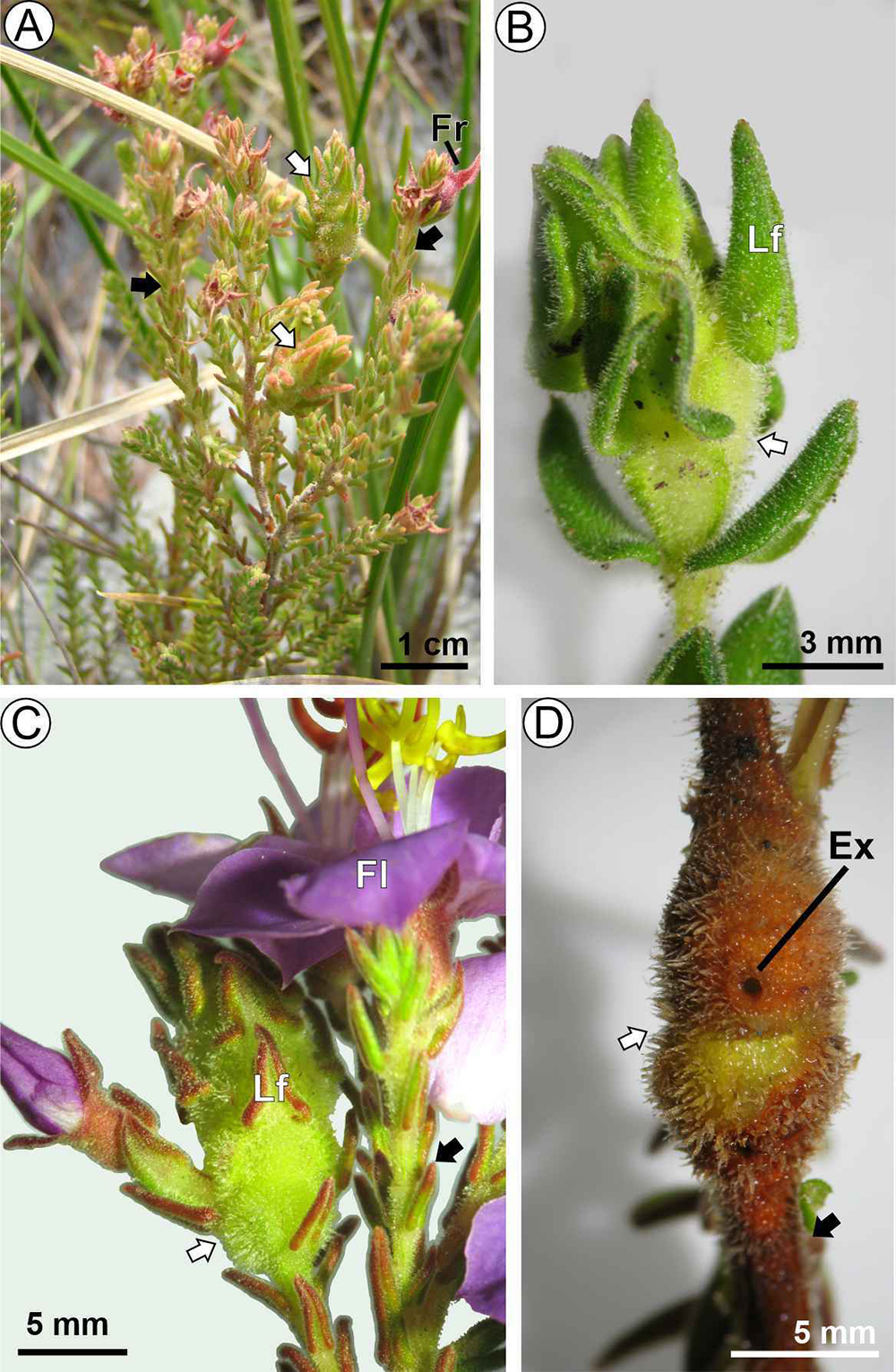
Distinct of leaves, the stems elongate predominantly in an anticlinal direction, which could involve the highly methyl-esterified homogalacturonans (HGs) and galactans (Hwang & Kokini, 1991; McCartney et al., 2003; Xu et al., 2011). Consequently, stem galls are good models to test the role of pectins in cell elongation and the fate of the cell wall composition because the functional alterations occur mainly in a group of cells. In this context, the nutritive cells derived from pith cells in the Lepidoptera-induced stem galls on Marcetia taxifolia (A.St.-Hil.) DC. (Melastomataceae) (Fig. 1 A ) (Ferreira & Isaias, 2013; Ferreira et al., 2015) are interesting models for examining how pectin dynamics contribute to the cellular adaptations that support gall development. Marcetia taxifolia is a plant adapted to the vegetation of Campos Rupestres (rocky fields) in Minas Gerais, Brazil. It is suited to extreme sunlight exposure and drought conditions during the day, cold nights, and foggy mornings (Alves et al., 2014; Oliveira et al., 2016). Based on studies on the same host plant-galling insect system, it is possible to establish parallels among cytological (Ferreira et al., 2015), histochemical peculiarities (Ferreira & Isaias, 2013), and cell wall functional composition of non-galled stems and lepidopteran galls. We expect that the cytological features of cell walls of gall nutritive tissues, with multivesicular bodies and thin and polylamellated cell walls (Ferreira & Isaias, 2013; Ferreira et al., 2015) are associated with a pectin composition that allows cell replication and nutrient availability to the gall inducer. We anticipate that the stress induced by the gall-inducing Lepidoptera alters the cell wall composition and dynamics of non-galled organs. Additionally, we expect the gall nutritive tissue to show high levels of HGs, galactans, and arabinans due to its hyperplasic characteristics.
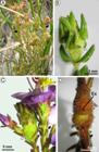
Stem galls on Marcetia taxifolia (Melastomataceae). A) Plant habit and stem galls. B) Gall in the growth and development phase (GGD). C) Gall in the maturation phase (GM). D) Gall in the senescence phase (GS). Black arrows: non-galled stems; White arrows = galls. Abbreviations: Ex = exit hole; Fl = flower; Fr = fruit. Lf = leaf.
Material and Methods
Sampling. Stems of Marcetia taxifolia (A.St.-Hil.) DC. (Melastomataceae) in primary growth (SP), secondary growth (SS), and fusiform stem galls induced by a microlepidopteran in the growth and development phase (GGD) (Fig. 1 B ), maturation (GM) (Fig. 1 C), and senescence (GS) (Fig. 1 D) were collected (n = 4 per category) from distinct individuals of the “crystal-pink” population (sensuGardoni et al., 2007) (19º17’52,5” S 043º35’27,8” W; 1321 m) at Serra do Cipó, Minas Gerais state, Brazil. The developmental phases were defined according to Ferreira & Isaias (2013), as follows: the GGD had 2-4 mm thickness, the GM was larger (4-6 mm), and the GS had the presence of an exit hole. A voucher of the fertile material (specimen with flowers) and with the Lepidoptera stem galls (with the gall inducer) was deposited in the BHCB herbarium under the number 161778.
Fixation and microtomy. Hemi-sectioned fragments of each sample were fixed on 2.5% glutaraldehyde and 4.5% formaldehyde in 0.1M, pH 7.2 PBS (phosphate buffered saline) (Karnovsky, 1965), dehydrated in n-butyl-series, and embedded in Paraplast® (Kraus & Arduin, 1997). The blocks were sectioned in a rotary microtome (12 µm) and fixed on the slides with Haupt adhesive (Haupt, 1930; Kraus & Arduin, 1997). The slides were dried on a 42º C hotplate, deparaffinized with butyl-acetate, and hydrated in ethyl-series (Kraus & Arduin, 1997). For anatomical analyses, the hydrated slides were stained with a 9:1 mixture of 0.5% astra blue and 0.5% safranin (Bukatsch, 1972), dehydrated in ethyl series followed by butyl acetate (Kraus & Arduin, 1997), and mounted in Acrilex® colorless varnish (Paiva et al., 2006). Another set of fixed plant fragments was dehydrated in ethyl series and embedded in glycol methacrylate Leica Historesin®. The historesin blocks were sectioned in rotary microtome (7 µm), and the slides were stained with toluidine O blue (O’Brien et al., 1964) and mounted in Entellan®.
Immunocytochemical analyses. Sections of Paraplast blocks were affixed in slides, deparaffinized, and hydrated. The hydrated slides were incubated for 30 min in 3% powder milk Molico® diluted in 0.1M PBS, pH 7.1, followed by incubation for two hours in monoclonal antibodies (MAbs) (Table 1) diluted in 3% powder milk/PBS (1:10). The MAbs were obtained from the Centre for Plant Sciences, University of Leeds, Leeds, UK. As a control, the slides were incubated in 3% powder milk in PBS without the MAbs. The slides were washed 5x in PBS, and incubated in a secondary antibody conjugated to FITC (fluorescein isothiocyanate) diluted in 3% powder milk/PBS (1:100) and kept in the dark for two hours. The slides were washed 5x in PBS, mounted with coverslips on 50% glycerin, and sealed with nail polish. The slides were analyzed under the Confocal Microscope Zeiss 510 META, Carl Zeiss, Germany, with 488 nm excitation of an argon laser and 505-530 nm emission filter.
Measurements of fluorescence and statistical analyses. The fluorescence peaks in the cell walls of distinct plant tissues were measured with the software MacBiophotonics ImageJ (Chomicki et al., 2014). The fluorescence intensities were calculated by the grayscale methodology (Gy = Gray value) along cell walls (from 5 cells each section; n = 3 samples) given by the software (Chomicki et al., 2014; Nogueira et al., 2024). The values obtained were compared by ANOVA in the SigmaStat® software, considering p ≤ 0.05 (Figs. S1-S3). Based on statistical analyses, the intensity of labeling was considered negative (-), weak (+), moderate (++), and intense (+++).
Results
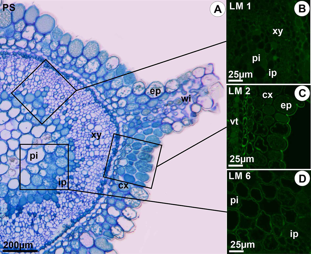
The stems of M. taxifolia on primary growth (SP) have an unstratified epidermis and 3-4 cell layers of homogeneous parenchyma on the cortex (Fig. 2 A ). The cortical layers have four wings containing chlorophyllian spongy parenchyma and small vascular traces (Fig. 2 A ). A continuous vascular cylinder, undivided in bundles, with phloem, xylem, and intraxylary phloem, surrounds a parenchymatic pith (Fig. 2 A ). Epitopes of extensins, recognized by LM1, were detected in all tissues of the SP (Table 2). These epitopes were intensely detected mainly in cell membranes of the cortical parenchyma and cell walls of the xylem, and moderately in the phloem and pith cell walls (Fig. 2 B ; Table 2). Epitopes of AGPs, recognized by LM2, were intensely detected in the cell walls and membranes of the xylem cells, moderately detected in the cortex, and weakly in the phloem and pith (Fig. 2 C ; Table 2). The epitopes of galactans, recognized by LM5, were weakly detected in the cell walls of all tissues except the phloem, where they were moderately labeled (Table 2). The epitopes of arabinans were weakly labeled by LM6 in all tissues except the xylem, where they were intensely labeled, mainly in inner cell wall layers of fibers and parenchymatic rays (Fig. 2 D ; Table 2). High methyl-esterified HGs, recognized by JIM7, were moderately detected in the cortex, phloem, xylem, and pith, while the low methyl-esterified HGs were moderately labeled by JIM5 only in the epidermis (Table 2).
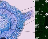
Distribution of extensins and some pectins in stems in primary growth (SP) in Marcetia taxifolia (Melastomataceae). A) Light micrograph with SP stained with toluidine O blue. B) Extensins marked by LM1 in intraxylary phloem (ip), pith (pi) and xylem (xy). C) Arabinogalactan-proteins (AGPs) marked by LM2 are shown in cell walls and membranes of the epidermis (ep) and cortical parenchyma (cx) of SP. D) Arabinans marked by LM6 are shown in the pith (pi) and intraxylary phloem (ip) of SP. Abbreviation: wi = one of the four wings that occurs in the stems of M. taxifolia.
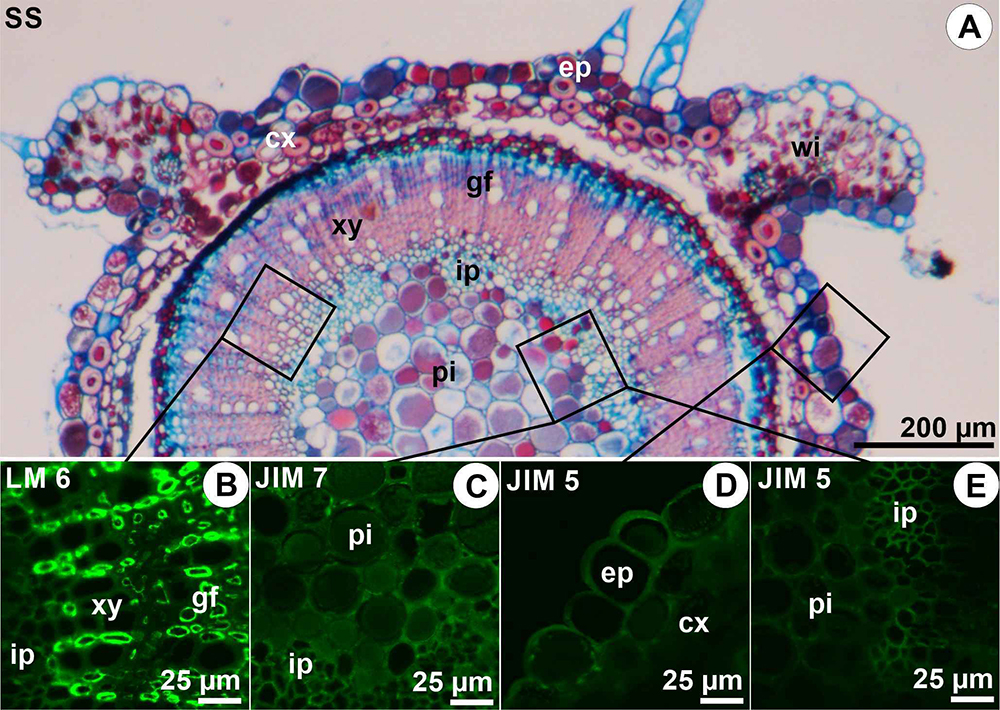
The secondary growth in the stems (SS) is established by a typical activity of the vascular cambium, generating secondary xylem with lignified parenchyma rays, and gelatinous fibers (Fig. 3 A ). The LM1 does not label the extensins in SS, while the LM2 intensely labeled the epitopes of AGPs in the xylem (in the middle lamellae), moderately in the cortex, and weakly in phloem and pith (Table 2). The epitopes of galactans, recognized by LM5, were moderately detected in the phloem, and weakly detected in the cortex (Table 2). The epitopes of arabinans recognized by LM6 were intensely detected in the xylem, mainly in gelatinous fibers, and weakly in the epidermis, cortex, phloem, and pith, especially in the inner layers of cell walls (Fig. 3 B ; Table 2). The epitopes of the high methyl-esterified HGs were intensely detected in the epidermis, cortex, and phloem, and moderately detected in the pith (Fig. 3 C ; Table 2), while the low methyl-esterified HGs were intensely detected in the phloem (Fig. 3 D ), moderately detected in the pith, and weakly detected in the epidermis, cortex (Fig. 3 E ), and xylem (Table 2). In the pith cells, the HGs were marked mainly in the inner cell wall layers (Fig. 3 C ).
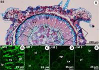
Distribution of some pectins in stems in secondary growh (SS) of Marcetia taxifolia (Melastomataceae). A) Light micrograph with SS stained with safranin and astra blue. B) Arabinans marked by LM6 in secondary xylem (xy), gelatinous fibers (gf), and ray parenchyma of SS. C) High methyl-esterified HGs (homogalacturonans) labeled by JIM7 in pith (pi) and intraxylary phloem (ip) of SS. D-E) Low methyl-esterified HGs (homogalacturonans) labeled by JIM5 in the epidermis (ep), intraxylary phloem (ip), and pith (pi) of SS. Abbreviation: wi = one of the four wings that occurs in the stems of M. taxifolia.
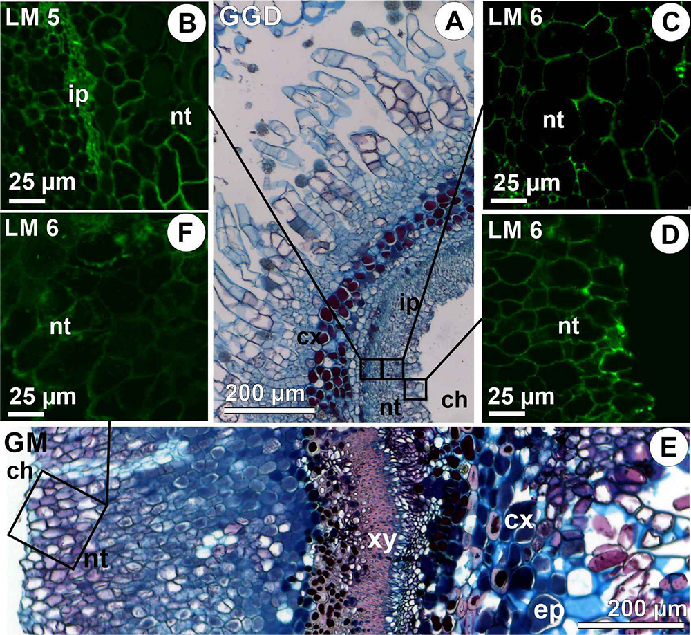
The stem galls are induced by a larva of an unidentified species of Lepidoptera in the pith of the stem in primary growth. The gall develops from cell hypertrophy and hyperplasia of the epidermis, cortical, vascular, and pith parenchyma (Fig. 4 A ). The larva lodges into a larval chamber within the pith cells, which are distinct from those of the stem, for they are hyperplasic in several layers, and redifferentiate into nutritive cells. The galls in growth and development phase (GGD) have an unstratified epidermis with dense and hypertrophied trichomes, a cortex with parenchymatic hypertrophic cells, a continuous vascular cylinder with phloem, xylem, and intraxylary xylem, and a hyperplasic nutritive tissue (Fig. 4 A ). The epitopes of extensins were moderately detected in the xylem of GGD, and weakly detected in the phloem and nutritive tissue (Table 2). The epitopes of AGPs were weakly detected only in the nutritive tissue (Table 2). The epitopes of galactans were intensely detected in phloem and moderately detected in the epidermis, cortex, xylem, and nutritive tissues (Table 2; Fig. 4 B ). The epitopes of arabinans were weakly detected by LM6 in all tissues (Fig. 4 C -D; Table 2). The epitopes of the high methyl-esterified HGs were moderately detected in the xylem, while those of the low methyl-esterified HGs were weakly detected in the epidermis and cortex (Table 2).
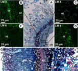
Distribution of some pectins in galls in the growth and development phase (GGD) and maturation (GM) induced by Lepidoptera on Marcetia taxifolia (Melastomataceae). A) Light micrograph with transversal section of GGD. B) Galactans marked by LM5 in intraxylary phloem (ip) and nutritive tissue. C-D) Arabinans marked by LM6 are shown in the nutritive tissue (nt) of GGD. E) Transversal section of GM stained with safranin and astra blue. F) Arabinans in the nutritive tissue (nt) of GM. Abbreviations: ch = larval chamber; cx = cortex; ep = epidermis; xy = xylem.
The galls in the maturation phase (GM) have vascular cambium activity, and lignification of some cortical cells, which are hypertrophied (Fig. 4 E ). The nutritive tissue is multilayered, with dividing cells into the outermost layers and vacuolated cells in the innermost layers in contact with the larva (Fig. 4 E ). On this phase, the epitopes of extensins were weakly detected only in the xylem, while the epitopes of AGPs were not detected in any cell type (Table 2). The epitopes of galactans were moderately detected in the nutritive tissue, and weakly detected in the epidermis, cortex, phloem, and xylem (Table 2). The epitopes of arabinans were weakly detected in the epidermis, phloem, xylem, and nutritive tissue (Fig. 4 F; Table 2). The high methyl-esterified HG epitopes were moderately detected only in nutritive tissue, while those of the low methyl-esterified HGs were intensely detected in the nutritive tissue, and weakly detected in the cortex, phloem, and xylem (Table 2).
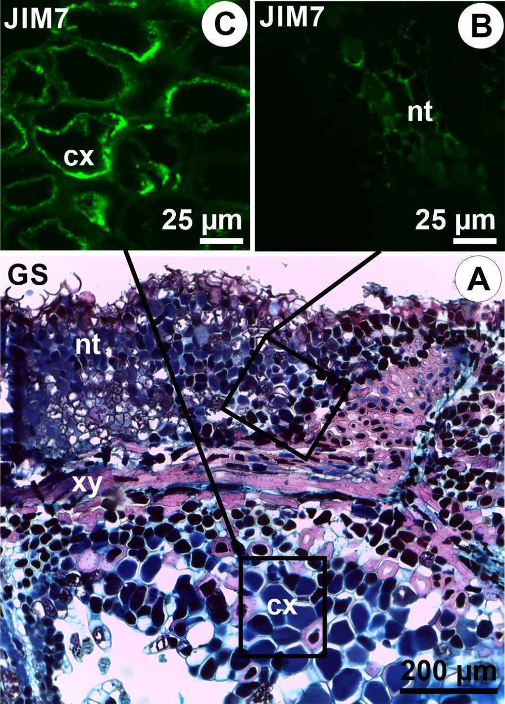
The galls reach senescence (GS) at the beginning of the lepidopteran pupal development, and the cells of the nutritive tissue enter autolysis (Fig. 5 A ). During this phase, the epitopes of extensins and AGPs were not detected. The epitopes of galactans were moderately detected in phloem and nutritive tissue, and weakly detected in the cortex and xylem (Table 2). The epitopes of arabinans were weakly detected in all tissues (Table 2). The high methyl-esterified HG epitopes were moderately detected in the cortex and nutritive tissue (Fig. 5 B ) and weakly detected in the epidermis and phloem (Fig. 5 C ; Table 2), while the epitopes of the low methyl-esterified HGs were detected only in the xylem (Table 2).
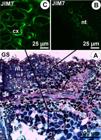
Distribution of some pectins in senescent galls (GS) of Marcetia taxifolia (Melastomataceae). A) Light micrograph with transversal section of GS stained with safranin and astra blue. B-C) High methyl-esterified HGs (homogalacturonans) labeled by JIM7 in the cytoplasm of cortical cells and cell walls of nutritive tissue in GS. Abbreviations: cx = cortex; nt = nutritive tissue; xy = xylem.
Discussion
Microlepidopteran galls on M. taxifolia induce hypertrophy in the epidermis and cortex, along with hyperplasia and redifferentiation of pith cells into nutritive tissues. These alterations lead to the stem swelling without drastic changes in tissue arrangements, in accordance with the results found by Ferreira & Isaias (2013). The cell walls of the nutritive tissues, derived from pith parenchyma in non-galled stems, show distinct composition, confirming previous descriptions of cell redifferentiation in M. taxifolia galls (Ferreira & Isaias, 2013). The walls of gall nutritive cells demonstrate a richer chemical profile than the other tissues, with a combination of arabinans, galactans, and low and high methyl-esterified HGs, similar to the secondary phloem in SS. The clear distinction in the pectin composition of the galls denotes the development of a new functional design, as previously proposed on the morphological basis for other insect galls (Shorthouse et al., 2005; Oliveira & Isaias, 2010; Oliveira et al., 2011; Ferreira & Isaias, 2014). The epitopes of arabinans were intensely detected in the xylem of non-galled stems, especially on gelatinous fibers, but they were weakly detected in galls. The immunocytochemical profiles of M. taxifolia stem in primary and secondary growth are markedly distinct from those of lepidopteran galls in growth and development, maturation, and senescent phases, demonstrating changes in functional profiles in galled condition. While the epitopes of arabinogalactan proteins (AGPs), extensins, and arabinans were more intensely detected in the non-galled stems, the galactans were more intensely detected in gall tissues, revealing changes toward cell wall reinforcement in hyperplasic conditions. The epitopes of HGs were widely labeled in the non-galled tissues, especially in the pith, but additional pectins were detected in the nutritive tissue, which enhances porosity and reinforces the cell walls, facilitating the flow of water and macromolecules toward the gall inducer (Silva et al., 2021), while also providing strength to the cell walls in response to the chewing habits of the lepidopteran larva. This reinforcement is crucial as it helps maintain the structural integrity of the tissue, allowing it to withstand the mechanical stress caused by feeding, which in turn supports the gall's overall health and functionality.
As expected, the nutritive cells of the Lepidoptera galls on M. taxifolia demonstrate a great variety of intensely detected pectins, as also observed in galls of Palaeomystella oligophaga Becker & Adamski (Lepidoptera) on Macairea radula (Bonpl.) DC. (Melastomataceae) (Santos et al., 2024). The meristematic nature of the storage nutritive tissue (Ferreira et al., 2015), which replaces the cells of the typical nutritive tissue, is associated with the galactan composition of the cell walls and with the presence of the multivesicular bodies associated with cell membranes (Ferreira et al., 2015). Distinct from other insect galls, the lepidopteran galls have an intense hyperplasia of nutritive cells also in the maturation phase, due to the voracious chewing habit of the larva (Ferreira et al., 2015; 2022). Therefore, the combination of galactans, arabinans, and high methyl-esterified HGs confer flexibility and stability to the hyperplasic cells in different gall systems (Carneiro et al., 2014; Teixeira et al., 2018). Moreover, this composition confers porosity to the cell walls (Zykwinska et al., 2005), allowing the free flow of macromolecules and water between the storage and the typical nutritive tissues of the lepidopteran galls (Santos et al., 2024). Such patterns were also observed in the nutritive tissues of mite and nematode galls (Ferreira et al., 2020), nutritive-like parenchyma of aphid galls (Nogueira et al., 2024), and senescent eriococcid galls (Silva et al., 2021), demonstrating the importance of this combination of pectins for the water and metabolite translocation in galls. In the case of Espinosa nothofagi (Pteromalidae, Hymenoptera) gall tissues, although no pectins or hemicelluloses were detected in the nutritive tissues by immunocytochemistry, the presence of galactans, arabinans, and HGs in the outer storage tissues was linked to facilitating nutrient flow to the nutritive cells (Guedes et al., 2025). Such combination can also explain the opposite gradients of carbohydrates and lipids in M. taxifolia galls, accumulated differentially in the two zones of the nutritive tissue (Ferreira & Isaias, 2013), suggesting a conversion of starch into reducing sugars, followed by their conversion into lipids, which are together considered the main food source of energy to the galling lepidopterans (Guedes et al., 2023; Martins et al., 2023).
The developmental changes in the cell walls, accessed by immunocytochemical analysis, and detected in the galls, may indicate new functional aspects not revealed by the histological analyses. The detection of galactans was more intense in the GGD than in the non-galled stems, which relates to cell wall stiffening during elongation and expansion (Hwang & Kokini, 1991; McCartney et al., 2003; Xu et al., 2011). The hyperplasic tissues in galls have galactan-rich cell walls (Carneiro et al., 2014; Teixeira et al., 2018), as herein observed in the GGD, for the epidermis, cortex, and nutritive tissues of M. taxifolia galls (Ferreira & Isaias, 2013). The maintenance of galactans in the nutritive tissues in the GM corroborates their importance in hyperplasic tissues, since nutritive cells still divide during gall maturation, but have shifted elongation patterns in comparison with the pith cells (Ferreira & Isaias, 2013). On the other hand, the high detection of the epitopes of galactans in the walls of phloem cells is enough to confer the stiffening and porosity of these walls, as proposed by Zykwinska et al. (2005), which have been related to the detection of galactans and arabinans in other galls.
Several studies have suggested that the low methyl-esterified HGs occur mainly in mature organs because the de-methyl-esterification releases the HG molecules sites that may bind to the calcium ions and confer rigidity to the plant cell walls (Knox et al., 1990; Sabba & Lulai, 2005; Mastroberti & Mariath, 2008; Formiga et al., 2013; Carneiro et al., 2014; Oliveira et al., 2014). The epitopes of HGs, in general, were slightly detected in the SP and the GGD, with a predominance of high methyl-esterified form. The loss of methyl-esterified groups in mature organs has been associated with the reinforcement of the cell walls in plant organs (Mastroberti & Mariath, 2008), including galls in the maturation phase (Formiga et al., 2013; Carneiro et al., 2014; Oliveira et al., 2014). This imbalance of HG forms may be associated with the defense against mechanical damage. However, in M. taxifolia stem galls, the epitopes of the HGs occur in weak intensity when compared to the non-galled stems, except for the nutritive cells. This peculiar chemical alteration of the lepidopteran stem galls on M. taxifolia in relation to the non-galled stems can be compensated by the presence of galactans, giving a distinct functional profile to the stem galls.
An increase of the high methyl-esterified HGs, both from SP to SS, and from GGD to GM, was also observed in the host leaves and associated galls on Baccharis dracunculifolia DC. (Asteraceae) (Oliveira et al., 2014) and Psidium myrtoides O.Berg (Myrtaceae) (Carneiro et al., 2014). The recycling of low methyl-esterified HGs and the production of new high methyl-esterified HGs in M. taxifolia likely maintain protection and rigidity in mature cell walls. Also, the detection of high methyl-esterified HGs in galls induced by Lepidoptera on M. taxifolia in higher intensity in senescent phase (GS) than in the GM and GGD may be indicative that there is a decrease in the activity of the PMEs due to the galling stress. The increase in the levels of high methyl-esterified HGs in senescent galls was also observed in other insect galls (Oliveira et al., 2014; Carneiro et al., 2014) and does not seem to be a consequence of the insect mode of feeding. The decline of the PMEs activity has been attributed to an increase of ethylene in ripening fruits (Awad & Young, 1980; Kanellis et al., 1989; Wegrzyn & MacRae, 1992; Pelloux et al., 2007), which is also related to ROS signaling in several plant processes (D’Haeze et al., 2003; Desikan et al., 2005). ROS and ethylene are important to induce the first steps of gall development and the maintenance of its structure (Isaias et al., 2015). The signaling of ROS followed by ethylene production can explain the detection of high methyl-esterified HGs in the senescent lepidopteran galls on M. taxifolia.
The cell walls of the primary growth stem (SP) of M. taxifolia have more intense detection of the epitopes of extensins and arabinogalactan-proteins (AGPs) than the SS and galls. The extensins may reinforce and confer elasticity to the cell walls during the growth and development phase, as well as control programmed cell death (PCD), and pathogens infection, as previously proposed (Showalter, 1993; Gao & Showalter, 1999; Chaves et al., 2002; Seifert & Roberts, 2007; Albersheim et al., 2010; Leroux et al., 2011). Primary stems have flexible and strong cell walls, for they are structures in the elongation process, which sustains new shoots and leaves. Thus, cell flexibility is important for the host organ elongation, without the collapse of the tissues. After the initiation of the secondary growth in the stems (SS), there is a weak expression of extensins, but maintenance of the AGPs in the secondary xylem. This chemical profile should be a consequence of the end of the cell elongation in the SS, where the xylem fibers and parenchyma cells seem to be alive. The AGPs, which were highly detected in these cells of the SS, seem to play a defensive role against PCD. Additionally, the arabinans with intense peaks in the cell walls of xylem cells in PS and SS, when linked to the RG-II, are responsible for the defense against drought stress (Moore et al., 2008). The presence of arabinans in the inner layers of gelatinous fibers of M. taxifolia supports the proposal of Evert (2006), for the G-layers of such type of cells can absorb a great quantity of water. The absence of gelatinous fibers in galls, along with the weaker detection of arabinans in xylem cells, may indicate alterations in this functional aspect within the galled portions. Additionally, the presence of arabinans in nutritive and storage tissue cells is crucial for maintaining water in gall cells (see Guedes et al., 2025), which is necessary for the continuity of cell divisions and growth. Further investigation is needed into potential compensatory mechanisms for the hydration of gall tissues, including possible changes in vascular cell histometry and density both inside and adjacent to the galls (Nobrega et al., 2023).
In summary, the galling activity of lepidopteran larvae on M. taxifolia stems induces significant alterations in cell structure and biochemical composition, particularly in the pectin profiles of stem tissues. The hypertrophy and hyperplasia of nutritive cells lead to increased accumulation of galactans, enhancing flexibility and structural reinforcement of the galled tissues. The accumulation of HGs, galactans, and arabinans in storage and nutritive cells facilitates water and nutrient flow, supporting gall development. However, the reduced detection of arabinans in galled xylem suggests a potential impairment of the host plant's water storage capabilities, which may negatively impact its resilience to environmental stressors. Further investigations are necessary to explore compensatory mechanisms in the hydration of gall tissues and their ecological implications.
Acknowledgments
Coordenação de Aperfeiçoamento de Pessoal de Nível Superior (CAPES, Brazil) (Finance Code 001). Conselho Nacional de Desenvolvimento Científico e Tecnológico (CNPq, Brazil) for the research grant to BGF (310520/2023-1), DCO (303691/2022-0), and RMSI (309713/2023-4). Fundação Carlos Chagas Filho de Amparo à Pesquisa do Estado do Rio de Janeiro (FAPERJ, Rio de Janeiro, Brazil) for financial support of the project (E-26/211.608/2021; E-26/210.705/2023; E-26/204.484/2024).
References
-
Agoda-Tandjawa G, Durand S, Gaillard C, Garnier C, Doublier JL. 2012. Rheological behaviour and microstructure of microfibrillated cellulose suspensions/low-methoxyl pectin mixed systems. Effect of calcium ions. Carbohydrate Polymers 87: 1045-1057. doi: 10.1016/j.carbpol.2011.08.021
» https://doi.org/10.1016/j.carbpol.2011.08.021 - Albersheim P, Darvill A, Roberts K, Sederoff R, Staehelin A. 2010. Plant Cell Walls. New York, Garland Science/Taylor and Francis Group.
-
Alves RJV, Silva NG, Oliveira JA, Medeiros D. 2014. Circumscribing campo rupestre - megadiverse Brazilian rocky montane savanas. Brazilian Journal of Biology 74: 355-362. doi: 1519-6984.23212
» https://doi.org/10.1590/1519-6984.23212 -
Awad M, Young RE. 1980. Avocado pectinmethylesterase activity in relation to temperature, ethylene, and ripening. Journal of the American Society for Horticultural Science 105: 638-641. doi: 10.21273/JASHS.105.5.638
» https://doi.org/10.21273/JASHS.105.5.638 -
Bragança GPP, Alencar CF, Freitas MSC, Isaias RMS. 2020. Hemicelluloses and associated compounds determine gall functional traits. Plant Biology 22: 981-991. doi: 10.1111/plb.13151
» https://doi.org/10.1111/plb.13151 -
Boanares D, Ferreira BG, Kozovits AR, Sousa HC, Isaias RMS, França MGC. 2018. Pectin and cellulose cell wall composition enables different strategies to leaf water uptake in plants from tropical fog mountain. Plant Physiology and Biochemistry 122: 57-64. doi: 10.1016/j.plaphy.2017.11.005
» https://doi.org/10.1016/j.plaphy.2017.11.005 - Bukatsch F. 1972. Bermerkungen zur Doppelfärbung Astrablau-Safranin. Mikrokosmos 61: 255.
-
Caffall KH, Mohnen D. 2009. The structure, function, and biosynthesis of plant cell all pectic polysaccharides. Carbohydrate Research 344: 1879-1900. doi: 10.1016/j.carres.2009.05.021
» https://doi.org/10.1016/j.carres.2009.05.021 -
Castilleux R, Plancot B, Gügi B et al 2020. Extensin arabinosylation is involved in root response to elicitors and limits oomycete colonization. Annals of Botany 125: 751-763. doi: 10.1093/aob/mcz068
» https://doi.org/10.1093/aob/mcz068 -
Carneiro RGS, Oliveira DC, Isaias RMS. 2014. Developmental anatomy and immunocytochemistry reveal the neo-ontogenesis of the leaf tissues of Psidium myrtoides (Myrtaceae) towards the globoid galls of Nothotrioza myrtoidis (Triozidae). Plant Cell Reports 33: 2093-2106. doi: 10.1007/s00299-014-1683-7
» https://doi.org/10.1007/s00299-014-1683-7 -
Chaves I, Regalado AP, Chen M, Ricardo CP, Showalter AM. 2002. Programmed cell death induced by (b-D-galactosyl)3 Yariv reagent in Nicotiana tabacum BY-2 suspension-cultured cells. Physiologia Plantarum 116: 548-553. doi: 10.1034/j.1399-3054.2002.1160414.x
» https://doi.org/10.1034/j.1399-3054.2002.1160414.x -
Chomicki G, Bidel LPR, Jay-Allemand C. 2014. Exodermis structure controls fungal invasion in the leafless epiphytic orchid Dendrophylax lindenii (Lindl.) Benth. ex Rolfe. Flora 209: 88-94. doi: 10.1016/j.flora.2014.01.001
» https://doi.org/10.1016/j.flora.2014.01.001 -
Cosgrove DJ. 2016. Plant cell wall extensibility: Connecting plant cell growth with cell wall structure, mechanics, and the action of wall-modifying enzymes. Journal of Experimental Botany 67: 463-476. doi: 10.1093/jxb/erv511
» https://doi.org/10.1093/jxb/erv511 -
D’Haeze W, De Rycke R, Mathis R et al 2003. Reactive oxygen species and ethylene play a positive role in lateral root base nodulation of a semiaquatic legume. PNAS 100: 11789-11794. doi: 10.1073/pnas.1333899100
» https://doi.org/10.1073/pnas.1333899100 -
Desikan R, Hancock JT, Bright J et al 2005. A role for ETR1 in hydrogen peroxide signaling in stomatal guard cells. Plant Physiology 137: 831-834. doi: 10.1104/pp.104.056994
» https://doi.org/10.1104/pp.104.056994 -
Douchiche O, Driouich A, Morvan C. 2010. Spatial regulation of cell-wall structure in response to heavy metal stress: cadmium-induced alteration of the methyl-esterification pattern of homogalacturonans. Annals of Botany 105: 481-491. doi: 10.1093/aob/mcp306
» https://doi.org/10.1093/aob/mcp306 - Evert RF. 2006. Esau's plant anatomy: Meristems, cells, and tissues of the plant body: their structure, function, and development. 3rd. edn. Hoboken, John Wiley & Sons.
-
Ferreira BG, Isaias RMS. 2013. Developmental stem anatomy and tissue redifferentiation induced by a galling Lepidoptera onMarcetia taxifolia(Melastomataceae). Botany 91: 752-760. doi: 10.1139/cjb-2013-0125
» https://doi.org/10.1139/cjb-2013-0125 -
Ferreira BG, Isaias RMS. 2014. Flora l-like destiny induced by a galling Cecidomyiidae on the axillary buds of Marcetia taxifolia (Melastomataceae). Flora 209: 391-400. doi: 10.1016/j.flora.2014.06.004
» https://doi.org/10.1016/j.flora.2014.06.004 -
Ferreira BG, Carneiro RGS, Isaias RMS. 2015. Multivesicular bodies differentiate exclusively in nutritive fast-dividing cells in Marcetia taxifolia galls. Protoplasma 252: 1275-1283. doi: 10.1007/s00709-015-0759-8
» https://doi.org/10.1007/s00709-015-0759-8 -
Ferreira BG, Álvarez R, Bragança GP, Alvarenga DR, Hidalgo NP, Isaias RMS. 2019. Feeding and other gall facets: patterns and determinants in galls structure. The Botanical Review 85: 78-106. doi: 10.1007/s12229-019-09207-w
» https://doi.org/10.1007/s12229-019-09207-w -
Ferreira BG, Bragança GP, Isaias RMS. 2020. Cytological attributes of storage tissues in nematode and eriophyid galls: Pectin and hemicellulose functional insights. Protoplasma 257: 229-244. doi: 10.1007/s00709-019-01431-w
» https://doi.org/10.1007/s00709-019-01431-w -
Ferreira BG, Moreira GRP, Isaias RMS, Carneiro RGS. 2022. Complex meristematic activity induced by Eucecidoses minutanus on Schinus engleri turns shoots into galls. American Journal of Botany 109: 209-225. doi: 10.1002/ajb2.1798
» https://doi.org/10.1002/ajb2.1798 -
Formiga AT, Oliveira DC, Ferreira BG et al 2013. The role of pectic composition of cell walls in the determination of the new shape-functional design in galls of Baccharis reticularia (Asteraceae). Protoplasma 250: 899-908. doi: 10.1007/s00709-012-0473-8
» https://doi.org/10.1007/s00709-012-0473-8 -
Formiga AT, Silveira FAO, Fernandes GW, Isaias RMS. 2015. Phenotypic plasticity and similarity among gall morphotypes on a superhost, Baccharis reticularia (Asteraceae). Plant Biology 17: 512-521. doi: 10.1111/plb.12232
» https://doi.org/10.1111/plb.12232 -
Gao M, Showalter AM. 1999. Yariv reagent treatment induces programmed cell death in Arabidopsis cell cultures and implicates arabinogalactan protein involvement. Plant Journal 19: 321-331. doi: 10.1046/j.1365-313x.1999.00544.x
» https://doi.org/10.1046/j.1365-313x.1999.00544.x -
Gardoni LCP, Isaias RMS, Vale FHA. 2007. Morfologia e anatomia foliar de três morfotipos de Marcetia taxifolia (A. St.-Hil.) DC. (Melastomataceae) na Serra do Cipó, MG. Revista Brasileira de Botânica 30: 487-500. doi: 10.1590/S0100-84042007000300013
» https://doi.org/10.1590/S0100-84042007000300013 -
Guedes LM, Costa EC, Isaias RMS, Sáez-Carillo K, Aguilera N. 2023. Structural patterns of Lepidoptera galls and the case of Andescecidium parrai (Cecidosidae) galls on Schinus polygama (Anacardiaceae). Journal of Plant Research 136: 715-728. doi: 10.1007/s10265-023-01472-6
» https://doi.org/10.1007/s10265-023-01472-6 -
Guedes LM, Aguilera N, Kuster VC, Carneiro RGS, Oliveira DC. 2025. Integrated insights into the cytological, histochemical, and cell wall composition features ofEspinosa nothofagi(Hymenoptera) gall tissues: implications for functionality. Protoplasma 262: 149-165. doi: 10.1007/s00709-024-01985-4
» https://doi.org/10.1007/s00709-024-01985-4 -
Haupt AW. 1930. A gelatin fixative for parafin sections. Stain Technology 5: 97-98. doi: 10.3109/10520293009115555
» https://doi.org/10.3109/10520293009115555 -
Hwang J, Kokini JL. 1991. Structure and rheological function of side branches of carbohydrate polymers. Journal of Texture Studies 22: 123-167. doi: 10.1111/j.1745-4603.1991.tb00011.x
» https://doi.org/10.1111/j.1745-4603.1991.tb00011.x -
Isaias RMS, Oliveira DC, Moreira AS, Soares GLG, Carneiro RGS. 2015. The imbalance of redox homeostasis in arthropod-induced plant galls: Mechanisms of stress generation and dissipation. Biochimica et Biophysica Acta 1850: 1509-1517. doi: 10.1016/j.bbagen.2015.03.007
» https://doi.org/10.1016/j.bbagen.2015.03.007 -
Jones L, Seymour GB, Knox JP. 1997. Localization of pectic galactan in tomato cell walls using a monoclonal antibody specific to (1→4) β-D-galactan. Plant Physiology 113: 1405-1412. doi: 10.1104/pp.113.4.1405
» https://doi.org/10.1104/pp.113.4.1405 -
Kanellis AK, Solomos T, Mattoo AK. 1989. Changes in sugars, enzymatic activities and acid phosphatase isoenzyme profiles of bananas ripened in air or stored in 2.5% O2 with and without ethylene. Plant Physiology 90: 251-258. doi: 10.1104/pp.90.1.251
» https://doi.org/10.1104/pp.90.1.251 - Karnovsky MJ. 1965. A formaldehyde-glutaraldehyde fixative of high osmolarity for use in electron microscopy. Journal of Cell Biology 27: 137-138.
-
Knox JP, Linstead PJ, King J, Cooper C, Roberts K. 1990. Pectin esterification is spatially regulated both within cell walls and between developing tissues of roots apices. Planta 181: 512-521. doi: 10.1007/BF00193004
» https://doi.org/10.1007/BF00193004 -
Knox JP. 1992. Cell adhesion, cell separation and plant morphogenesis. The Plant Journal 2: 137-141. doi: 10.1111/j.1365-313X.1992.00137.x
» https://doi.org/10.1111/j.1365-313X.1992.00137.x - Kraus JE, Arduin M. 1997. Manual básico de métodos em morfologia vegetal. Seropédica, Editora da Universidade Federal Rural do Rio de Janeiro.
-
Lahaye M, Falourd X, Laillet B, Le Gall S. 2020. Cellulose, pectin and water in cell walls determine apple flesh viscoelastic mechanical properties. Carbohydrate Polymers 232: 115768. doi: 10.1016/j.carbpol.2019.115768
» https://doi.org/10.1016/j.carbpol.2019.115768 -
Leroux O, Knox JP, Masschaele B et al 2011. An extensin-rich matrix lines the carinal canals in Equisetum ramosissimum, which may function as water-conducting channels. Annals of Botany 108: 307-319. doi: 10.1093/aob/mcr161
» https://doi.org/10.1093/aob/mcr161 - Lev-Yadun S. 2003. Stem cells in plants are differentiated too. Current Topics in Plant Biology 4: 93-102.
-
Lionetti V, Cervone F, Bellincampi D. 2012. Methyl esterification of pectin plays a role during plant-pathogen interactions and affects plant resistance to diseases. Journal of Plant Physiology 169: 1623-1630. doi: 10.1016/j.jplph.2012.05.006
» https://doi.org/10.1016/j.jplph.2012.05.006 -
Liu X, Cui H, Zhang B, Song M, Chen S, Xiao C, Tang Y, Liesche J. 2021. Reduced pectin content of cell walls prevents stress-induced root cell elongation in Arabidopsis Journal of Experimental Botany 72: 1073-1084. doi: 10.1093/jxb/eraa533
» https://doi.org/10.1093/jxb/eraa533 -
Marín-Rodriguez MC, Orchard J, Seymour GB. 2002. Pectate lyases, cell wall degradation and fruit softening. Journal of Experimental Botany 53: 2115-2119. doi: 10.1093/jxb/erf089
» https://doi.org/10.1093/jxb/erf089 -
McCartney L, Steele-King CG, Jordan E, Knox JP. 2003. Cell wall pectin (1-4)-D-galactan marks the acceleration of cell elongation in the Arabidopsis seedling root meristem. Plant Journal 33: 447-454. doi: 10.1046/j.1365-313x.2003.01640.x
» https://doi.org/10.1046/j.1365-313x.2003.01640.x -
Martini VC, Moreira ASFP, Kuster VC, Oliveira DC. 2019. Galling insects as phenotype manipulators of cell wall composition during the development of galls induced on leaves of Aspidosperma tomentosum (Apocynaceae). South African Journal of Botany 127: 226-233. doi: 10.1016/j.sajb.2019.09.006
» https://doi.org/10.1016/j.sajb.2019.09.006 -
Martins GS, Santos MG, Reis A, Ferreira BG. 2023. Alterations induced by Tortrimosaica polypodivora on the stems of Microgramma vacciniifolia: Simple or complex galls? Rodriguésia 74: e00532023. doi: 10.1590/2175-7860202374070
» https://doi.org/10.1590/2175-7860202374070 -
Mastroberti AA, Mariath JEA. 2008. Imunocytochemistry of the mucilage cells of Araucaria angustifolia (Bertol.) Kuntze (Araucariaceae). Brazilian Journal of Botany 31: 1-13. doi: 10.1590/S0100-84042008000100002
» https://doi.org/10.1590/S0100-84042008000100002 -
Micheli F. 2001. Pectin methylesterases: Cell wall enzymes with important roles in plant physiology. Trends in Plant Science 6: 414-419. doi: 10.1016/s1360-1385(01)02045-3
» https://doi.org/10.1016/s1360-1385(01)02045-3 -
Moore JP, Farrant JM, Driouich A. 2008. A role for pectin-associated arabinans in maintaining the flexibility of the plant cell wall during water deficit stress. Plant Signaling & Behavior 3: 102-104. doi: 10.4161/psb.3.2.4959
» https://doi.org/10.4161/psb.3.2.4959 -
Nobrega LP, Silva JB, Luna BN, Ferreira BG. 2021. Modulation of anatomical adaptations of leaves of Avicennia schaueriana (Acanthaceae) by a galling Meunieriella (Cecidomyiidae). Flora 274: e151750. doi: 10.1016/j.flora.2020.151750
» https://doi.org/10.1016/j.flora.2020.151750 -
Nobrega LP, Sá-Haiad B, Ferreira BG. 2023. Mechanisms of hydraulic conductivity in the leaf galls of Meunieriella sp. (Cecidomyiidae) in Avicennia schaueriana (Acanthaceae): Does vascularization explain the preferred sites of induction? Plant Biology 25: 198-207. doi: 10.1111/plb.13490
» https://doi.org/10.1111/plb.13490 -
Nogueira RM, Freitas MSC, Picoli EAT, Isaias RMS. 2024. Implications of cell wall immunocytochemical profiles on the structural and functional traits of root and stem galls induced by Eriosoma lanigerum on Malus domestica Protoplasma 261: 911-926. doi:10.1007/s00709-024-01939-w
» https://doi.org/10.1007/s00709-024-01939-w -
O’Brien TP, Feder N, McCully ME. 1964. Polychromatic Staining of Plant Cell Walls by Toluidine Blue O. Protoplasma 59: 368-373. doi: 10.1007/BF01248568
» https://doi.org/10.1007/BF01248568 -
Oliveira DC, Isaias RMS. 2010. Redifferentiation of leaflet tissues during midrib gall development in Copaifera langsdorffii (Fabaceae). South African Journal of Botany 76: 239-248. doi: 10.1016/j.sajb.2009.10.011
» https://doi.org/10.1016/j.sajb.2009.10.011 -
Oliveira DC, Magalhães TA, Carneiro RGS, Alvim MN, Isaias RMS. 2010. Do Cecidomyiidae galls of Aspidosperma spruceanum (Apocynaceae) fit the pre-established cytological and histochemical patterns? Protoplasma 242: 81-93. doi: 10.1007/s00709-010-0128-6
» https://doi.org/10.1007/s00709-010-0128-6 -
Oliveira DC, Carneiro RGS, Magalhães TA, Isaias RMS. 2011. Cytological and histochemical gradients on two Copaifera langsdorffii Desf. (Fabaceae) - Cecidomyiidae gall systems. Protoplasma 248: 829-837. doi: 10.1007/s00709-010-0258-x
» https://doi.org/10.1007/s00709-010-0258-x -
Oliveira DC, Magalhães TA, Ferreira BG et al 2014. Variation in the Degree of pectin methylesterification during the development of Baccharis dracunculifolia kidney-shaped gall. PLoS One 9: e94588. doi: 10.1371/journal.pone.0094588
» https://doi.org/10.1371/journal.pone.0094588 -
Oliveira RS, Abrahão A, Pereira C et al 2016. Ecophysiology of campos rupestres plants. In: Fernandes GW (ed.). Ecology and Conservation of Mountaintop Grasslands in Brazil. Switzerland, Springer International Publishing. p. 227-272. doi: 10.1007/978-3-319-29808-5_11
» https://doi.org/10.1007/978-3-319-29808-5_11 -
Paiva JGA, Fank-De-Carvalho SM, Magalhães MP, Graciano-Ribeiro D. 2006. Verniz vitral incolor 500®: uma alternativa de meio de montagem economicamente viável. Acta Botanica Brasilica 20: 257-264. doi: 10.1590/S0102-33062006000200002
» https://doi.org/10.1590/S0102-33062006000200002 -
Pelloux J, Rustérucci C, Mellerowicz EJ. 2007. New insights into pectin methylesterase structure and function. Trends in Plant Sciences 12: 267-277. doi: 10.1016/j.tplants.2007.04.001
» https://doi.org/10.1016/j.tplants.2007.04.001 -
Rezende UC, Cardoso JCF, Kuster VC, Goncalves LA, Oliveira DC. 2019. How the activity of natural enemies changes the structure and metabolism of the nutritive tissue in galls? Evidence from the Palaeomystella oligophaga (Lepidoptera) - Macairea radula (Metastomataceae) system. Protoplasma 256: 679-679. doi: 10.1007/s00709-018-1321-2
» https://doi.org/10.1007/s00709-018-1321-2 -
Sabba RP, Lulai EC. 2005. Immunocytological Analysis of Potato Tuber Periderm and Changes in Pectin and Extensin Epitopes Associated with Periderm Maturation. Journal of the American Society for Horticultural Science 130: 936-942. doi: 10.21273/JASHS.130.6.936
» https://doi.org/10.21273/JASHS.130.6.936 -
Santos PD, Bragança GPP, Kuster VC, Rocha DI, Oliveira DC. 2024. Cell wall epitope distribution in the functional compartments of galls induced by Palaeomystella oligophaga (Lepidoptera) in Macairea radula (Melastomataceae). Australian Journal of Botany 72: BT23064. doi: 10.1071/BT23064
» https://doi.org/10.1071/BT23064 -
Seifert GJ, Roberts K. 2007. The Biology of Arabinogalactan Proteins. Annual Review of Plant Biology 58: 137-161. doi: 10.1146/annurev.arplant.58.032806.103801
» https://doi.org/10.1146/annurev.arplant.58.032806.103801 -
Showalter AM. 1993. Structure and Function of Plant Cell Wall Proteins. Plant Cell 5: 9-23. doi: 10.1105/tpc.5.1.9
» https://doi.org/10.1105/tpc.5.1.9 -
Shorthouse JD, Wool D, Raman A. 2005. Gall-inducing insects - Nature’s most sophisticated herbivores. Basic and Applied Ecology 6: 407-411. doi: 10.1016/j.baae.2005.07.001
» https://doi.org/10.1016/j.baae.2005.07.001 -
Silva AFM, Lana LG, Kuster VC, Oliveira DC. 2021. Chemical composition of cell wall changes during developmental stages of galls on Matayba guianensis (Sapindaceae): Perspectives obtained by immunocytochemistry analysis. The Science of Nature 108: 16. doi: 10.1007/s00114-021-01732-2
» https://doi.org/10.1007/s00114-021-01732-2 -
Smallwood M, Yates EA, Willats WG, Martin H, Knox JP. 1996. Immunochemical comparison of membrane associated and secreted arabinogalactan-proteins in rice and carrot. Planta 198: 452-459. doi: 10.1007/BF00620063
» https://doi.org/10.1007/BF00620063 -
Teixeira CT, Oliveira DC, Kuster VC, Isaias RMS. 2018. Immunocytochemical demonstration of cell wall components related to tissue compartments in the globoid galls induced by Clinodiplosis sp. (Cecidomyiidae) on Croton floribundus Spreng. (Euphorbiaceae). Botany 96: 9-18. doi: 10.1139/cjb-2017-0123
» https://doi.org/10.1139/cjb-2017-0123 -
Vecchi C, Menezes NL, Oliveira DC, Ferreira BG, Isaias RMS. 2013. The redifferentiation of nutritive cells in galls induced by Lepidoptera on Tibouchina pulchra (Cham.) Cogn. reveals predefined patterns of plant development. Protoplasma 250: 1363-1368. doi: 10.1007/s00709-013-0519-6
» https://doi.org/10.1007/s00709-013-0519-6 -
Wegrzyn TF, MacRae EA. 1992. Pectinesterase, polygalacturonase, and β-galactosidase during softening of ethylene-treated kiwifruit. HortScience 27: 900-902. doi: 10.21273/HORTSCI.27.8.900
» https://doi.org/10.21273/HORTSCI.27.8.900 -
Willats WGA, Marcus SE, Knox JP. 1998. Generation of monoclonal antibody specific to (1-5)-α-L-arabinan. Carbohydrate Research 308: 149-152. doi: 10.1016/s0008-6215(98)00070-6
» https://doi.org/10.1016/s0008-6215(98)00070-6 -
Willats WGA, Limberg G, Buchholt HC et al 2000. Analysis of pectic epitopes recognised by hybridoma and phage display monoclonal antibodies using defines oligossaccharides, polysaccharides, and enzymatic degradation. Carbohydrate Research 327: 309-320. doi: 10.1016/s0008-6215(00)00039-2
» https://doi.org/10.1016/s0008-6215(00)00039-2 -
Wu H-C, Bulgakov VP, Jinn T-L. 2018. Pectin Methylesterases: Cell Wall Remodeling Proteins Are Required for Plant Response to Heat Stress. Frontiers in Plant Sciences 9: 1612. doi: 10.3389/fpls.2018.01612
» https://doi.org/10.3389/fpls.2018.01612 -
Xu C, Zhao L, Pan X, Samaj J. 2011. Developmental localization and methylesterification of pectin epitopes during somatic embryogenesis of banana (Musa spp. AAA). PLoS One 6: e22992. doi: 10.1371/journal.pone.0022992
» https://doi.org/10.1371/journal.pone.0022992 -
Zykwinska AW, Ralet MCJ, Garnier CD, Thibault JFJ. 2005. Evidence for In Vitro Binding of Pectin Side Chains to Cellulose. Plant Physiology 139: 397-407. doi: 10.1104/pp.105.065912
» https://doi.org/10.1104/pp.105.065912
Supplementary Materials
The following online material is available for this article: