 To love and to drought: a cohesive relationship between desiccation-tolerant Cecidomyiidae galls and the poikilohydric Pleopeltis minima (Polypodiaceae)
ABSTRACT
The structure and function of fern galls are poorly studied, especially those occurring in poikilohydric ferns such as Pleopeltis minima (Polypodiaceae), which hosts globoid Cecidomyiidae galls on fronds. We expected that anatomical and histochemical features related to poikilohydry would be expressed in galls, potentially enhancing features that support gall inducer survival. We examined non-galled pinnae (NGP) and galled pinnae (GP) of P. minima from anatomical, histochemical, and histometrical perspectives. The NGP are covered on the abaxial surface by peltate scales with unlignified, pectin-thickened cell walls on the shield, which contain mucilage and polyphenols. The GP are covered by similar but larger scales on the adaxial and abaxial surfaces. This feature, potentiated in galls, is related to water absorption and reduction of water loss. The NGP mesophyll is dorsiventral, with parenchyma cells featuring thickened pectocellulosic walls. In galls, an outer storage tissue also has pectin-rich, thickened walls that also contain mucilage, increasing hydrophilic capacity. Nutritive cells surrounding the larval chamber accumulate metabolites that nourish the gall inducer, along with phenolics, which may mitigate free radical damage. Pleopeltis minima galls exhibit adaptations tied to poikilohydry, making them a valuable model for studying the physiological and biochemical aspects of galls in poikilohydric ferns.
Keywords:
fern galls; mucilage; pectins; poikilohydry; scales
Introduction
Many organisms survive dehydration‒rehydration cycles through desiccation tolerance mechanisms (Bartels, 2005). Some plants use various physiological and biochemical strategies to withstand extreme desiccation and restore normal metabolic functions after rehydration (Živković et al., 2005). Desiccation tolerance occurs to varying degrees across all the plant groups. For example, in angiosperms, seeds may tolerate desiccation for varying periods (Tweddle et al., 2003). However, tolerance in adult organisms is more common in bryophyte gametophytes, lichens, and some fern sporophytes (Bartels, 2005). These species retake normal metabolic activity even after suffering substantial metabolic reduction and dehydration stress, which may represent a loss of up to 95 % of their cellular water (Alpert & Oliver, 2002; Luttge et al., 2011; John & Hasenstein, 2017). The “resurrection” ability or poikilohydry evolved several times within fern and lycophyte lineages, allowing them to thrive as drought-adapted plants (Kessler & Siorak, 2007).
Several epiphytic ferns have been reported as gall hosts (Santos & Maia, 2018; Santos et al., 2019). Galls are newly formed plant structures that result from anatomical and chemical changes induced by species-specific herbivores, primarily insects and mites (Mani, 1964; Isaias et al., 2014). Despite their ecological importance, the diversity of insect‒plant interactions, especially those involving galler insects, remains understudied in ferns (Santos & Maia, 2018), as do their life cycles, anatomy, and physiology (Santos et al., 2019). Gall-inducing insects trigger tissue reorganization and cellular redifferentiation events through chemical and mechanical stimuli (Isaias et al., 2015; Ferreira et al., 2019), with effects varying by insect species and their host plants (Mani, 1964; Ferreira et al., 2019; Teixeira et al., 2022; Isaias et al., 2024). These insects regulate host plant traits, inducing anatomical and histochemical changes (Mani, 1964; Hartley, 1998; Kuster et al., 2020) that protect against natural enemies and harsh xeric conditions such as hygrothermal stress (Price et al., 1987; Fernandes & Price, 1992; Stone & Schönrogge, 2003). Gall tissues are typically organized into outer and inner layers. The outer layers usually consist of a dermal system and storage tissues rich in starch and secondary metabolites, which protect the gall inducers from abiotic stress and support tissue function. Inside, nutritive tissues contain cells loaded with proteins, lipids, and/or reducing sugars. These substances not only serve as food for the gall-inducing organisms but also act as antioxidant molecules (Bragança et al., 2017; Ferreira et al., 2017; 2019; Kuster et al., 2020; Marquesine et al., 2025). Also, the presence of some secondary metabolites is important for avoiding increased oxidative stress in galls, especially under stressful abiotic conditions (Guedes et al., 2022; 2024). These general characteristics of tissue compartmentalization are consistent across galls on diverse host plant taxa, including the less-studied galls on ferns (Bragança et al., 2023; Martins et al., 2023). Specifically, galls induced by Cecidomyiidae often exhibit such clear tissue compartmentalization, with scraping-sucking larvae typically disrupting nutritive cells on inner gall layers to feed on reducing sugars, proteins, and sometimes lipids (Bronner, 1992; Oliveira & Isaias, 2010; Oliveira et al., 2011; Ferreira et al., 2019). However, the morphophysiological constraints of host plants determine specific gall characteristics, such as types of primary and secondary metabolite accumulation and structural features (Ferreira et al., 2017; 2019; Kuster et al., 2020).
While gall development is complex in any host plant species (Ferreira et al., 2019; Isaias et al., 2024), it becomes particularly intriguing in poikilohydric plants, which are adapted to withstand extreme dehydration. Resurrection ferns, such as those in the genus Pleopeltis Humb. & Bonpl. ex Willd. (Hietz, 2010), possess remarkable structural adaptations to survive severe water loss. However, unlike bryophytes and filmy ferns (Hymenophyllaceae), they may have anatomical adaptations to control intense water loss (Aros-Mualin & Kessler, 2024), such as thick-textured leaves, cuticles, and sunken stomata, which are occasionally complemented by a dense indument layer (Stuart, 1968; John & Hasenstein, 2017; Voytena et al., 2014). In addition, the dense scale indumentum in Pleopeltis spp. is important for rapid water absorption, which is relevant to the rehydration of leaves when rainwater is available (Voytena et al., 2014). In situations where extreme desiccation occurs, sugars and amino acids such as proline and glycine betaine accumulate and act as osmolytes, preserving the cell structure (Hietz, 2010; Voytena et al., 2014). Secondary metabolites, such as phenolics, accumulate in desiccation-tolerant plants to protect the photosynthetic apparatus from oxidative stress (Oliver et al., 2020). To avoid photodamage during desiccation stress, the leaves may curl, roll, or wilt, and changes in pigment contents are common (Alpert, 2000). The interactions among desiccation-tolerant ferns and gall-inducing insects need to be investigated to determine which anatomical and chemical modifications allow galls to endure the same drastic drought-rehydration cycles as their host plants do. The interaction between gall-inducing Cecidomyiidae (Diptera) and Pleopeltis ferns (Santos et al., 2019) offers a compelling model for investigating this phenomenon. Although species of Cyatheaceae, Hymenophyllaceae, and Polypodiaceae have been reported as hosts of globoid galls induced by similar Cecidomyiidae (Santos & Maia, 2018), only Pleopeltis hirsutissima (Raddi) de la Sota and Pleopeltis minima (Bory) J.Prado & R.Y.Hirai are poikilohydric species (Hietz, 2010; Prado & Hirai, 2010). This makes Pleopeltis minima an excellent model for studying gall tolerance to desiccation, which could represent an additional adaptive strategy for the gall inducer.
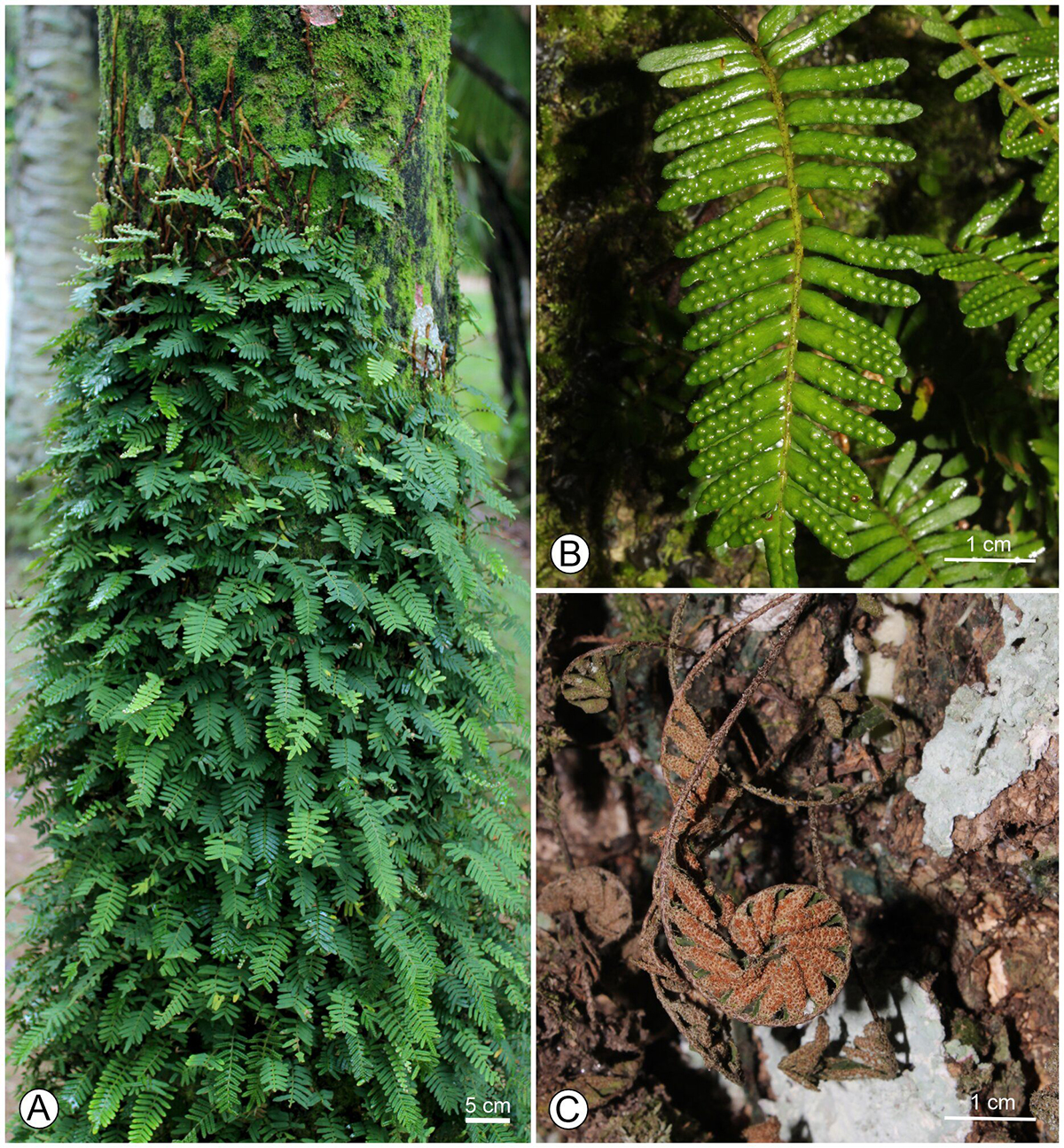
Here, we examine globoid leaf galls on Pleopeltis minima (Fig. 1 A ), an epiphytic fern from the Atlantic Forest and Cerrado (Souza & Salino, 2021), to explore the structural strategies enabling this specialized interaction to thrive in a poikilohydric system. Pleopeltis minima has a long-creeping stem covered by clathrate scales (Souza & Salino, 2021) and displays turgid, dark green pinnae during high water availability (GS Martins, unpublished data) (Fig. 1 B ). When water becomes scarce, the fronds curl to reduce the exposed laminar surface, revealing a brownish, scaly abaxial epidermis (Fig. 1 C ), which reflects solar radiation and minimizes evaporation (Pessin, 1924; Starnecker & Winkler, 1982; Hernández et al., 2012). Our study aimed to analyze anatomical and histochemical changes in gall formation versus non-galled pinnae in P. minima. We expected to find preserved dermal structures for water absorption and reduced desiccation, as well as outer tissue compartments with specialized tissues for water retention in galls. We also expected outer gall tissues to accumulate starch and protective secondary metabolites, and inner nutritive tissue compartments that are important for gall inducer feeding.
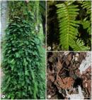
Pleopeltis minima in the Jardim Botânico do Rio de Janeiro, Rio de Janeiro, Brazil. A. Natural habitat, attached to phorophyte bark. B. Turgid frond. C. Desiccated frond.
Material and methods
Sampling and collection. A naturally growing population of P. minima was monitored in the arboretum of the Jardim Botânico do Rio de Janeiro (JBRJ), Rio de Janeiro, Brazil (22° 58’ 14’’ S and 43° 13’ 18’’ W). The cultivated arboretum harbors dozens of Atlantic Forest native epiphytic species, such as P. minima, that spontaneously grow due to the continuum between the garden and the Tijuca National Park (Reis et al., 2023). The area has a tropical climate, with a dry season in the winter (< 60 mm rainfall from May to October), a mean annual temperature of ca. 24 °C, and annual precipitation is 1,171 mm (Macedo et al., 2020). According to the Köppen-Geiger classification system, the climate is categorized as type Aw (Beck et al., 2018).
For the current study, we collected galled and non-galled individuals of P. minima monthly over 12 months (October 2023-September 2024), encompassing both dry and rainy seasons, from five phorophytes of the following species: Dimocarpus longan Lour. (Sapindaceae), Mangifera indica L. (Anacardiaceae), Roystonea oleracea (Jacq.) O.F.Cook (Arecaceae), Latania loddigesii Mart. (Arecaceae), and Adansonia digitata L. (Malvaceae). The voucher specimens, bearing galls, were deposited at the Instituto de Pesquisas Jardim Botânico do Rio de Janeiro Herbarium (RB), under the following collection numbers and barcodes: Martins 07 (RB1584479), 08 (RB1584480), 09 (RB1584481), 10 (RB1584482), 11 (RB1584483), 12 (RB1584484).
Anatomical analysis. We collected non-galled pinnae (NGP) and galled pinnae with mature galls from at least five distinct individuals, each growing on separate phorophytes, across the entire study period. All samples were taken from completely expanded, mature leaves that showed no signs of senescence. Mature galls were defined as those measuring > 1.9 mm in height and > 0.9 mm in width, containing a Cecidomyiidae larva and lacking an exit hole on the adaxial surface. The NGP samples were collected from the same frond as the galls on the pinna (GP), from a corresponding pinna, serving as a direct and developmentally comparable control for the frond tissue. Fragments of NGP and mature galls (n=5 per category, from distinct individuals) were carefully rehydrated if desiccated prior to fixation. This procedure ensured that all anatomical analyses were performed on material in a standardized hydrated state. The samples were fixed in Karnovsky solution (4 % paraformaldehyde and 5 % glutaraldehyde in phosphate buffer, pH 7.2) (Karnovsky, 1965). The samples were then dehydrated in an ethanol series (70 %, 80 %, 90 %, 99.5 %), embedded in a Leica Historesin following the manufacturer’s protocols, and transversely sectioned (5-7μm) using a Spencer rotary microtome. The sections were stained with toluidine blue (0.05 % in acetate buffer, pH 4.6) (Kraus & Arduin, 1997, modified), mounted in Entellan®, examined under an Olympus CX31 light microscope, and photographed using the attached camera.
For scanning electron microscopy (SEM) analysis, samples from NGP and GP (both adaxial and abaxial surfaces; n = 3 per category) were fixed, dehydrated in ethanol series, and subjected to critical point drying in a Bal-Tec CPD 030 Critical Point Dryer (New York, Columbia Nano Initiative, USA) with 20 CO2 exchanges. The fragments were then mounted on stubs and sputter-coated with 18 nm gold using a Leica EM SCD050 (Leica Microsystems, Wetzlar, Germany). These procedures were performed at the Unidade de Microscopia Multiusuário Padrón Lins (UniMicro), Instituto de Microbiologia Paulo de Góes (IMPG), Universidade Federal do Rio de Janeiro (UFRJ). Scanning electron microscopy analysis was conducted using a Zeiss EVO 10 microscope (ZEISS Microscopy, Jena, Germany) at Centro Nacional de Biologia Estrutural e Bioimagem (CENABIO), UFRJ. For morphological classification according to Lellinger (2002), scales were carefully removed from NGP and GP under a stereomicroscope using tweezers and needles and then slide-mounted (Lino et al., 2023).
Histochemical analysis. Transverse sections of NGP and GP (n = 5), obtained from fresh material using a razor blade and from material embedded in Historesin® and sectioned with a microtome, were subjected to the following histochemical tests to compare the accumulation of primary and secondary metabolites: Lugol for starch detection (Johansen, 1940), Sudan IV for lipophilic substances (Jensen, 1962), Fehling’s reagent for reducing sugars (Sass, 1951), 10 % aqueous ferric chloride for phenolic compounds (Johansen, 1940), Coomassie brilliant blue for proteins (Dunn, 1933), ruthenium red for pectins (Johansen, 1940), and tannic acid-ferric chloride for mucilage (Pizzolato & Lillie, 1973). The non-galled pinnae scales and gall scales were detached and subsequently treated with ruthenium red for pectins (Johansen, 1940), tannic acid-ferric chloride for mucilage (Pizzolato & Lillie, 1973), 10 % aqueous ferric chloride for polyphenols (Johansen, 1940), and the Maule test for lignins (Patten et al., 2007). All samples were mounted on slides with 50 % glycerol and sealed with colorless nail polish (Kraus & Arduin, 1997). As negative controls, blank sections and designated controls for each test were used. These samples were analyzed and photographed under a light microscope.
Histometry analysis. Transverse sections of NGP and GP (n = 5 for each category) mounted on slides and stained with toluidine blue were analyzed using ImageJ 1.34a software (Abramoff et al., 2004) for histometric comparisons. The number of layers was quantified for palisade parenchyma and spongy parenchyma in the NGP and for the outer storage tissue and typical nutritive tissue (sensuFerreira et al., 2017) in the GP (3 transects per sample, totaling 15 per tissue). The cell area, height (anticlinal axis), and width (periclinal axis) were measured for the epidermis, palisade parenchyma, spongy parenchyma, outer storage tissue, and typical nutritive tissue (5 cells per section, 5 sections per tissue, totaling 125 cells per tissue). Detached scales (from NGP and GP) were measured (length and width) using ImageJ 1.34a software for comparison.
Statistical analyses. For histometric comparisons, data normality and homoscedasticity were assessed using the Kolmogorov‒Smirnov and Lavene tests, respectively, in SigmaStat® software. Parameters that met the assumptions of normality and homoscedasticity were compared using Student’s t-test (reported as t-values) in SigmaStat® software (Systat Software, Inc.). If assumptions were still not met, the Mann-Whitney test (reported as U-values) was used for comparison. Differences were considered statistically significant at p ≤ 0.05.
Results
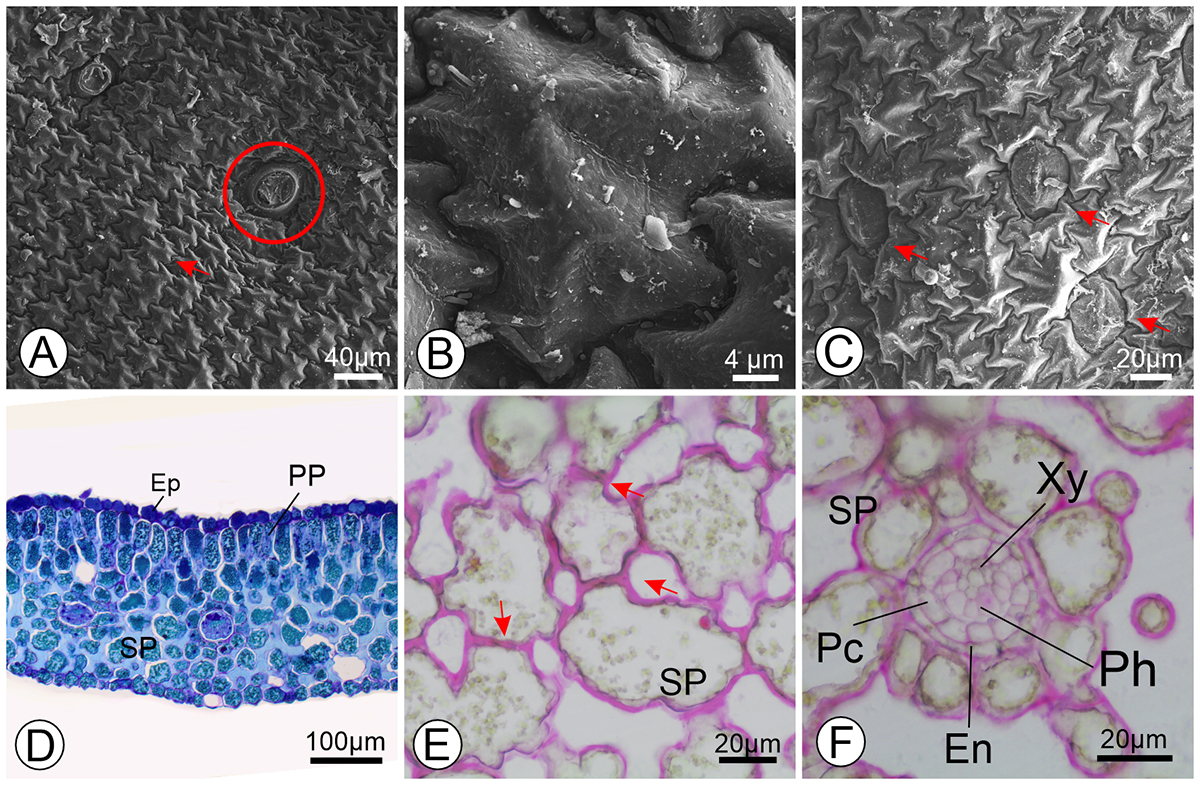
Anatomy. In the frontal view of the adaxial surface, the epidermal cells of the NGP of P. minima have sinuous anticlinal walls and slightly projected outer periclinal walls (Fig. 2 A-B ). The abaxial surface of the epidermis of the pinna lamina is covered by peltate scales, which are not observed on the adaxial surface (Fig. 2B-C). The pinna lamina is amphistomatic, with anomocytic stomata (Fig. 2 C ). The NGP epidermis is unstratified on both surfaces (Fig. 2 D ) and covered by a thin cuticle. The pinna mesophyll is dorsiventral, with a two-layered palisade parenchyma with cells with pectin-thickened walls in the adaxial portion (Fig. 2 D ). The spongy parenchyma adjacent to the palisade parenchyma is composed of two to four layers of cells with pectocellulosic thickenings and arm-like projections (Fig. 2 E ). The vascular bundles are collateral and are surrounded by a pericycle and an endodermal layer (Fig. 2 F ).
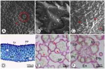
Anatomy of Pleopeltis minima pinna. A-C. Scanning electron microscopy (SEM) of the adaxial surface of the epidermis. A. Pinna epidermal cells (arrow) and stomata (circle) on the adaxial surface. B. Epidermal cells of the adaxial surface with sinuous anticlinal walls. C. Anomocytic stoma (arrow). D. Overview of pinna anatomy in transverse section, stained with toluidine blue. E-F. Pinna mesophyll in transverse section, stained with ruthenium red. E. Arm-like projections (arrows) of the spongy parenchyma cells. F. Vascular bundle in transverse section. Abbreviations: En = endodermis; Ep = epidermis; Pc = pericycle; Ph = phloem; PP = palisade parenchyma; SP = spongy parenchyma; Xy = xylem.
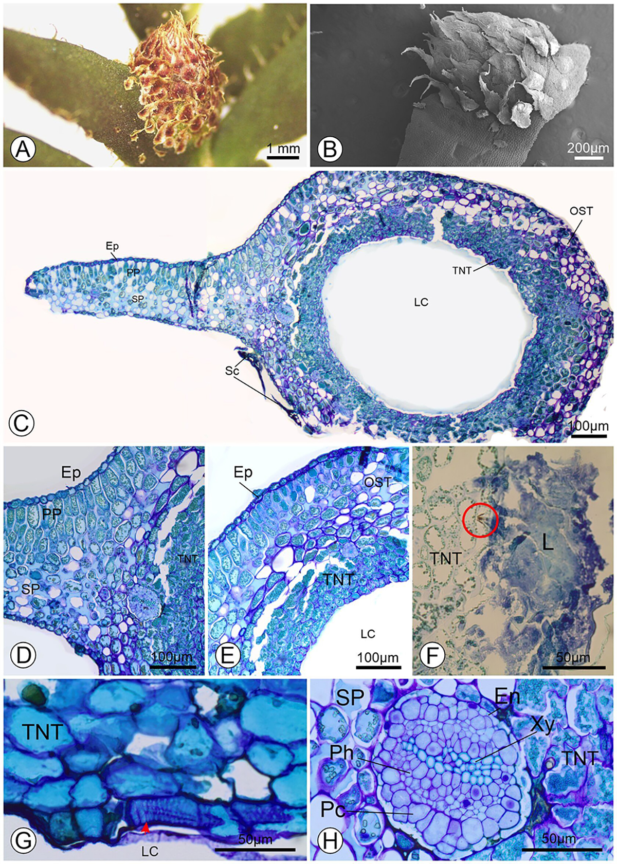
The gall has a globoid shape and is adaxially projected in the galled pinnae. The gall is covered by an unstratified epidermis with scales on both the adaxial and abaxial surfaces (Fig. 3 A-B ). Remarkably, galls maintain their shape even in severely dehydrated and twisted pinnae. Beneath the gall epidermis, we observed an outer storage tissue with four to nine layers of cells with thickened pectic walls (Fig. 3 C ). The outer storage tissue in the adaxial portion appears continuous with the palisade parenchyma, which extends laterally within the gall and connects to non-galled regions in the pinna transverse sections (Fig. 3 C-D ). In the inner layers, below the outer storage tissue, the typical nutritive tissue comprises four to seven layers of densely cytoplasmic cells (Fig. 3 E ). The typical nutritive tissue region is continuous to the spongy parenchyma in non-galled portions of the pinna (Fig. 3 E ). The typical nutritive tissue differs visually from spongy parenchyma due to its reduced intercellular spaces and its cells contain vacuoles with dense floccular phenolic inclusions (Fig. 3 E ). Some sections revealed that the gall-inducing larvae were in direct contact with the typical nutritive tissue cells, apparently feeding on the cellular contents (Fig. 3 F ). We observed cellulose thickenings in their tangential walls (Fig. 3 G ). Amphicribral vascular bundles are located between the outer storage tissue and typical nutritive tissue and are surrounded by a pericycle and an endodermis (Fig. 3 H ). Remarkably, galls apparently maintain their shape even in severely dehydrated and twisted pinnae.
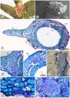
Anatomy and morphology of galls induced by Cecidomyiidae on pinnae of Pleopeltis minima. A. Globoid gall on pinna. B. Globoid galls on the pinna covered by scales under a scanning electron microscope. C. Overview of gall anatomy in transverse section. D. Pinna to gall transition in transverse section. E. Outer storage tissue and typical nutritive tissue in transverse section. F. Gall inducer sucking (circle) contents of cells of typical nutritive tissue in transverse section. G. Helical thickening of cellulose in the cells (arrowhead) of typical nutritive tissue in transverse section. H. Vascular bundle of the gall in transverse section. Abbreviations: En = endodermis; Ep = epidermis; L = larva; LC = larval chamber; OST = outer storage tissue; Pc = pericycle; Ph = phloem; PP = palisade parenchyma; Sc = scale; SP = spongy parenchyma; TNT = typical nutritive tissue; Xy = xylem.
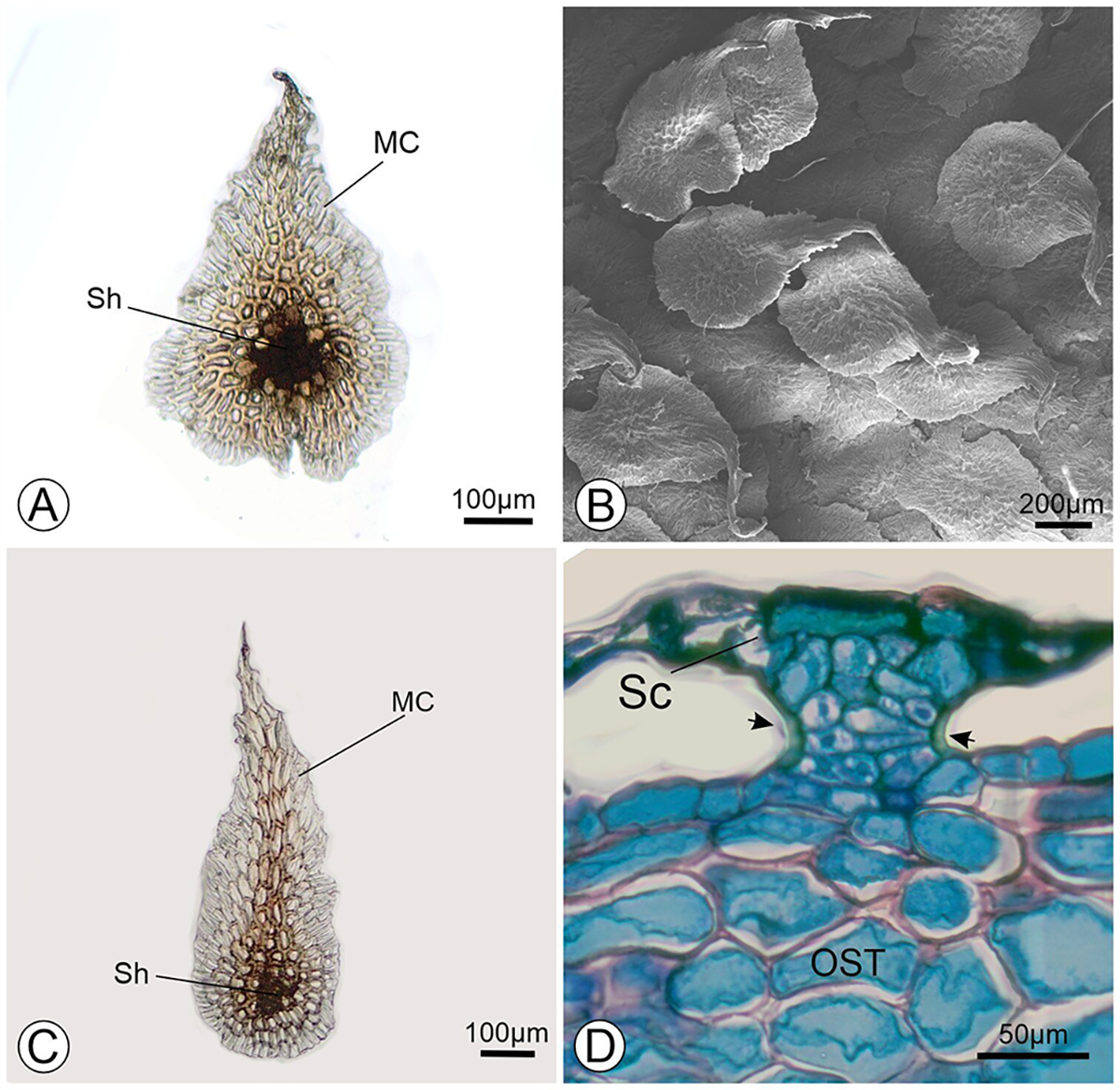
The scales are peltate, bicoloured, and clathrate (Fig. 4 A, C ), with their pedicel inserted among the ordinary epidermal cells (Fig. 4 D ) in both NGP (Fig. 4 A ) and GP (Fig. 4 C ). The NGP scales cover only the abaxial surface of the pinnae lamina (Fig. 4 B ). In frontal view, the central portion of the shield is composed of brownish thick-walled cells with densely pigmented lumina (Fig. 4 A, C ). Marginally, the cells are elongated with thinner translucent walls (Fig. 4 A, C ). The lower part of the central portion of the shield connects to ordinary epidermal cells via a peduncle that supports the scale and is composed of living cells with nuclei, as observed in transverse sections of NGP (Fig. 4 D ). Notably, although the gall is projected to the adaxial surface, where the lamina normally lacks scales, the gall scales develop extensively on this gall surface, completely covering the gall ordinary epidermal cells (Figs. 3A-B).
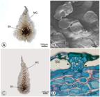
Scales of Pleopeltis minima. A. Pinna scale. B. Abaxial epidermis of the pinna with scales by scanning electron microscopy. C. Gall scale. D. Insertion of the gall scale among the ordinary epidermal cells, with the lateral walls of peduncle cells reacting positively to lignin for toluidine blue (arrows). Note the nuclei in the peduncle cells, indicating that they are alive. Abbreviations: Sh = shield; MC = marginal cells; OST = outer storage tissue; Sc = scale.
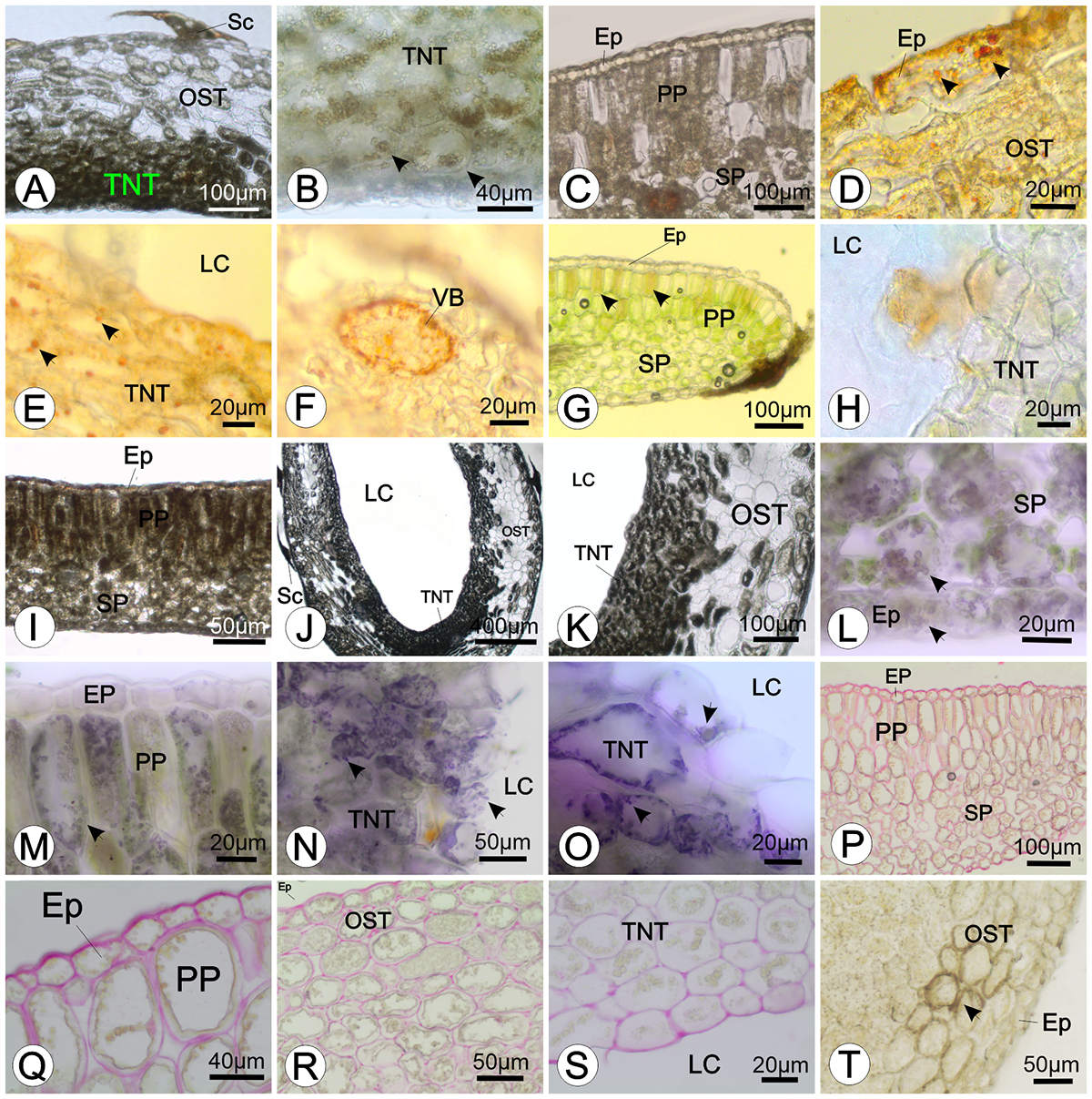
Histochemistry. Starch grains were moderately detected in the palisade parenchyma and spongy parenchyma of NGP. In the GP, starch moderately occurred in the outer storage tissue cells (Fig. 5 A ) and visually more intensely in the cells of the typical nutritive tissue, except for the innermost layer (Fig. 5 A-B ). Lipophilic compounds were not detected in NGP (Fig. 5 C ), except for cell wall thickenings in the endodermis. In the GP, lipid droplets were observed in the epidermis, outer storage tissue (Fig. 5 D ), and typical nutritive tissue cells (Fig. 5 E ), whereas lipophilic compounds were found in the endodermal cell wall thickenings (Fig. 5F). The presence of reducing sugars was positive in the vacuoles of the palisade parenchyma cells and some epidermal cells of the pinnae (Fig. 5 G ). In galls, we observed strong reducing sugar accumulation in typical nutritive tissue, particularly in the cells surrounding the larval chamber (Fig. 5 H ). The phenolic compounds were visually intensely stained by ferric chloride in palisade parenchyma and scales of NGP (Fig. 5 I ). In galls, typical nutritive tissue cells exhibited intense ferric chloride reactivity (Fig. 5 J ), whereas some cells of the outer storage tissue exhibited positive reactions in vacuoles and cell walls (Fig. 5 K ), corresponding to floccular inclusions in the vacuoles (Fig. 3 E ). Proteins were localized in the cytoplasm of the epidermis and parenchyma cells in NGP (Fig. 5 L-M ). More intense protein accumulation occurred in the typical nutritive tissue cells of GP (Fig. 5 N-O ). All the cell walls reacted positively to ruthenium red, indicating the presence of pectins in both NGP (Fig. 5 P-Q ) and GP (Fig. 5 R-S ), with cells having very thick walls. Unlike those of pinnae, the outer storage tissue cell walls showed positive staining for mucilage with tannic acid-ferric chloride reaction (Fig. 5 T ).
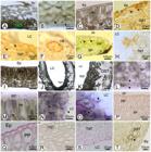
Histochemistry of non-galled pinnae and galls of Pleopeltis minima in transverse sections. A. Starch grains accumulated in the tissues of the galls. B. Starch grains (arrows) in typical nutritive tissue. C. Non-galled pinnae epidermis, negative reaction for lipids. D. Lipid droplets (arrows) in the gall epidermis. E. Lipid droplets accumulated in the typical nutritive tissue of the gall. F. Endodermis of the vascular bundle stained to lipophilic compounds. G. Vacuoles of the pinna epidermis and palisade parenchyma cells accumulating reducing sugars (arrows). H. Reducing sugars detected in the typical nutritive tissue of galls. I. Pinna with accumulation of polyphenols in the epidermis, palisade parenchyma, and spongy parenchyma. J. Polyphenols observed in cells of the outer storage tissue, typical nutritive tissue, and scales of the galls. K. Cells of the typical nutritive tissue containing polyphenols. L. Proteins (arrows) found in the abaxial epidermis and spongy parenchyma. M. Proteins (arrow) found in the adaxial epidermis and palisade parenchyma. N-O. Proteins (arrows) accumulated in the typical nutritive tissue cells. P. Cell walls of the pinna with pectic thickenings. Q. Details of the thickened walls of the epidermis and palisade parenchyma. R. Gall tissues stained for pectins. S. Details of cells with pectic thickening in the typical nutritive tissue. T. Mucilage (arrow) found in the outer storage tissue. Stainings: A-B) Lugol; C-F) Sudan IV; G-H) Fehling’s test; I-K) Ferric chloride; L-O) Coomassie brilliant blue; P-S) ruthenium red; T) tannic acid-ferric chloride test. Abbreviations: Ep = epidermis; LC = larval chamber; OST = outer storage tissue; PP = palisade parenchyma; Sc = scale; SP = spongy parenchyma; TNT = typical nutritive tissue; VB = vascular bundles.
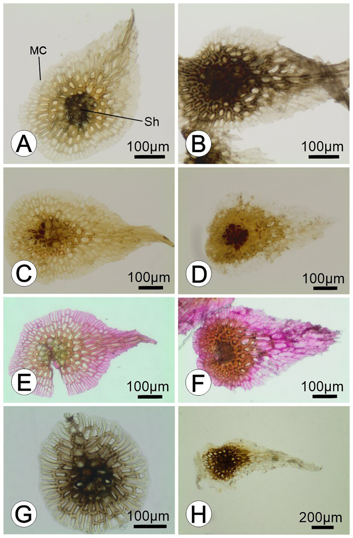
The pinnae scales (Fig. 6 A ) revealed intense tannic acid-ferric chloride staining for mucilage in the cell walls of the shield central portion, but not in the translucent marginal cells of the shield. The scales of galls (Fig. 6 B ) showed visually stronger reactions to mucilage (Table 1). Lignins were not detected in the shields of gall and pinna scales (Table 1) (Fig. 6 C-D ). The secondary cell walls of the central cells of both the gall and pinna scale shields tested positive for ruthenium red, indicating the presence of pectin (Fig. 6 E-F ) (Table 1). Although we detected polyphenols in gall and pinna scales (Table 1), greater labeling occurred in the shield cell walls (Fig. 6 G-H ).
Comparative histochemistry of pinna scales (PS) and scales on galls (GS) on Pleopeltis minima (Polypodiaceae). (-) = negative reaction; (+) = moderately detected; (++) = intensely detected.

Histochemistry of the scales of non-galled pinnae and on the galls of Pleopeltis minima. A. Mucilage accumulated in the cells of the shield of the pinna on the scale. B. Mucilage reaction in the cells of the scales over the galls. C. Absence of lignin deposition in shield and marginal cells of pinna scales. D. Absence of lignin deposition in shield and marginal cells of the gall scale. E. Pectic cell wall thickening in the scales of the pinnae. F. Pectic cell wall thickening in the scales over the galls. G. Polyphenols observed in the cells of the pinna on scales. H. Polyphenols found in the cells of the scales over the galls. Stainings: A-B) Acid tannic-ferric chloride test. C-D) Maule test. E-F) Ruthenium red. G-H) ferric chloride. Abbreviations: Sh = shield; MC = marginal cells.
Histometry. Histometric analysis revealed significant differences between NGP and GP (Table 2). Compared with pinna scales, gall scales were approximately 45 % greater in average length (t = 3.924; P < 0.001) and approximately 15 % greater in average width (U = 283.0; P = 0.026). In transverse sections, the GP epidermis exhibited an approximately 40 % reduction in cell height compared with that of the NGP epidermis (t = 6.603; P < 0.001), a 25 % reduction in width (t = 3.653; P = 0.001), and a 30 % reduction in area (t = 2.805; P = 0.011) (Table 2). The cell height, width, area, and number of cell layers differed significantly between the palisade parenchyma in the NGP and the outer storage tissue in the GP (Table 2). Compared with the palisade parenchyma cells of the NGP, the outer storage tissue cells demonstrated a height reduction of approximately 45 % (U = 5.0; P < 0.001) but an increase in width of approximately 60 % (U = 225.0; P < 0.001), as well as an increase in area of 27 % (U = 169.0; P = 0.020). Compared with that in the palisade parenchyma, the number of cell layers in the outer storage tissue increased by approximately 240 % (U = 225.0; P < 0.001), and the thickness in the outer storage tissue was approximately 120 % greater than that in the palisade parenchyma (U = 225.0; P < 0.001). The height (t = -0.489; P = 0.629) and area (U = 44.0; P = 0.573) of the typical nutritive tissue cells did not significantly differ from those of the spongy parenchyma cells, whereas they were 30 % smaller in width (t = 3.844; P < 0.001). The number of cell layers in typical nutritive tissue was approximately 60 % greater than that in spongy parenchyma (U = 220.5; P < 0.001), but the tissue thickness was similar between typical nutritive tissue and spongy parenchyma (t = -1.663; P = 0.107) (Table 2).
Histometrical parameters of pinna scales (PS) and gall scales (GS) and transverse sections of non-galled pinnae (NGP) and Cecidomyiidae induced pinna galls (GP) in Pleopeltis minima (Polypodiaceae). The data are presented as the means ± standard deviations. Values with different letters within a single row are significantly different (P ≤ 0.05).
Discussion
The galls induced on the poikilohydric fern P. minima revealed maintenance and an increase in structural and chemical strategies to avoid structural damage during desiccation periods. Compared to NGP, the neoformation of larger scales and their occurrence on the gall adaxial surface revealed potentiated mechanisms of water absorption (Oliveira et al., 2017; Prats & Brodersen, 2021). The accumulation of mucilage in the cell walls of the outer storage tissue also supported our hypothesis regarding the presence of external compartments in the gall with water storage capacity (Oliveira et al., 2017). Similarly, polyphenol accumulation in the outer gall tissue was both expected and observed. In addition, other metabolites, such as sugars, proteins, and starch, also accumulate in typical nutritive tissue to support the feeding of the gall inducer and the antioxidant mechanisms in stress situations, which has also been reported in other gall systems (Bronner, 1992; Oliveira & Isaias, 2010; Oliveira et al., 2011; 2014; Marquesine et al., 2025).
Galls on a poikilohydric fern vs. typical Cecidomyiidae galls. Histometric comparisons revealed that gall formation in P. minima occurs through hyperplasia of the outer storage parenchyma, with a reduction in the size but proliferation of layers, a morphogenetic phenomenon that also occurs in Lepidoptera-induced galls on M. vacciniifolia (Martins et al., 2023), although in other gall systems in angiosperms, this compartment usually suffers not only hyperplasia, but also cell hypertrophy (Ferreira & Isaias, 2013; Ferreira et al., 2019; Marquesine et al., 2025). The development of an outer storage tissue is common in galls, revealing an intermediate level of complexity (Ferreira et al., 2019). The outer gall storage tissue is commonly reported in nematode, acari, and insect galls and contains vacuolated cells and starch reserves that maintain gall metabolism (Ferreira et al., 2019), which aligns with our detection of starch, phenolics, and proteins, and unlike palisade parenchyma, mucilage in cell walls. Similar responses occur in other gall systems, where outer cells adopt defensive functions by accumulating terpenoid, alkaloid, and phenolic substances (Bragança et al., 2017). However, we propose that mucilage in the outermost tissue of the gall may also play a protective role, given its hydrophilic properties, particularly in preventing excessive water loss (Gregory & Baas, 1989; Shin et al., 2021).
The galling Cecidomyiidae feed directly on typical nutritive tissue by sucking contents from ruptured nutritive cells (Bragança et al., 2023). This tissue shows metabolic activity, as evidenced by the dense cytoplasm, evident nucleus and nucleoli, and accumulation of sugars (Ferreira et al., 2017; 2019), while maintaining tissue thickness compared with the spongy parenchyma, confirming hyperplasia as further evidence of gall formation. This is a common developmental process reported in galls (Mani, 1964; Ferreira et al., 2019), driven by intense larval feeding activity (Ferreira & Isaias, 2013). We observed reducing sugars and protein accumulation in typical nutritive tissue, which directly nourishes Cecidomyiidae larvae. The presence of starch in the outer typical nutritive tissue layers and reducing sugars in the innermost layer is a pattern observed in Cecidomyiidae galls as a product of starch hydrolysis mediated by the larval salivary secretions (Bronner, 1992; Oliveira et al., 2014; Marquesine et al., 2025). Reducing sugars are also related to the mitigation of the high oxidative stress in plants due to their inherent chemical structure, which allows them to act as reducing agents that can directly scavenge reactive oxygen species and chelate pro-oxidant metal ions (Ende & Peshev, 2013). This role is particularly relevant in attenuating the stress caused by the feeding activity of the galler on these cells (Isaias et al., 2015). In addition, lipids and proteins in typical nutritive tissue have also been observed in other Cecidomyiidae gall systems (Bronner, 1992; Oliveira & Isaias, 2010; Oliveira et al., 2011), highlighting the role of host plant potentials in gall histochemical profiles (Ferreira & Isaias, 2014). Moreover, the mobilization of sugars and proteins in desiccation-tolerant plants that act as osmoregulators is already well known, as they are capable of protecting membranes and macromolecules from oxidation and mechanical damage caused by cell shrinkage due to water scarcity (Stuart, 1968; Hietz, 2010; Moraes et al., 2014).
The detection of polyphenols in typical nutritive tissue cells may be related to tissue meristematic function, given the role of phenolics in regulating cell growth (Bedetti et al., 2014), in addition to their known defensive functions against pathogens (Feeny, 1976; Tempel, 1981). Additionally, the accumulation of phenolics in the innermost gall tissues may be related to the antioxidant role of phenolic compounds in protecting cells against the harmful effects of free radicals (Aboul-Enein et al., 2007; Isaias et al., 2015), especially considering that this host plant may experience stress during periods of desiccation (Lagoria et al., 2018; John & Hasenstein, 2017). As in the outer gall layers, in the typical nutritive tissue, we detected cellulose helicoidal thickenings, which were also observed by Lagoria et al. (2018) in P. macrocarpa roots. We may attribute such features to other adaptations to water absorption and storage, due to the hydrophilic properties of the primary cell walls.
Structural adaptations related to poikilohydry. Compared with those of other Polypodiaceae species, the anatomical characteristics of P. minima pinnae, including fewer palisade parenchyma layers (Tejero-Díez et al., 2009), intercellular spaces, and arm-shaped thickenings in spongy parenchyma, appear to protect chloroplasts against desiccation and UV radiation (Hevly, 1963; Tejero-Díez et al., 2009; Hernández et al., 2012). The vascular bundles immersed in the spongy parenchyma of P. minima NGP did not present a circumendodermal band, as was observed in the stem of the gall-host fern Microgramma vacciniifolia (Langsd. & Fisch.) Copel (Polypodiaceae) (Martins et al., 2023). Most likely, the absence of a circumendodermal band around the endodermis and vascular bundles may be related to frond curling during dehydration, facilitating frond movement-a hypothesis supported by Lagoria et al. (2018) in P. macrocarpa (Bory ex Willd.) Kaulf (Polypodiaceae). In addition to these protective features, some structural features contribute to water absorption. Pectin thickenings present in the mesophyll of the non-galled pinnae are important for a quick water absorption in periods of water availability (Boanares et al., 2018; Shin et al., 2021), a feature potentiated in gall outer storage parenchyma, where also mucilage was detected.
Along with paraphyses-free sori, the peltate scale is a delimiting character of Pleopeltis and is associated with atmospheric moisture uptake (Smith & Tejero-Díez, 2014; Lagoria et al., 2018). The scales on the fronds of P. minima share similar anatomical characteristics with those of galls. However, the neogenesis of scales in the P. minima gall adaxial epidermis may be considered a complex modification, especially since the dermal system is considered more ontogenetically conserved than other plant tissue systems are (Glover, 2000; Ferreira et al., 2019).
The scales of P. minima present lignin in the external walls of their pedicel cells, a component that can confer mechanical resistance and reduce water loss (Hernández et al., 2012). On the other hand, the mucilage detected in the cell walls of the central portion of the scale shield in galls and NGP is an important feature related to water absorption (Oliveira et al., 2017), which is important for poikilohydry. The mucilage is a hydrophilic compound detected in other ferns, such as in the secretory head of glandular trichomes (Oliveira et al., 2017) and glandular scales (Lino et al., 2023). The maintenance of the shape of the galls, even in dehydrated fronds, is probably due to the stronger reactions to mucilage in the outer storage tissue of the galls than in NGP and larger gall scale dimensions, which may reduce transpiration rates and reflect excessive light while serves as the main route to water absorption (Voytena et al., 2014; Aros-Mualin & Kessler, 2024).
Despite the presence of mucilage in the pedicel cell walls of scales, we found pectic cell walls in the shield and marginal cells of the scale. In Pleopeltis polypodioides (L.) E.G.Andrews & Windham, scales create a hydrophilic surface that increases water absorption, serving as the main rehydration pathway (Prats & Brodersen, 2021). As suggested by these authors for P. polypodioides, without the scales on the fronds and galls of P. minima, the relatively hydrophobic epidermis of the foliage would be exposed, which could reduce the water uptake capacity of the frond.
Poikilohydric adaptations in both leaves and galls may play crucial physiological roles under conditions of climate change and intensified drought (Boanares et al., 2021). The structural strategies for the desiccation tolerance of P. minima are conserved in galls, creating a microhabitat favorable for gall maintenance and gall inducer survival. The neogenesis of scales is induced by the galling organism and may represent a protective mechanism against desiccation that allows efficient water uptake and ensures survival during periods of water and heat stress.
Conclusions and perspectives. Pleopeltis minima appears to be a promising model for advancing the anatomical and chemical study of galls in poikilohydric species. Our findings support the hypothesis that P. minima galls exhibit enhanced desiccation tolerance traits, including neoformation and hypertrophy of hydrophilic scales, which likely facilitate water absorption, and accumulation of mucilage in outer gall tissues that are specialized for water retention. These structural and chemical modifications may allow the maintenance of gall function during drought stress. In addition to the expected responses in gall formation, such as tissue hyperplasia and the development of a typical nutritive tissue accumulating sugars (common in Cecidomyiidae systems), lipids and proteins were also observed, although less frequently reported in such systems. Our results contribute to future studies on galls in poikilohydric ferns under different environmental conditions and interactions with other organisms.
Acknowledgments
We thank Jardim Botânico do Rio de Janeiro for granting collection permission in the cultivated areas
References
- Abramoff MD, Magalhães PJ, Ram SJ. 2004. Image processing with ImageJ. Biophotonics International 11: 36-42.
- Aboul-Enein HY, Kruk I, Kładna A, Lichszteld K, Michalska T. 2007. Scavenging effects of phenolic compounds on reactive oxygen species. Biopolymers 86: 222-230.
- Alpert P. 2000. The discovery, scope, and puzzle of desiccation tolerance in plants. Plant Ecology 151: 5-17.
- Alpert P, Oliver MJ. 2002. Drying without dying. In: Desiccation and survival in plants: Drying without dying. United Kingdom, CABI: Digital Library. p. 3-43.
- Aros-Mualin D, Kessler M. 2024. Untangling poikilohydry and desiccation tolerance: Evolutionary and macroecological drivers in ferns. Annals of Botany 134: 1139-1150.
- Bartels D. 2005. Desiccation tolerance studied in the resurrection plant Craterostigma plantagineum Integrative and Comparative Biology 45: 696-701.
-
Beck HE, Zimmermann NE, McVicar TR, Vergopolan N, Berg A, Wood EF. 2018. Present and future Koppen-Geiger climate classification maps at 1-km resolution. Scientific Data 30: 10.1038/sdata.2018.214.
» https://doi.org/10.1038/sdata.2018.214 - Bedetti CS, Modolo LV, Isaias RMS. 2014. The role of phenolics in the control of auxin in galls of Piptadenia gonoacantha (Mart.) MacBr. (Fabaceae: Mimosoideae). Biochemical Systematics and Ecology 55: 53-59.
- Boanares D, Ferreira BG, Kozovits AR, Sousa HC, Isaias RMS, França MGC. 2018. Pectin and cellulose cell wall composition enables different strategies to leaf water uptake in plants from tropical fog mountain. Plant Physiology and Biochemistry 122: 57-64.
- Boanares D, Lemos- Filho JP, Isaias RMS, França MGC. 2021. Photosynthetic heat tolerance in plants with different foliar water-uptake strategies. American Journal of Botany 108: 811-819.
- Bragança GP, Oliveira DC, Isaias RMS. 2017. Compartmentalization of metabolites and enzymatic mediation in nutritive cell of Cecidomyiidae galls on Piper arboretum Aubl. (Piperaceae). Journal of Plant Studies 6: 11-22.
- Bragança GPP, Costa EC, Arriola IA, Sanin D, Isaias RMS. 2023. As soft as silk: Structural and chemical traits can help with the identification of Niphidium crassifolium (Polypodiaceae) gall inducers. Rodriguesia 74: e00572023.
- Bronner R. 1992. The role of nutritive cells in the nutrition of cynipids and cecidomyiids. In: Shorthouse JD, Rohfritsch - (eds.). Biology of insect-induced galls. Oxford, Oxford University Press. p. 118-140.
- Dunn MJ. 1933. Gel Electrophoresis: Proteins. Oxford, Bios Scientific Publishers.
- Ende WV, Peshev D. 2013. Sugars as antioxidants in plants. In: Tuteja N, Gill S (ed.). Crop Improvement Under Adverse Conditions. New York, Springer. p. 285-307.
- Feeny P. 1976. Plant apparency and chemical defense. In: Wallace JW, Mansell RL (eds.). Biochemical Interaction Between Plants and Insects. Cham, Springer. p.1-39.
- Fernandes GW, Price PW. 1992. The adaptive significance of insect gall distribution: Survivorship of species in xeric and mesic habitats. Oecologia 90: 14-20.
- Ferreira BG, Isaias RMS. 2013. Developmental stem anatomy and tissue redifferentiation induced by a galling Lepidoptera on Marcetia taxifolia (Melastomataceae). Botany 91: 752-760.
- Ferreira BG, Isaias RMS. 2014. Floral-like destiny induced by a galling Cecidomyiidae on the axillary buds of Marcetia taxifolia (Melastomataceae). Flora 209: 391-400.
- Ferreira BG, Álvarez R, Avritzer SC, Isaias RMS. 2017. Revisiting the histological patterns of storage tissues: Beyond the limits of gall-inducing taxa. Botany 95: 173-184.
- Ferreira BG, Álvarez R, Bragança GP, Alvarenga DR, Hidalgo PN, Isaias RMS. 2019. Feeding and other gall facets: Patterns and determinants in gall structure. Botanical Review 85: 78-106.
- Glover BJ. 2000. Differentiation in plant epidermal cells. Journal of Experimental Botany 51: 497-505.
- Gregory M, Baas P. 1989. A survey of mucilage cells in vegetative organs of the dicotyledons. Israel Journal of Botany 38: 125-174.
- Guedes LM, Torres S, Sáez-Carillo K, Becerra J, Pérez CI, Aguilera N. 2022. High antioxidant activity of phenolic compounds dampens oxidative stress in Espinosa nothofagi galls induced on Nothofagus obliqua buds. Plant Science 314: 111-114.
- Guedes LM, Aguilera N, Torres S, Gavilán E, Rosales N. 2024. Gall inducer Dasineura sp. alters the polyphenol profile and antioxidant activity of Peumus boldus stems. Tree Physiology 44: tpae024.
- Hartley SE. 1998. The chemical composition of plant galls: Are levels of nutrients and secondary compounds controlled by the gall-former? Oecologia 113: 492-501.
- Hernández MA, Rodolfo VO, Lucrecia TC, Mata M. 2012. Preferencia de hospedero, orientación y rasgos morfológicos-anatómicos de Pleopeltis minima (Polypodiaceae) en un ambiente urbano. Liloa 49: 105-117.
- Hevly RH. 1963. Adaptations of Cheilanthoid Ferns to Desert Environments. Journal of the Arizona Academy of Science 2: 164-175.
- Hietz P. 2010. Fern adaptations to xeric environments. In: Mehltreter K, Walker LR, Sharpe JM (eds.). Fern Ecology. Cambridge, Cambridge University Press. p. 140-176.
- Isaias RMS, Oliveira DC, Carneiro RGS, Kraus JE. 2014. Developmental anatomy of galls in the neotropics: Arthropod stimuli versus host plant constraints. In: Fernandes GW, Santos JC (eds.). Neotropical Insect Galls. Dordrecht, Springer. p. 15-34.
- Isaias RMS, Oliveira DC, Moreira ASFP, Soares GLG, Carneiro RGS. 2015. The imbalance of redox homeostasis in arthropod-induced plant galls: Mechanisms of stress generation and dissipation. Biochimica et Biophysica Acta 1850: 1509-1517.
- Isaias RMS, Kraus JE, Costa EC, Carneiro RGS. 2024. The anatomy of neotropical galls and the untold lessons about the morphogenetical potentialities of plants. Rodriguésia 75: e01542023.
- Jensen WA. 1962. Botanical Histochemistry. San Francisco, W.H. Freeman.
- Johansen DA. 1940. Plant Microtechnique. New York, McGraw-Hill.
- John SP, Hasenstein KH. 2017. The role of peltate scales in desiccation tolerance of Pleopeltis polypodioides Planta 245: 207-220.
- Karnovsky MJ. 1965. A formaldehyde-glutaraldehyde fixative of high osmolality for use in electron microscopy. Journal of Cell Biology 27: 137-138.
- Kessler M, Siorak Y. 2007. Desiccation and rehydration experiments on leaves of 43 pteridophyte species. American Fern Jornal 96: 175-185.
- Kraus JE, Arduin M. 1997. Manual Básico de Métodos em Morfologia Vegetal. Seropédica, Editora da Universidade Federal Rural do Rio de Janeiro.
- Kuster VC, Rezende UC, Cardoso JCF, Isaias RMS, Oliveira DC. 2020. How Galling Organisms Manipulate the Secondary Metabolites in the Host Plant Tissues? A Histochemical Overview in Neotropical Gall Systems. In: Mérillon JM, Ramawat K (eds.). Co-Evolution of Secondary Metabolites - Reference Series in Phytochemistry. Cham, Springer : 823-842.
- Lagoria MLA, Ávila G, Neira DA, Rios NF, Prado J, Hernández MA. 2018. Morphoanatomical and histochemical characteristics of the epiphytic fern Pleopeltis macrocarpa (Polypodiaceae). Brazilian Journal of Botany 41: 739-750.
- Lellinger DB. 2002. A modern multilingual glossary for taxonomic pteridology. Washington, American Fern Society
- Lino V, Reis A, Canaveze Y, Farah MS, Sylvestre LS, Ferreira BG. 2023. Beyond the physical protection: What can Asplenium glandular scales secrete? Flora 305: 152338.
- Luttge U, Beck E, Bartels D. 2011. Plant Desiccation Tolerance. Berlin, Springer.
- Macedo TM, Barros CF, Lima HC et al 2020. Climate signals in tree rings of Paubrasilia echinata (Leguminosae‐Caesalpinioidea) from the Atlantic Forest of Brazil. Trees 34: 337-347.
- Mani MS. 1964. Ecology of Plant Galls. Dordrecht, Springer .
- Marquesine RR, Canaveze Y, Ferreira BG. 2025. Ontogenetic differences in sun and shade galls of Clinodiplosis profusa on Eugenia uniflora leaves and the cytological antioxidant mechanisms in gall cells. Protoplasma 262: 15-34.
- Martins GS, Santos MG, Reis A, Ferreira BG. 2023. Alterations induced by Tortrimosaica polypodivora on the stems of Microgramma vacciniifolia: Simple or complex galls? Rodriguesia 74: e00532023.
- Moraes MG, Oliveira AAQ, Santos MG. 2014. Sugars in ferns and lycophytes growing on rocky outcrops from the southeastern Brazilian coast. Bioscience Journal 30: 1882-1884.
- Oliveira DC, Isaias RMS. 2010. Cytological and histochemical gradients induced by a sucking insect in galls of Aspidosperma australe Arg. Muell (Apocynaceae). Plant Science 178: 350-358.
- Oliveira DC, Carneiro RGS, Magalhães TA, Isaias RMS. 2011. Cytological and histochemical gradients on two Copaifera langsdorffii Desf. (Fabaceae) Cecidomyiidae gall systems. Protoplasma 248: 829-837.
- Oliveira DC, Moreira ASFP, Isaias RMS. 2014. Functional gradients in insect gall tissues, studies on neotropical host plants. In: Fernandes GW, Santos JC (eds.). Neotropical Insect Galls. Dordrecht, Springer . p. 35-49.
- Oliveira CS, Salino A, Paiva EAS. 2017. Colleters in Thelypteridaceae: Unveiling mucilage secretion and its probable role in ferns. Flora 228: 65-70.
- Oliver MJ, Farrant JM, Hilhorst HW, Mundree S, Williams B, Bewley JD. 2020. Desiccation tolerance: Avoiding cellular damage during drying and rehydration. Annual Review of Plant Biology 71: 435-460.
- Patten AM, Jourdes M, Brown EE, Laborie MP, Davin LB, Lewis NG. 2007. Reaction tissue formation and stem tensile modulus properties in wild-type and p-coumarate-3-hydroxylase downregulated lines of alfalfa, Medicago sativa (Fabaceae). American Journal of Botany 94: 912-925.
- Pessin LJ. 1924. A physiological and anatomical study of the leaves of Polypodium polypodioides American Journal of Botany 11: 370-381.
- Pizzolato TD, Lillie RD. 1973. Mayer’s tannic acid-ferric chloride stain for mucins. Journal of Histochemistry and Cytochemistry 21: 56-64.
- Prado J, Hirai RY. 2010. A new combination in Pleopeltis and Some Nomenclatural Notes Related to Illustrations Validating Fern Names. American Fern Journal 100: 189-194.
- Prats KA, Brodersen CR. 2021. Desiccation and rehydration dynamics in the epiphytic resurrection fern Pleopeltis Plant Physiology 187: 1501-1518.
- Price PW, Fernandes W, Waring GL. 1987. Adaptive nature of insect galls. Environmental Entomology 16: 15-24.
- Reis A, Oliveira MIR, Bial M, Mynssen CM. 2023. Exploring the diversity of ferns and lycophytes in the Jardim Botânico do Rio de Janeiro. Rodriguésia 74: e00682023.
- Santos MG, Maia VC. 2018. A synopsis of fern galls in Brazil. Biota Neotropica 18: e20180513.
- Santos MG, Hanson P, Maia VC, Mehltreter K. 2019. A review of galls on ferns and lycophytes. Environmental Entomology 48: 53-60.
- Sass JE. 1951. Botanical microtechnique. 2nd. edn. Ames, The Iowa State College Press.
- Shin Y, Chane A, Jung M, Lee Y. 2021. Recent advances in understanding the roles of pectin as an active participant in plant signaling networks. Plants 10: 1712.
- Smith AR, Tejero-Díez D. 2014. Pleopeltis (Polypodiaceae), a redefinition of the genus and nomenclatural novelties. Botanical Sciences 92: 43-58.
- Souza FS, Salino A. 2021. Pleopeltis (Polypodiaceae) in Brazil. Phytotaxa 512: 213-256.
- Starnecker G, Winkler S. 1982. Zur Ökologie epiphytischer Farne in Südbrasilien II. Anatomische und physiologische Anpassungen. Flora 172: 57-68.
- Stone GN, Schönrogge K. 2003. The adaptive significance of insect gall morphology. Trends in Ecology & Evolution 18: 512-522.
- Stuart TS. 1968. Revival of respiration and photosynthesis in dried leaves of Polypodium polypodioides Planta 83: 185-206.
- Teixeira CT, Kuster VC, Carneiro RGS, Cardoso JCF, Isaias RMS. 2022. Anatomical profiles validate gall morphospecies under similar morphotypes. Journal of Plant Research 135: 593-608.
- Tejero-Díez JD, Mickel JT, Smith AR. 2009. A Hybrid Phlebodium (Polypodiaceae, Polypodiophyta) and Its Influence on the Circumscription of the Genus. American Fern Journal 99: 109-116.
- Tempel AS. 1981. Field studies of the relationship between herbivore damage and tannin concentration in bracken (Pteridium aquilinum Kuhn). Oecologia 51: 97-106.
- Tweddle JC, Dickie JB, Baskin CC, Baskin JM. 2003. Ecological aspects of seed desiccation sensitivity. Journal of Ecology 91: 294-304.
- Voytena APL, Minardi BD, Barufi JB, Santos M, Randi AM. 2014. Pleopeltis pleopeltifolia (Polypodiopsida, Polypodiaceae), a poikilochlorophyllous desiccation-tolerant fern: Anatomical, biochemical and physiological responses during water stress. Australian Journal of Botany 62: 647-656.
- Živković T, Quartacci MF, Stevanovic B, Marinone F, Navari-Izzo F. 2005. Low-molecular weight substances in the poikilohydric plant Ramonda serbica during dehydration and rehydration. Plant Science 168: 105-111.
-
Data Availability
The datasets generated during and/or analyzed during the current study are available in the Scielo Data repository, https://doi.org/10.48331/SCIELODATA.DDZNNR.
-
Funding Information
We thank Fundação Carlos Chagas Filho de Amparo à Pesquisa do Estado do Rio de Janeiro (FAPERJ, Brazil) for financial support of the project (E-26/210.736/2024; E-26/210.705/2023; E-26/211.608/2021) and for the grant to BGF (E-26/204.484/2024) . Conselho Nacional de Desenvolvimento Científico e Tecnológico (CNPq, Brazil) for the research grant to BGF (310520/2023-1), MGS (311469/2025-6), MSc scholarship to GSM (131726/2023-4), and the Ph.D. scholarship to AR (140089/2023-3) PROCIÊNCIA (Programa de Incentivo à Produção Científica, Técnica e Artística da UERJ) for the research grant to MGS.
The datasets generated during and/or analyzed during the current study are available in the Scielo Data repository, https://doi.org/10.48331/SCIELODATA.DDZNNR.