 Morphological and molecular identification of a new halotolerant cyanobacterial strain, Limnospira fusiformis TL03, isolated from Telamine Lake in the northwest of Algeria
ABSTRACT
This study examines a new strain, Limnospira fusiformis TL03, isolated from Lake Telamine in northwest Algeria. The strain was morphologically identified using a light microscope and molecularly characterized using its 16S rRNA gene sequence. The effect of salinity on growth and photosynthetic pigments was studied using a spectrophotometric method; different sodium chloride concentrations (0, 5, 15, 30, 45, and 60 g/l) in blue-green medium (BG11) cultured uniformly as homogenized cell suspension were tested. A morphological examination confirmed that the isolated strain belonged to the Limnospira genus. It has trichome lengths ranging from 210-2027 µm, as well as pitch and coil diameters ranging from 30-137 µm and 20-60 µm, respectively. The data from the 16S rRNA gene sequence analysis confirmed that the isolated strain was Limnospira fusiformis TL03 with 100% sequence similarity to Limnospira fusiformis SAG 85.79. The results also indicate that this strain can grow at various salt concentrations, with the highest optical density values (1.58 ± 0.014 and 1.56 ± 0.003) obtained in cultures containing 15 g/L and 5 g/L NaCl, respectively.
Keywords:
Identification; Isolation; Limnospira fusiformis TL03; Salt stress; Tolerance
Introduction
The demand for cyanobacteria and microalgae has increased rapidly in recent years due to their numerous potential applications in fields such as nutrition, biopharmacy, and renewable energy. Extremophile cyanobacteria and microalgae are gaining popularity due to their ability to grow in harsh environments, allowing for outdoor cultivation with minimal contamination risks (Hirooka et al., 2014; Varshney et al., 2015; D’Alessandro & Antoniosi Filho, 2016; Sydney et al., 2019).
Among the phytoplankton microorganisms, the phylum Cyanobacteria is the most ancient, being one of the most versatile and ecologically successful groups, living in a variety of environments (Abed et al., 2009), including oceans, freshwater, soil, bare rocks, ice shelves, hot springs, and hypersaline to alkaline lakes. They can also grow in environments with high metal concentrations and low water availability, such as desert regions, by forming endolithic communities (Sanchez-Baracaldo et al., 2005; Thajuddin & Subramanian, 2005; Rastogi & Sinha, 2009). Among the numerous cyanobacteria species, we highlight Limnospira (= Arthrospira), also known commercially as Spirulina.
Limnospira is a newly established genus with three validated species that were reclassified from Arthrospira based on morphological, molecular, and ecological differences (Nowicka-Krawczyk et al., 2019). Arthrospira fusiformis, Arthrospira maxima, and Arthrospira indica were reclassified to Limnospira fusiformis, Limnospira maxima, and Limnospira indica respectively (Nowicka-Krawczyk et al., 2019). Limnospira and Arthrospira are cyanobacteria (blue-green algae), multicellular, photosynthetic prokaryotes, and filamentous. They have a high protein content that ranges from 55 to 70% of dry weight and contains all essential amino acids (Babadzhanov et al., 2004; Volkmann et al., 2008; Sotiroudis & Sotiroudis, 2013; Da Silva et al., 2019; Jung et al., 2019). They also contain all the B vitamins, as well as vitamins C, E, D, and provitamin A (Mukhopadhyay, 2015; Jung et al., 2019). Among the numerous cyanobacteria species, we highlight Limnospira (= Arthrospira), also known commercially as Spirulina.
Limnospira and Arthrospira have numerous advantages and can be used in a variety of applications, including human food, animal feed, aquaculture, bioenergy, wastewater treatment, cosmetics, bioplastics, and agriculture. Limnospira and Arthrospira have anti-inflammatory properties (Wu et al., 2016; Aladaileh et al., 2020), as well as antibacterial and antifungal properties (Borowitzka, 1995; Furmaniak et al., 2017; Falaise, 2019; Jung et al., 2019). The extract of A. maxima cultured in deep sea water effectively suppressed the expression of the Bcl-2 gene in A549 human lung adenocarcinoma cells, as well as inhibited various human cancer cells (Choi et al., 2013), stimulating the immune system and lowering hyperlipidemia and obesity (Jiménez et al., 2003; Costa et al., 2004; Ghaeni & Roomiani, 2016).
Arthrospira extracts and compounds may be useful in preventing cardiovascular disease (Memije-Lazaro et al., 2018). They reduce both blood pressure and plasma lipid concentration (Furmaniak et al., 2017). Several international organizations, including the World Health Organization (WHO), Food and Agriculture Organization (FAO), and United Nations Children’s Fund (UNICEF), have recognized Limnospira and Arthrospira as dietary supplements (FAO Fisheries and Aquaculture Circular. No. 1034, Joint FAO/WHO Expert Committee on Food Additives - JECFA, 86th meeting 2018) (Cho et al., 2020).
Arthrospira spp. was recommended by the WHO for inclusion in the diet of National Aeronautics and Space Administration (NASA) astronauts in space because it is an ideal and compact food for space travel (Koyandea et al., 2019). Spirulina was declared the best food for the future by the United Nations World Food Conference, and it is gaining popularity today (Pulz & Gross, 2004). In comparison to other cyanobacteria and microalgae, Arthrospira production is the most widespread in the world (Villaró et al., 2022). The efficiency of dry mass production reached 90,000 tons in 2010 and remained there until the last FAO report in 2016 (Furmaniak et al., 2017). The main populations of Limnospira and Arthrospira have been reported in 30 countries worldwide: 11 in Africa, nine in Asia, five in America, and five in Europe (Sili et al., 2012).
Numerous studies of Limnospira fusiformis habitat have revealed that it is more commonly found in alkaline environments and can survive in a wide range of salinities (Kebede, 1997; Sudhir & Murthy, 2004; Thajuddin & Subramanian, 2005; Dadheech et al., 2010; Asulabh et al., 2012; Benavente-Valdés et al., 2016; Costa et al., 2016; Cellamare et al., 2018). Arthrospira was observed for the first time in Algeria in 1996 at Tamanrasset Pond in the Southern region of this country (Fox, 1996), and it was also found in Telamine Lake in Northwest Algeria, where the latter genus will be studied for the first time in this publication.
Telamine Lake is characterized by high alkalinity and, depending on the season, middle to high salinity. This lake has a high biodiversity of plants and phytoplankton (Hadjadj, 2018). The purpose of this study was to isolate a strain of Limnospira fusiformis TL03 from Telamine Lake and investigate its morphological and molecular characteristics, as well as the effect of sodium chloride concentration on its growth, chlorophyll, and carotenoid content.
Materials and Methods
Sampling site description and Limnospira fusiformis TL03 isolation
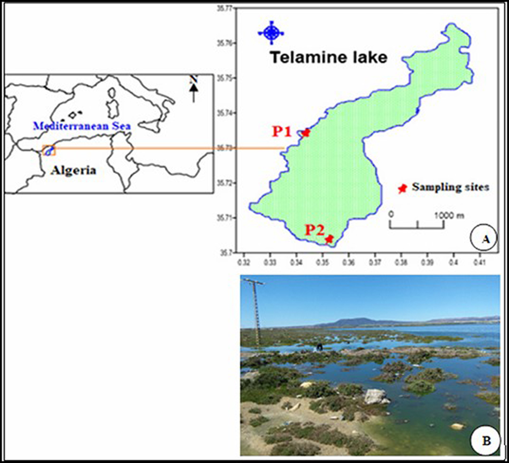
Telamine brackish water Lake is located in northwest Algeria, approximately 20 km East of Oran city and exactly northwest of the Arzew salt flats. It is located at 35º 42′ 32′′ North and 00º 22′ 30′′ West, and it is approximately 8.5 km long and 0.5 to 1.5 km wide (Fig. 1 A ). The Telamine Lake has a surface area of 2.399 ha and a perimeter of 20 km. The majority of its water is alkaline, with a pH range of 7.43 to 8.95 (Hadjadj, 2018). Telamine Lake is a protected wetland (Ramsar list on 12/12/2004) due to its high animal and plant biodiversity.
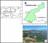
(A) Site description and geographic location of Telamine Lake. (B) Phytoplankton bloom in Telamine Lake (April 2018).
A phytoplankton bloom (Fig. 1 B ) of cyanobacteria (Arthrospira sp., Phormidium sp., and Microcystis sp.), diatoms (Navicula sp.), and chlorophyte (Chlorella sp.) were also observed. Water samples were collected from Telamine Lake about 30 cm below the surface using a phytoplankton net with a mesh size of 50 µm. Samples were collected at two locations: p1 (35°43’53.9 “N 0°23’17.7” W) and p2 (35°42’12.9 “N 0°23’38.2” W). The samplings were completed at the end of April 2018. The samples were immediately transported to the laboratory for analysis. The microbiology laboratory at the National Center for Research and Development of Fisheries and Aquaculture (CNRDPA), Bou Ismail, Algeria, handled the samples and isolated the cyanobacteria. We performed centrifugation at 1000 rpm for 2 min as a first treatment step to remove unwanted microorganisms and solid residues from a sample, then one hundred microliters of the sample were spread aseptically on the surface of Petri dishes containing BG11 (Allen & Stanier, 1968) sterile solid media with different NaCl concentrations (0, 5, 15, and 25 g/l), and the pH was adjusted to 9.5. The plates were incubated at room temperature and illuminated 24 hours a day with 1 Klux light-emitting diodes (LEDs, ABALIGHT 18 W, 220V, 50 HZ). Individual cyanobacteria colonies formed on the surface of the agar medium were picked up and streaked in new, similar solid medium under aseptic conditions. This procedure was repeated several times until pure isolates were obtained.
Description and morphological measurements of the isolated strain
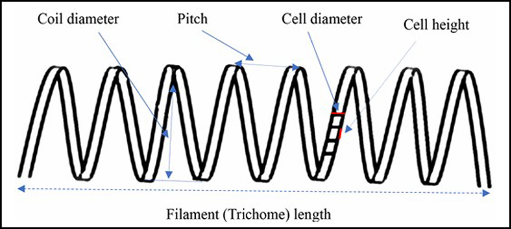
Using a Zeiss microscope connected to a camera (CMEX1, DC 1300C), the isolates were observed and examined directly to try to detect the Limnospira strains morphologically similar to the TL03-like isolate, as described in the literature (Komárek & Anagnostidis, 2005; Sili et al., 2012; Nowicka-Krawczyk et al., 2019). The morphological properties of 610 filaments (trichomes) were measured using the method depicted in Fig. (2). The diameter and the length of the cell (Kaggwa et al., 2013) were measured at 1000 x magnification, the coil diameter and pitch at 400 x magnification, and the filament length at 100 x.
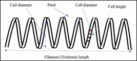
Schematic diagram for cell and filament morphological measurements of Arthrospira (Kaggwa et al., 2013).
Molecular identification
DNA extraction
Two mL of cyanobacterium culture were mixed with 2 µL of Poloxamer (https://www.sigmaaldrich.com/catalog/product/sial/p2164009?lang=fr®ion=FR). After 10 min of centrifugation at 11,000 g, the supernatant was removed, and the DNA was extracted according to the kit’s instructions: http://www.mn-net.com/media/pdf/5b/d0/d9/Instruction-NucleoSpin-Tissue.pdf.
PCR amplification and sequencing
The PCR amplification (polymerase chain reaction) for 16S rRNA gene fragments was carried out using the universal bacterial primers 27F (5′- AGA-GTT-TGA-TCC-TGG-CTC-AG -3′) and 1492R (5′- GGT-TAC-CTT-GTT-ACG-ACT-T -3′) (Lane, 1991). The PCR reaction mixture contained 15 μl of Phusion, 0.9 µl DMSO, 0.75 µl of each primer, 11.6 µL of deionized water, and 1 µl of DNA genome for a total volume of 30 µl of the reaction mixture. The PCR conditions were as follows: 1 cycle at 98 °C for 30 s, followed by 35 cycles of 98 °C for 30 s, 58 °C for 30 s, 72 °C for 1 min, and a final extension at 72 °C for 10 min. PCR was used to amplify the 16SrRNA gene sequence from isolated genomic DNA using the Applied Biosystems GeneAmp PCR 9700 Thermal Cycler. PCR products were examined on 0.8% agarose gel stained with ethidium bromide, and the migration was performed in an electrophoresis chamber. The lecture was done on an ImageQuant Las 4000 (GE HealthCare).
The PCR products were purified using Exosap-it PCR product clean-up (https://www.thermofisher.com/fr/fr/home/life-science/sequencing/sanger-sequencing/sanger-sequencing-kits-reagents/exosap-it-pcr-product-cleanup.html) and reamplified using primers specific to cyanobacteria: forward primer 359F (GGG GAA TYT TCC GCA ATG GG) and reverse primer 781R(GAC-TAC-WGG-GGT-ATC-TAA-TCC-CWT-T) (Nübel et al., 1997; Lau et al., 2005).
Exosap mixture for 1 primer contains 2 µl of the PCR gene product, 1 µl ExoSap-IT, and 2 µl Eau millliQ, incubated at 37 °C for 4 min and 80 °C for 1 min and directly sequenced in both directions with primers 27F, 359F, 1492R, 781R. The sequencing was performed by Macrogen (https://dna.macrogen-europe.com/eng/). The sequences were assembled in the software Geneious.
Phylogenetic analysis
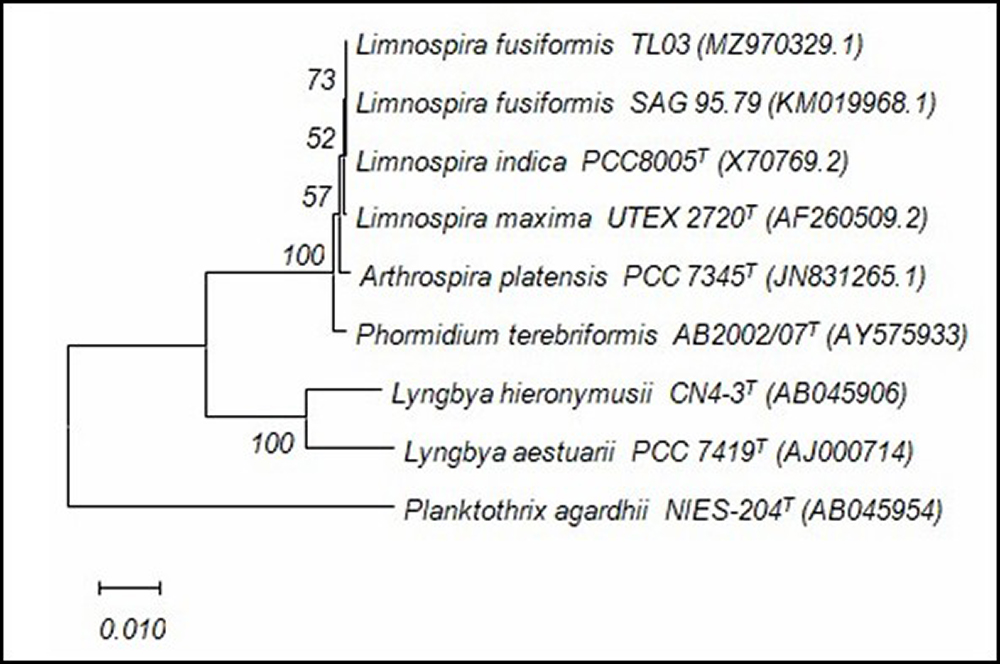
Using the Basic Local Alignment Search Tool (BLAST), the 16S rRNA gene sequences were aligned with reference sequences obtained from the National Center for Biotechnology Information database (NCBI database, http://www.ncbi.nlm.nih.gov/). The alignments were manually checked and edited in the BioEdit sequence alignment editor (version 7.2.6.1). The neighbor-joining (NJ) algorithm and bootstrap resampling (1000 replicates) were used for phylogenetic analysis and tree construction. Next to the branches is the percentage of replicate trees (numbers at nodes) in which the associated taxa clustered together in the bootstrap test (Felsenstein, 1985). The MEGA package database was used to build the phylogenetic tree, and the ClustalW algorithm with the MEGA version was used (Kumar et al., 2018). The evolutionary distances were calculated using the Maximum Composite Likelihood method (Tamura et al., 2004) and are in base substitutions per site (Bar, 0.01 nucleotide substitution per nucleotide position). The tree was established using Planktothrix agardhii NIES-204T as an outgroup. The 16S rRNA gene sequences were entered into the GenBank database.
GenBank accession numbers and taxonomic data for all species included in the 16S rRNA gene sequence alignments in the present study are listed in Table 1.
taxonomy ID, and NCBI GenBank accession number of taxa in 16S rRNA gene sequence alignments in this study.
Culturing conditions
The isolated strain was maintained and cultured in BG11 medium (Allen & Stanier, 1968), containing the following nutrients in g/l: NaNO3, 1.5; K2HPO4, 0.04; MgSO4.7H2O, 0.075; CaCl2.2H2O, 0.036; citric acid, 0.006; ferric ammonium citrate, 0.006; Na2EDTA, 0.001; Na2CO3, 0.02; and 1 ml of trace element mix was added (containing in g/l: H3BO3, 2.86; MnCl2.4H2O, 1.81; ZnSO4.7H2O, 0.222; NaMoO4.2H2O, 0.39; CuSO4.5H2O, 0.079; Co(NO3)2.6H2O, 0.0494). The pH was adjusted to 9.5. The media were autoclaved at 121º C for 20 min. The isolated strain was maintained in a liquid BG11 medium containing 5 g/l of NaCl and subcultured in the same fresh medium every month. The experiment on the effect of salinity on L. fusiformis TL03 was done in 500-ml Erlenmeyer flasks with 250 ml of fresh BG11 medium and different NaCl concentrations (0, 5, 15, 25, 35, 45, and 60 g/l). Before sterilizing at 121 °C for 20 min, the pH of all media was adjusted to 9.5. All culture media were uniformly inoculated by adding 25 mL of homogenized cell culture (stock culture) with an initial optical density of 0.220 ± 0.002 at 750 nm.
This experiment uses BG11 with 15 g/l NaCl, the same salinity as its natural habitat, as a control. The cultures were incubated in a culture chamber at a temperature of 27 ± 2 °C, with continuous illumination provided by light-emitting diodes (LEDs) with an intensity of (2.7 ± 0.2 Klux). A Mini Light Meter UT383 was used to measure the intensity of the light. The culture was continuously agitated with an air pump to keep cells suspended and prevent biomass cell agglomeration. Four replicates were used in the experiments. The cultures were kept under these conditions for 20 days.
Growth and pigment measurements
Every four days, a sample of the homogeneous suspensions of cyanobacterial culture was taken from each Erlenmeyer flask and analyzed for growth, chlorophyll a, chlorophyll b, and total carotenoids. Optical density at 750 nm was used to calculate cell growth every four days during the incubation period. Other than that, chlorophyll (a, b) and carotenoids were measured in accordance with Wellburn (1994). Volumes of 3 ml of each culture medium’s homogenized cell suspension were centrifuged at 3800 rcf for 10 min (Centurion Scientific Ltd, C2 series Benchtop Centrifuges). After centrifugation at the same speed, the pellets were washed twice with 3 ml of distilled water. The supernatant was removed, and the cells were suspended in 3 mL of pure methanol (99.9%) in the dark at 4 °C for 24 h (until colorless pellets). After centrifugation, the aliquots were read at three different wavelengths (447, 653, and 666 nm) using a Jasco V-630 Spectrophotometer. Wellburn (1994) provided the equation for calculating chlorophylls and carotenoids.
Chlorophyll a (µg/ml) = 15.66x (A666) - 7.34x (A653)
Chlorophyll b (µg/ml) =27.05 x (A653) - 11.21 x (A666)
Total carotenoids (μg/ml) = 1000 × (A470 - 2.86 × Chl a - 129.2 × Chl b)/221
Statistical analyses
The data from this study were statistically analyzed using IBM SPSS software (version 25) (SPPS I 2013). To verify if there were statistically significant differences in the growth of L. fusiformis TL03 under different concentrations of sodium chloride, the data of each parameter were subjected to One-Way ANOVA and Tukey’s HSD (Honest Significant Difference) tests using SPSS with a 95% confidence interval.
Results
Morphological characteristics of the isolated strain
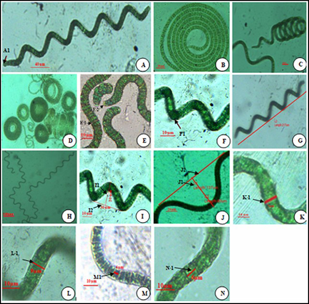
Based on the microscopic examination of the strain isolated from the current study, we recorded several characteristics and morphological features, among which a blue-green color, filamentous form, and unbranched and free-floating trichomes. The filaments are motile, gliding along their axis. The filaments are divided by the cross-wall of the cell into multiple cylindrical cells arranged in trichomes in an open helix with a regular helical coil, gradually narrowing slightly towards the ends (Fig. 3 A ). Morphological modification from loosely coiled trichomes to tightly reversed coils was observed. The isolated strain showed various degrees of coiling, from loosey coiled S-type (Fig. 3 A ) to intermediate C-type (Fig. 3 B -C) to highly coiled H-type (Fig. 3 D )
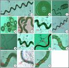
A-N Microscopic view of various morphological features of the isolated Limnospira fusiformis TL03 strain. (A.1) Trichome type C with attenuation at the end (100 X). (B-C) Tightly coiled trichome (400 X). (D) Tightly coils H shape (100X). (E.1) Cell cross wall. (E2 2) Caliptra at trichome end (1000 X). (F1) Gas vacuole (1000 X). (G-H) Smallest and tallest trichome with loose coils (100 X). (I1) Small coil diameter (400 X). (I2). Small pitch (400 X). (J1) Large coil diameter (400 X). (J2). Large pitch (400 X). (K1-L1). Cell diameter. (M1. N1) (1000 X) Cell length (1000 X).
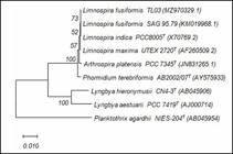
Phylogenetic tree based on 16S rRNA gene sequences showing the relationship between strain TL03 and related type-strains. The evolutionary distances were computed using the Maximum Composite Likelihood method and are in the units of the number of base substitutions per site (Bar, 0.01 nt substitution per nt position). The evolutionary history was inferred using the Neighbor-Joining method. The optimal tree is shown. The percentage of replicate trees (numbers at the nodes) in which the associated taxa clustered together in the bootstrap test (1000 replicates) are shown next to the branches. This analysis involved nine nucleotide sequences.
Cell cross-walls (Fig. 3 E1 ), calyptras at the end of the trichome (Fig. 3 E2 ), and gas vacuoles (Fig. 3 F1 ) were visible under a light microscope.
The results of the measurements of 610 filament morphology are recorded in Table 1 and accompanied by photographs of the morphological features of this cyanobacterium (Fig. 3). The results indicate that the length, diameter, and pitch of the trichome range from 210 to 2027 µm (Fig. 3G-H), 20 to 60 µm (Fig. 3 I1 -J1), and 30 to 137 µm (Fig. 3 I2 -J2), respectively The diameters and lengths of the trichome’s cells range from 7 to 9 µm (Fig.3K1-L1) and 4 to 6 µm (Fig. 3 M1 -N1), respectively.
Molecular characteristics of the isolated strain
The molecular studies based on the 16S rRNA gene allowed us to identify the newly isolated strain as Limnospira fusiformis TL03 with the accession number MZ970329. The partial sequence (1,411 bp length) of the Limnospira fusiformis TL03 16S ribosomal RNA gene was compared to all partial sequences presented in NCBI. MegaBlast searches revealed that the isolated strain matched with 100% similarity to Limnospira fusiformis SAG 85.79 (accession number: KM019968.1) (Fig. 4).
Effects of different NaCl concentrations on the growth of the isolated L. fusiformis TL03
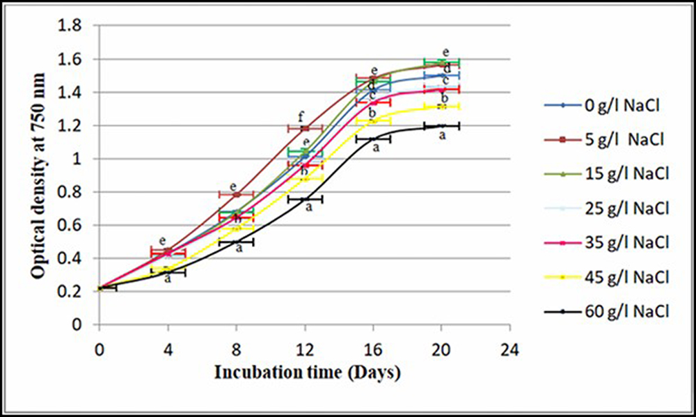
Figure 5 depicts the growth of L. fusiformis TL03 from the start to the end of the experiment for all treatments. The L. fusiformis TL03 strain demonstrated a high level of salt tolerance. The general trend of the curve shows that the highest optical density values for all treatments occurred on the 20th day, whereas the concentration of NaCl had a significant effect on cell growth across all treatment groups (P < 0.05). The maximum optical densities (1.580 ± 0.014 and 1.560 ± 0.003) were obtained in both cultures containing 15 g/l and 5 g/l NaCl, respectively, seven times greater than the initial optical density. In contrast, the lowest optical densities (1.314 ± 0.006 and 1.197 ± 0.003) were recorded in cultures containing 45 and 60 g/l NaCl, respectively.
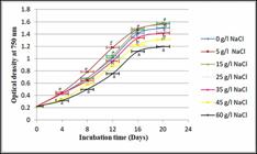
Effect of different NaCl concentrations on the growth of L. fusiformis TL03. Data represent mean values ± SEM (n = 4).
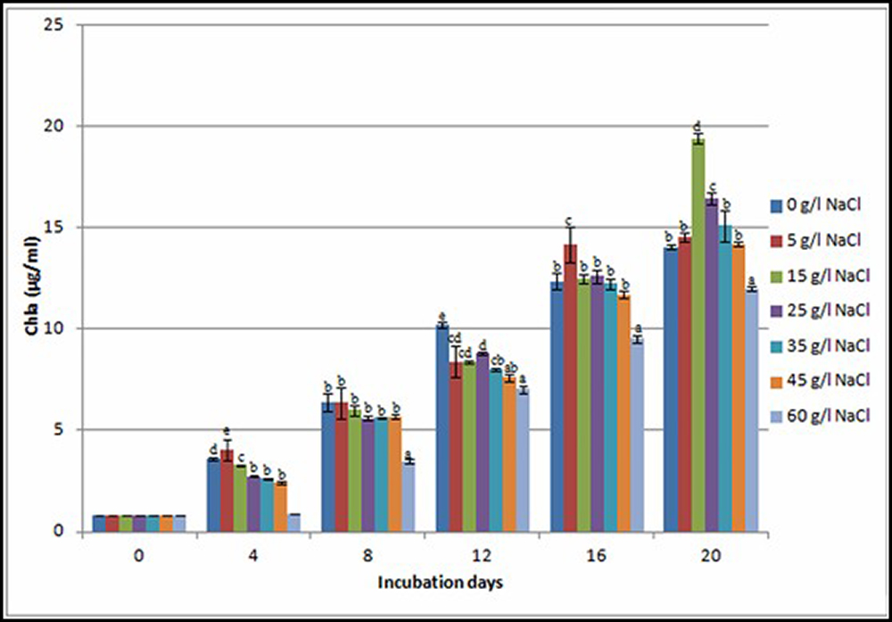
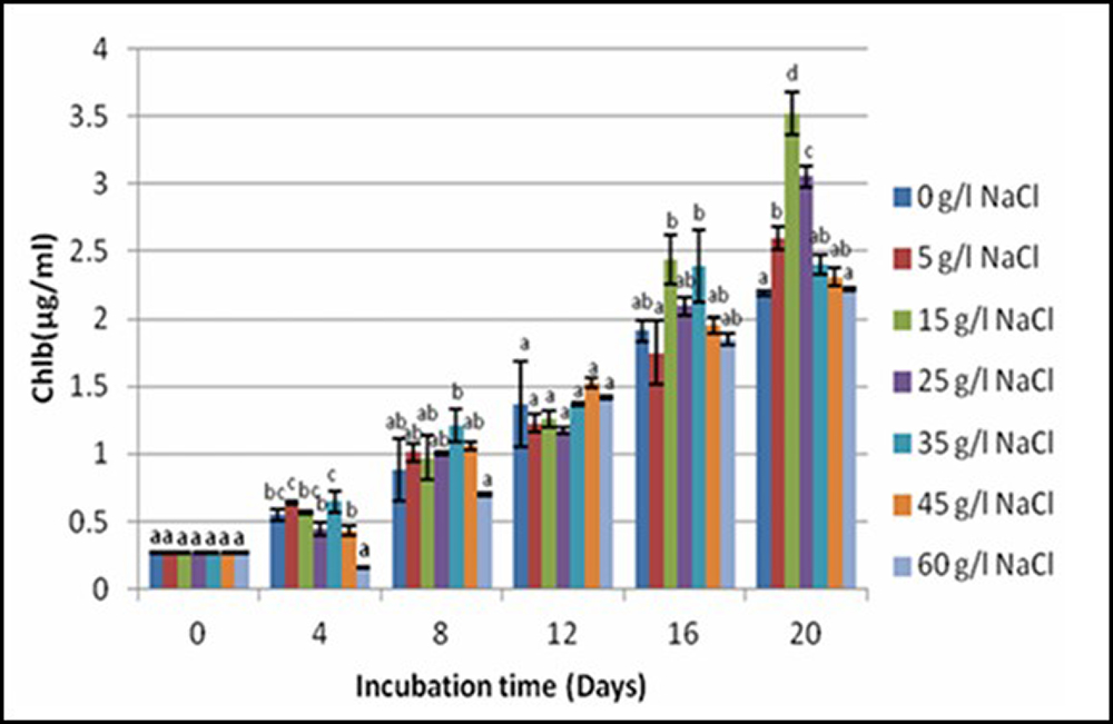
These results show that when the concentration of sodium chloride increases, the growth of L. fusiformis TL03 slows down. We also remarked that L. fusiformis TL03 continued to grow even under NaCl starvation conditions. The results of chlorophyll a content in the L. fusiformis TL03 strain subjected to salt stress for 20 days showed a gradual increase among all NaCl concentrations tested (Fig. 6). By comparing the trend of the curves of the effect of salinity on growth and the concentration of chlorophyll, it becomes clear that they follow a similar trend. Growth and chlorophylls (a, b) were negatively affected at the highest salt concentrations. Nevertheless, the TL03 strain was still able to grow until the 20th day, which was the end of the experiment. The highest concentrations of chlorophyll a (Fig. 6) and chlorophyll b (Fig. 7) in all tested cultures were observed at the end of the experiment (20th day). The maximum concentrations of chlorophyll a (19.42 ± 0.25 µg/ml) and chlorophyll b (3.52 ± 0.16 µg/ml) were obtained in BG11 containing 15 g/l NaCl. The lowest amounts of Chl a (12 ± 0.08 µg/ml) and Chl b (2.22 ± 0.01 µg/ml) were registered in BG11, which contained a high NaCl concentration (60 g/l).
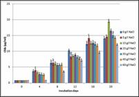
Chlorophyll a amount of L. fusiformis TL03 exposed to different NaCl concentrations. Different letters indicate a significant difference (P< 0.5). Error bar represents standard error (n = 4).
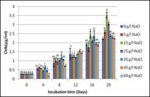
Chlorophyll b amount of L. fusiformis TL03 strain exposed to different NaCl concentrations. Different letters indicate a significant difference (P< 0.5). Error bar represents standard error (n = 4).
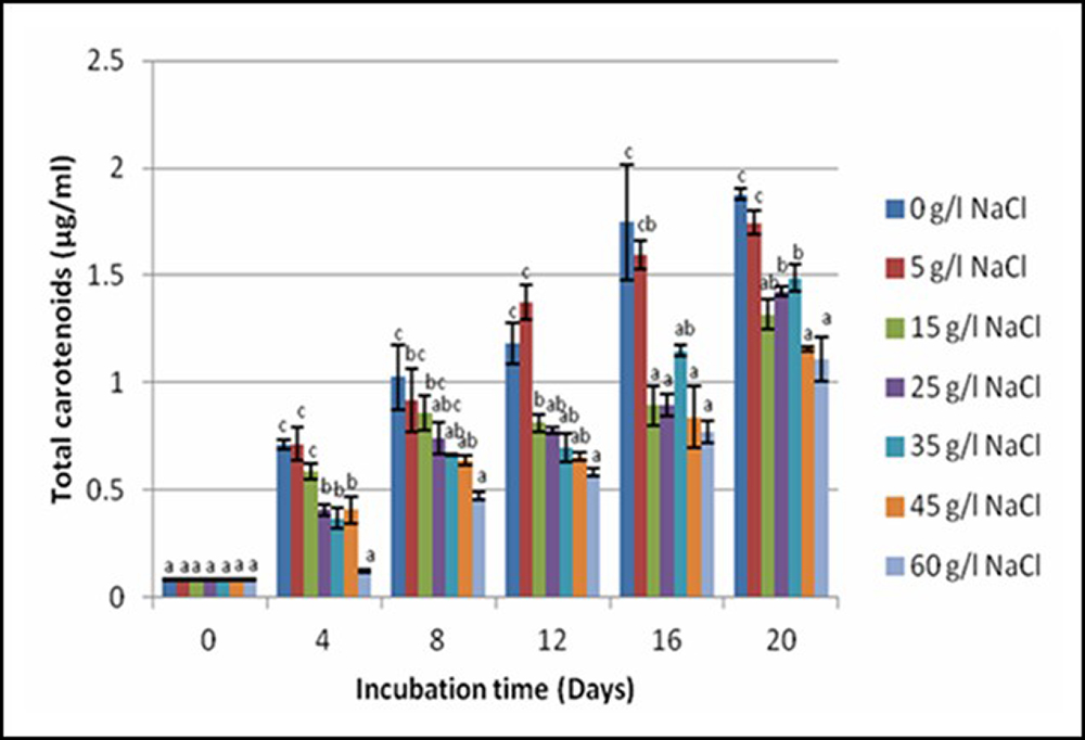
The variation of total carotenoids in the L. fusiformis TL03 strain under different NaCl concentrations demonstrated a continuing increase in all treatment groups, reaching a maximum on the 20th day of the experiment, with a significant difference for all treatment media (P < 0.05) (Fig. 8). The medium without NaCl yielded the highest number of carotenoids, a 23-fold increase over the initial value. At the same time, carotenoid concentration ranged from the highest value of 1.88 ± 0.02 µg/ml in BG11 without NaCl to the lowest value of 1.10 ± 0.01 µg/ml under high salt stress (60 g/l). The total carotenoid concentration was lower in the TL03 strain cultured at 15 g/l NaCl (control) compared to those cultured at 25 and 35 g/l and was higher compared to those cultured in higher salinity culture media (45 and 60 g/l). On the fourth day of the experiment, the maximum concentrations of carotenoids (0.71 ± 0.02 and 0.71 ± 0.08 µg/ml) were observed in BG11 and BG11 containing 5 g/l of NaCl, respectively.
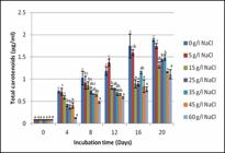
Total carotenoids amount of L. fusiformis TL03 strain exposed to different NaCl concentrations. Different letters indicate a significant difference (P< 0.5). Error bar represents standard error (n = 4).
Discussion
The presence of numerous cyanobacteria and microalgae, dominated by the L. fusiformis TL03 strain, was revealed by microscopic examination of the sample. This finding is consistent with the findings of Krienitz et al. (2005) and Sili et al. (2012), who found that in alkaline saline lakes in tropical and subtropical regions, the alkaliphilic filamentous cyanobacterium Arthrospira is frequently the dominant taxon and is responsible for permanent or seasonal blooms. In this study, we used the traditional method based on morphological and molecular characteristics to identify the genus of the isolated strain. The arrangement of its multicellular cylindrical trichomes in an open helix, usually of relatively large diameter, sometimes attenuated at the ends, is the main taxonomic criteria for the differentiation of both genus Arthrospira and Limnospira species (Sili et al., 2012).
A morphological study of the isolated strain revealed that there are several morphotypes that most likely belong to the same species. During successive cultures in liquid media, this isolated strain maintained a spiral shape with varying degrees of helicity. The results obtained by measuring trichome length, coil diameter, and pitch, as well as cell diameter and length (Table 2), differ slightly from those reported by Jung et al. (2021). Cell diameters of Arthrospira platensis grown in a flat-type bioreactor ranged from 5.25 ± 1.26 to 7.5 ± 2.9 µm, and coil diameters ranged from 20 to 60 µm.
Range of dimensional variation of different measurements of morphology features of Limnospira fusiformis TL03
Many studies have been conducted to describe the morphological characteristics of A. fusiformis and A. maxima. The diameter and length of the cells of the latter species range from 7 to 9 μm and 5 to 7 μm, respectively (Sili et al., 2012). They form open regular spirals with coil diameters ranging from 40 to 60 µm and pitches ranging from 70 to 80 µm. Coils have regular screw-like shapes at the ends that are slightly attenuated (Gardner, 1917; Fott & Karim, 1973; Komárek & Anagnostidis, 2005; Sili et al., 2012). Rout et al. (2015) isolated the Arthrospira strain NPS-011, which has the same cell trichome length and diameter as previously studied strains. This species has a variable helix pitch ranging from 80 to 150 µm and a helix diameter ranging from 30 to 70 μm. The results for filament length, coil diameter, pitch, cell diameter, and length for our isolate (Table 2) show that they are almost in agreement with those reported in several previous studies (Sanchez et al., 2003; Komárek & Anagnostidis, 2005; Rout et al., 2015; Cellamare et al., 2018; Roussel et al., 2023).
Sili et al. (2012) noted that the Arthrospira helix architecture (pitch and diameter) is highly dependent on growth and environmental conditions under laboratory and mass cultivation conditions. Dhiab et al. (2007) found that increasing the concentration of sodium chloride in the culture medium caused Arthrospira platensis to change its trichome shape from spiral to straight, as well as its physiological behavior. The salinity and anion content of the culture medium, according to Kebede (1997), influence the helicity degree of Arthrospira fusiformis. The authors observed that higher salinity culture media had the longest pitches, with very lax helices in media containing high concentrations of SO4 2- compared to those containing the same concentrations of HCO₃⁻ and Cl⁻.
The phylogenetic trees matched the classifications based on the morphological characteristics of the isolates in this study very well. The data from the 16S rRNA gene sequence analysis confirmed that the isolated strain Tl03 (MZ215991) belongs to Limnospira fusiformis, with a 100% similarity to L. fusiformis SAG 85.79 (KM019968.1). Despite the high salinity of the culture medium (60 g/l), the L. fusiformis TL03 strain showed tolerance and continued growth in culture media with varying salinity rates. These findings are consistent with those reported by Reed & Stewart (1985), Kebede (1997), and Moisander et al. (2002), who found that A. platensis did not stop growing even at 88 g/l salinity. According to Dadheech et al. (2010), Arthrospira lives in alkaline waters with pH ranging from 8 to 11 and salinity ranging from 1.1 to 300 g/l.
When compared to other media, the L. fusiformis TL03 isolates grew the best in both media containing 5 and 15 g/l NaCl. The salinity of the BG11, which contains 15 g/l NaCl, is thought to be similar to that of our strain’s original habitat (Telamine Lake). BG11 with 5 g/l NaCl has the same salinity concentration as when the strain was grown and adapted before being used in this experiment. In terms of optical density, chlorophylls (a, b) levels increased slowly in BG11 containing up to 15 g/l NaCl (control) during incubation compared to a lower salinity medium. These findings differ from those of several previous studies. According to Ayachi et al. (2007), Spirulina platensis (A. platensis) grew the fastest at a NaCl concentration of 1 g/l. Meanwhile, at 60 g/l NaCl, the first five days are latent, followed by a decreasing growth phase until the end of the experiment.
Phang (2002), on the other hand, described an immediate cessation of A. platensis growth when exposed to high NaCl concentrations. Slower growth usually follows this stage. Sudhir et al. (2005) discovered that a 47 g/l NaCl concentration significantly reduced A. platensis’s photosystem II (PS II)-mediated oxygen evolution activity. Sharma et al. (2014) reported that increasing the NaCl concentration reduced growth and completely inhibited A. platensis chlorophyll a biosynthesis. The total carotenoid content of L. fusiformis TL03 grown in a low-salinity medium was significantly higher than in the control medium but lower in higher-salinity media (45 and 60 g/l). This fact could be explained as follows: exposing L. fusiformis TL03 to hypersalinity and a slightly higher salinity compared to the Arthrospira origin environment induced carotenoids biosynthesis, but the biosynthesis was lower at a higher salinity rate.
This study reported the identification and isolation of a novel strain of cyanobacteria, L. fusiformis TL03, from Telamine Lake in northwest Algeria. This strain was first discovered by Fox (1996) in Tamanrasset Pond, located in the southern region of Algeria. Through the examination of morphological characteristics and comparing the 16S rRNA gene sequence of the isolated strain with other Arthrospira or Spirulina strains, we verified that the strain in question is indeed a member of the Limnospira genus. The strain was designated as Limnospira fusiformis TL03 and was submitted to the GenBank database with the accession number MZ215991.1.
The strain exhibited the ability to acclimate to varying levels of salinity, ranging from 0 to 60 g/l NaCl. This adaptability enables its cultivation using seawater, brackish water from lakes, and untapped subterranean saline water sources in Algeria. Additionally, it thrives in alkaline aquatic environments, such as alkaline lakes, where its exposure to contamination from microalgae and other microorganisms is minimized in outdoor cultivation systems such as ponds, basins, tanks, circular ponds, and raceway ponds. This strain exhibits elongated trichomes measuring up to 2027 μm in length, featuring spiral shapes that facilitate efficient harvesting through the filtration method. This method is particularly advantageous for commercial production due to its cost-effectiveness compared to alternative harvesting techniques. Due to its adaptation to the local climatic conditions, this strain can be cultivated in outdoor ponds.
Acknowledgments
We would like to express our gratitude to our colleague, Mr. Kamel BOUDJEMA, as well as all those who played a role, whether direct or indirect, in the successful completion of this research paper.
References
-
Abed RM, Dobretsov S, Sudesh K. 2009. Applications of cyanobacteria in biotechnology. Journal of Applied Microbiology 106: 1-12. doi: 10.1111/j.1365-2672.2008.03918.x.
» https://doi.org/10.1111/j.1365-2672.2008.03918.x. -
Aladaileh SH, Khafaga AF, Abd El-Hack ME et al 2020. Spirulina platensis ameliorates the sub-chronic toxicities of lead in rabbits via anti-oxidative, anti-inflammatory, and immune stimulatory properties. Science of the Total Environment 701: 134879. doi: 10.1016/j.scitotenv.2019.134879.
» https://doi.org/10.1016/j.scitotenv.2019.134879. -
Allen MM, Stanier RY. 1968. Growth and division of some unicellular blue-green algae. Microbiology 51: 199-202. doi: 10.1099/00221287-51-2-199.
» https://doi.org/10.1099/00221287-51-2-199. - Asulabh KS, Supriya G, Ramachandra TV. 2012. Effect of salinity concentrations on growth rate and lipid concentration in Microcystis sp., Chlorococcum sp., and Chaetoceros sp. In: National Conference on Conservation and Management of Wetland Ecosystems, 6-9 November 2012, Kottayam/Kerala. p 1-7.
-
Ayachi S, El Abed A, Dhifi W, Marzouk B. 2007. Chlorophylls, proteins, and fatty acids amounts of Arthrospira platensis growing under saline conditions. Pakistan Journal of Biological Sciences 10: 2286-2291. doi : 10.3923/pjbs.2007.2286.2291.
» https://doi.org/10.3923/pjbs.2007.2286.2291. -
Babadzhanov AS, Abdusamatova N, Yusupova FM, Faizullaeva N, Mezhlumyan LG, Malikova MK. 2004. Chemical Composition of Spirulina platensis Cultivated in Uzbekistan. Chemistry of Natural Compounds 40: 276-279. doi: 10.1023/B:CONC.0000039141.98247.e8.
» https://doi.org/10.1023/B:CONC.0000039141.98247.e8. -
Benavente-Valdés JR, Aguilar C, Contreras-Esquivel JC, Méndez-Zavala A, Montañez J. 2016. Strategies to enhance the production of photosynthetic pigments and lipids in Chlorophyceae species. Biotechnology Reports 10: 117-125. doi: 10.1016/j.btre.2016.04.001.
» https://doi.org/10.1016/j.btre.2016.04.001. -
Borowitzka MA. 1995. Microalgae as sources of pharmaceuticals and other biologically active compounds. Journal of Applied Phycology 7: 3-15. doi:10.1007/BF00003544.
» https://doi.org/10.1007/BF00003544. -
Cellamare M, Duval C, Drelin Y et al 2018. Characterization of phototrophic microorganisms and description of new cyanobacteria isolated from the saline-alkaline crater-lake Dziani Dzaha (Mayotte, Indian Ocean). FEMS Microbiology Ecology 94: fiy108. doi: 10.1093/femsec/fiy108.
» https://doi.org/10.1093/femsec/fiy108. -
Cho JA, Baek SY, Cheong SH, Kim MR. 2020. Spirulina enhances bone modeling in growing male rats by regulating growth-related hormones. Nutrients 12: 1187. doi: 10.3390/nu12041187.
» https://doi.org/10.3390/nu12041187. -
Choi WY, Kang DH, Lee HY. 2013. Enhancement of immune activation activities of Spirulina maxima grown in deep-sea water. International Journal of Molecular Sciences 14: 12205-12221. doi: 10.3390/ijms140612205.
» https://doi.org/10.3390/ijms140612205. -
Costa JAV, Colla LM, Duarte Filho PF. 2004. Improving Spirulina platensis biomass yield using a fed-batch process. Bioresource Technology 92: 237-241. doi:10.1016/j.biortech.2003.09.013.
» https://doi.org/0.1016/j.biortech.2003.09.013. -
Costa NB, Kolman MA, Giani A. 2016. Cyanobacteria diversity in alkaline saline lakes in the Brazilian Pantanal wetland: A polyphasic approach. Journal of Plankton Research 38: 1389-1403. doi: 10.1093/plankt/fbw066.
» https://doi.org/10.1093/plankt/fbw066. -
D’Alessandro EB, Antoniosi Filho NR. 2016. Concepts and studies on lipid and pigments of microalgae: A review. Renewable and Sustainable Energy Reviews 58: 832-841. doi: 10.1016/j.rser.2015.12.162.
» https://doi.org/10.1016/j.rser.2015.12.162. -
Da Silva SC, Fernandes IP, Barros L et al 2019. Spray-dried Spirulina platensis as an effective ingredient to improve yogurt formulations: Testing different encapsulating solutions. Journal of Functional Foods 60: 103427. doi: 10.1016/j.jff.2019.103427.
» https://doi.org/10.1016/j.jff.2019.103427. -
Dadheech PK, Ballot A, Casper P et al 2010. Phylogenetic relationship and divergence among planktonic strains of Arthrospira (Oscillatoriales, Cyanobacteria) of African, Asian and American origin deduced by 16S-23S ITS and phycocyanin operon sequences. Phycologia 49: 361-372. doi: 10.2216/09-71.1.
» https://doi.org/10.2216/09-71.1. -
Dhiab RB, Ouada HB, Boussetta H, Franck F, Elabed A, Brouers M. 2007. Growth, fluorescence, photosynthetic O2 production and pigment content of salt adapted cultures of Arthrospira (Spirulina) platensis Journal of Applied Phycology 19: 293-301. doi: 10.1007/s10811-006-9113-z.
» https://doi.org/10.1007/s10811-006-9113-z. - Falaise C. 2019. Valorisation des activités biologiques de la diatomée marine Haslea ostrearia PhD Thesis, Universite Maine, French.
-
Felsenstein J. 1985. Confidence limits on phylogenies: An approach using the bootstrap. Evolution 39: 783-791. doi: 10.2307/2408678.
» https://doi.org/10.2307/2408678. - Fott B, Karim AG. 1973. Spirulina plankton community in a lake in Jebel Marra, Sudan. Archiv für Protistenkunde 115: 408-418.
- Fox RD. 1996. Spirulina: production & potential. Aix-en-Provence, Edisud.
-
Furmaniak MA, Misztak AE, Franczuk MD, Wilmotte A, Waleron M, Waleron KF. 2017. Edible cyanobacterial genus Arthrospira: Actual state of the art in cultivation methods, genetics, and application in medicine. Frontiers in Microbiology 8: 2541. doi: 10.3389/fmicb.2017.02541.
» https://doi.org/10.3389/fmicb.2017.02541. - Gardner NL. 1917. New Pacific coast marine I. University of California Publications in Botany 6: 377-416.
-
Ghaeni M, Roomiani L. 2016. Review for application and medicine effects of Spirulina, microalgae. Journal of Advanced Agricultural Technologies 3: 114-117. doi: 10.18178/joaat.3.2.114-117.
» https://doi.org/10.18178/joaat.3.2.114-117. - Hadjadj M. 2018. Suivi des indicateurs de pollution dans une zone aride à semi-aride: Diagnostic des environs de Gdyel suite aux dépôts des déchets ménagers et rejet d'eau usée au niveau du lac Télamine. Magister Thesis, Université d’Oran, Algeria.
-
Hirooka S, Higuchi S, Uzuka A, Nozaki H, Miyagishima SY. 2014. Acidophilic green alga Pseudochlorella sp. YKT1 accumulates high amount of lipid droplets under a nitrogen-depleted condition at a low-pH. PLoS One 9: e107702. doi: 10.1371/journal.pone.0107702.
» https://doi.org/10.1371/journal.pone.0107702. -
Jiménez C, Cossı́o BR, Labella D, Niell FX. 2003. The feasibility of industrial production of Spirulina (Arthrospira) in Southern Spain. Aquaculture 217: 179-190. doi: 10.1016/s0044-8486(02)00118-7.
» https://doi.org/10.1016/s0044-8486(02)00118-7. -
Jung CH, Braune S, Waldeck P, Küpper JH, Petrick I, Jung F. 2021. Morphology and Growth of Arthrospira platensis during Cultivation in a Flat-Type Bioreactor. Life 11: 536. doi: 10.3390/life11060536.
» https://doi.org/10.3390/life11060536. -
Jung F, Krüger-Genge A, Waldeck P, Küpper JH 2019. Spirulina platensis, a super food. Journal of Cellular Biotechnology 5: 43-54. doi: 10.3233/JCB-189012.
» https://doi.org/10.3233/JCB-189012. -
Kaggwa MN, Burian A, Oduor SO, Schagerl M. 2013. Ecomorphological variability of Arthrospira fusiformis (Cyanoprokaryota) in African soda lakes. Microbiology Open 2: 881-891. doi: 10.1002/mbo3.125.
» https://doi.org/10.1002/mbo3.125. -
Kebede E. 1997. Response of Spirulina platensis (Arthrospira fusiformis) from Lake Chitu, Ethiopia, to salinity stress from sodium salts. Journal of Applied Phycology 9: 551-558. doi: 10.1023/A:1007949021786.
» https://doi.org/10.1023/A:1007949021786. - Komárek J, Anagnostidis K. 2005. Cyanoprokaryota. 2. Teil: Oscillatoriales. In: Büdel B, Gärtner G, Krienitz L, Schagerl M (eds.). Süßwasserflora von Mitteleuropa, Bd. 19/2. München, Elsevier GmbH. p 341-353.
-
Kumar S, Stecher G, Li M, Knyaz C, Tamura K. 2018. MEGA X: Molecular evolutionary genetics analysis across computing platforms. Molecular Biology and Evolution 35: 1547-1549. doi: 10.1093/molbev/msy096.
» https://doi.org/10.1093/molbev/msy096. -
Koyandea AK, Chewa KW, Rambabub K, Taoc Y, Chud DT, Showa PL. 2019. Microalgae: A potential alternative to health supplementation for humans. Food Science and Human Wellness 8: 16-24. doi: 10.1016/j.fshw.2019.03.001.
» https://doi.org/10.1016/j.fshw.2019.03.001. - Krienitz L, Ballot A, Casper P, Kotut K, Pflugmacher S, Wiegand C. 2005. Toxic cyanobacteria and massive deaths of flamingos: What we can learn from Arthrospira mass-cultivation. Phycologia 44: 59-59.
- Lane D. 1991. 16S/23S rRNA sequencing. In: Stackebrandt E, Goodfellow M (eds.). Nucleic acids techniques in bacterial systematics. New York, Wiley. p. 115-147.
-
Lau E, Nash CZ, Vogler DR, Cullings KW. 2005. Molecular diversity of cyanobacteria inhabiting coniform structures and surrounding mat in a Yellowstone hot spring. Astrobiology 5: 83-92. doi: 10.1089/ast.2005.5.83.
» https://doi.org/10.1089/ast.2005.5.83. -
Memije-Lazaro IN, Blas-Valdivia V, Franco-Colín M, Cano-Europa E. 2018. Arthrospira maxima (Spirulina) and C-phycocyanin prevent the progression of chronic kidney disease and its cardiovascular complications. Journal of Functional Foods 43: 37-43. doi: 10.1016/j.jff.2018.01.013.
» https://doi.org/10.1016/j.jff.2018.01.013. -
Moisander PH, McClinton E, Paerl HW. 2002. Salinity effects on growth, photosynthetic parameters, and nitrogenase activity in estuarine planktonic cyanobacteria. Microbial Ecology 43: 432-442. doi: 10.1007/s00248-001-1044-2.
» https://doi.org/10.1007/s00248-001-1044-2. -
Mukhopadhyay CD. 2015. Engineering Spirulina for Enhanced Medicinal Application. In: Algal Biorefinery: An Integrated Approach. New York, Springer. p. 235-252. doi: 10.1007/978-3-319-22813-6_11.
» https://doi.org/10.1007/978-3-319-22813-6_11. -
Nowicka-Krawczyk P, Mühlsteinová R, Hauer T. 2019. Detailed characterization of the Arthrospira type species separating commercially grown taxa into the new genus Limnospira (Cyanobacteria). Scientific Reports 9: 1-11. doi: 10.1038/s41598-018-36831-0.
» https://doi.org/10.1038/s41598-018-36831-0. -
Nübel U, Garcia-Pichel F, Muyzer G. 1997. PCR primers to amplify 16S rRNA genes from cyanobacteria. Applied and Environmental Microbiology 63: 3327-3332. doi: 10.1128/aem.63.8.3327-3332.1997.
» https://doi.org/10.1128/aem.63.8.3327-3332.1997. - Phang SM. 2002. Arthrospira Stizenb. ex Gomont. Plant Resources of South-East Asia 15: 88-94.
-
Pulz O, Gross W. 2004. Valuable products from biotechnology of microalgae. Applied Microbiology and Biotechnology 65: 635-648. doi: 10.1007/s00253-004-1647-x.
» https://doi.org/10.1007/s00253-004-1647-x. -
Rastogi RP, Sinha RP. 2009. Biotechnological and industrial significance of cyanobacterial secondary metabolites. Biotechnology Advances 27: 521-539. doi: 10.1016/j.biotechadv.2009.04.009.
» https://doi.org/10.1016/j.biotechadv.2009.04.009. -
Reed RH, Stewart WDP. 1985. Osmotic adjustment and organic solute accumulation in unicellular cyanobacteria from freshwater and marine habitats. Marine Biology 88: 1-9. doi: 10.1007/bf00393037.
» https://doi.org/10.1007/bf00393037. -
Roussel T, Halary S, Duval C et al 2023. Monospecific renaming within the cyanobacterial genus Limnospira (Spirulina) and consequences for food authorization. Journal of Applied Microbiology 134: 1-15. doi: 10.1093/jambio/lxad159.
» https://doi.org/10.1093/jambio/lxad159. -
Rout NP, Khandual S, Gutierrez-Mora A, Ibarra-Montoya JL, Vega-Valero G. 2015. Divergence in three newly identified Arthrospira species from Mexico. World Journal of Microbiology and Biotechnology 31: 1157-1165. doi: 10.1007/s11274-015-1865-7.
» https://doi.org/10.1007/s11274-015-1865-7. -
Sanchez‐Baracaldo P, Hayes PK, Blank CE. 2005. Morphological and habitat evolution in the Cyanobacteria using a compartmentalization approach. Geobiology 3: 145-165. doi: 10.1111/j.1472-4669.2005.00050.x.
» https://doi.org/10.1111/j.1472-4669.2005.00050.x. - Sanchez M, Bernal-Castillo J, Rozo C, Rodríguez I. 2003. Spirulina (Arthrospira): An edible microorganism: A review. Universitas Scientiarum 8: 7-24.
-
Sharma G, Kumar M, Ali MI, Jasuja ND. 2014. Effect of carbon content, salinity and pH on Spirulina platensis for phycocyanin, allophycocyanin and phycoerythrin accumulation. Microbial and Biochemical Technology 6: 202-206. doi: 10.4172/1948-5948.1000144.
» https://doi.org/10.4172/1948-5948.1000144. -
Sili C, Torzillo G, Vonshak A. 2012. Arthrospira (Spirulina). In: Ecology of Cyanobacteria II. Dordrecht, Springer. p. 677-705. doi: 10.1007/978-94-007-3855-3_25.
» https://doi.org/10.1007/978-94-007-3855-3_25. -
Sotiroudis TG, Sotiroudis GT. 2013. Health aspects of Spirulina (Arthrospira) microalga food supplement. Journal of the Serbian Chemical Society 78: 395-405. doi: 10.2298/JSC121020152S.
» https://doi.org/10.2298/JSC121020152S. - SPSS I. 2013. IBM SPSS statistics for windows. New York/Armonk, IBM SPSS.
-
Sudhir PR, Pogoryelov D, Kovács L, Garab G, Murthy SD. 2005. The effects of salt stress on photosynthetic electron transport and thylakoid membrane proteins in the cyanobacterium Spirulina platensis BMB Reports 38: 481-485. doi: 10.5483/bmbrep.2005.38.4.481.
» https://doi.org/10.5483/bmbrep.2005.38.4.481. -
Sudhir P, Murthy SDS. 2004. Effects of salt stress on basic processes of photosynthesis. Photosynthetica 42: 481-486. doi: 10.1007/S11099-005-0001-6.
» https://doi.org/10.1007/S11099-005-0001-6. -
Sydney EB, Schafranski K, Barretti BRV et al 2019. Biomolecules from extremophile microalgae: From genetics to bioprocessing of a new candidate for large-scale production. Process Biochemistry 87: 37-44. doi: 10.1016/j.procbio.2019.09.012.
» https://doi.org/10.1016/j.procbio.2019.09.012. -
Tamura K, Nei M, Kumar S. 2004. Prospects for inferring very large phylogenies by using the neighbor-joining method. Proceedings of the National Academy of Sciences 101: 11030-11035. doi: 10.1073/pnas.0404206101.
» https://doi.org/10.1073/pnas.0404206101. - Thajuddin N, Subramanian G. 2005. Cyanobacterial biodiversity and potential applications in biotechnology. Current Science 89: 47-57.
-
Varshney P, Mikulic P, Vonshak A, Beardall J, Wangikar PP. 2015. Extremophilic micro-algae and their potential contribution in biotechnology. Bioresource Technology 184: 363-372. doi: 10.1016/j.biortech.2014.11.040.
» https://doi.org/10.1016/j.biortech.2014.11.040. -
Villaró S, Morillas-España A, Acién G, Lafarga T. 2022. Optimisation of operational conditions during the production of Arthrospira platensis using pilot-scale raceway reactors, protein extraction, and assessment of their techno-functional properties. Foods 11: 2341. doi: 10.3390/foods11152341.
» https://doi.org/10.3390/foods11152341. -
Volkmann H, Imianovsky U, Oliveira JL, Sant'Anna ES. 2008. Cultivation of Arthrospira (Spirulina) platensis in desalinator wastewater and salinated synthetic medium: Protein content and amino-acid profile. Brazilian Journal of Microbiology 39: 98-101. doi: 10.1590/S1517-838220080001000022.
» https://doi.org/10.1590/S1517-838220080001000022. -
Wellburn AR. 1994. The spectral determination of chlorophylls a and b, as well as total carotenoids, using various solvents with spectrophotometers of different resolution. Journal of Plant Physiology 144: 307-313. doi: 10.1016/S0176-1617(11)81192-2.
» https://doi.org/10.1016/S0176-1617(11)81192-2. -
Wu Q, Liu L, Miron A, Klímová B, Wan D, Kuča K. 2016. The antioxidant, immunomodulatory, and anti-inflammatory activities of Spirulina: An overview. Archives of Toxicology 90: 1817-1840. doi: 10.1007/s00204-016-1744-5.
» https://doi.org/10.1007/s00204-016-1744-5.